Review
. 2017 Feb;94(2):246-259.
doi: 10.1097/OPX.0000000000000997.

Fundus Autofluorescence in Age-related Macular Degeneration

Affiliations
Review

Fundus Autofluorescence in Age-related Macular Degeneration

Angelica Ly et al. Optom Vis Sci. 2017 Feb.

Abstract

: Fundus autofluorescence (FAF) provides detailed insight into the health of the retinal pigment epithelium (RPE). This is highly valuable in age-related macular degeneration (AMD) as RPE damage is a hallmark of the disease. The purpose of this paper is to critically appraise current clinical descriptions regarding the appearance of AMD using FAF and to integrate these findings into a chair-side reference. A wide variety of FAF patterns have been described in AMD, which is consistent with the clinical heterogeneity of the disease. In particular, FAF imaging in early to intermediate AMD has the capacity to reveal RPE alterations in areas that appear normal on funduscopy, which aids in the stratification of cases and may have visually significant prognostic implications. It can assist in differential diagnoses and also represents a reliable, sensitive method for distinguishing reticular pseudodrusen. FAF is especially valuable in the detection, evaluation, and monitoring of geographic atrophy and has been used as an endpoint in clinical trials. In neovascular AMD, FAF reveals distinct patterns of classic choroidal neovascularization noninvasively and may be especially useful for determining which eyes are likely to benefit from therapeutic intervention. FAF represents a rapid, effective, noninvasive imaging method that has been underutilized, and incorporation into the routine assessment of AMD cases should be considered. However, the practicing clinician should also be aware of the limitations of the modality, such as in the detection of foveal involvement and in the distinction of phenotypes (hypo-autofluorescent drusen from small areas of geographic atrophy).


Figures

FIGURE 1
FIGURE 1
Imaging results from a 43-year-old Caucasian female with a normal macula. Note that the normal fundus displays diffuse, homogenous autofluorescence with large retinal blood vessels and the optic disc appearing as shadows of hypo-autofluorescence. The central macula shows a gradual reduction in autofluorescence approaching the fovea, due to macular pigment. A, Optomap composite ultra-widefield image representing approximately 200 degrees of the fundus. B, Corresponding ultra-widefield fundus autofluorescence (FAF) image showing the potential of this device to detect more widespread disease than narrower fields. C, Retinal photograph cropped to size. D, FAF image obtained using the Heidelberg Retinal Angiograph 2 with a 30-degree field of view, which was the primary method of image acquisition in 49/64 articles included in this review. UWFI, ultra-widefield image; FAF, fundus autofluorescence; CFP, color fundus photograph.
FIGURE 2
FIGURE 2
The appearance of drusen subtypes using fundus autofluorescence. All images are presented in pairs. A–B, Drupelets (small drusen) and (C–D) medium-sized drusen causing a negligible effect on the fundus autofluorescence image. E–F, Large, soft indistinct drusen, which appear as well-defined patches of hyper-autofluorescence. G–H, Drusen varying in size from small to large, showing their variable effects. I–J, Reticular pseudodrusen, appearing as multiple, clustered, regularly networked, round areas of low-contrast hypo-autofluorescence. Abbreviations as in Fig. 1.
FIGURE 3
FIGURE 3
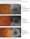
Page one of a chair-side reference chart designed to aid in the differential diagnosis of AMD phenotypes using fundus autofluorescence (FAF) imaging. Conclusions regarding the prognostic utility of FAF imaging in intermediate AMD are still equivocal, and this should be considered as a limitation of this clinical chart. The phenotypes in intermediate AMD pictured may be associated with a higher risk of conversion to choroidal neovascularization. Further details are described under section 4. Stratification of early to intermediate AMD phenotypes using FAF. CFP, color fundus photograph; FAF, fundus autofluorescence.
FIGURE 4
FIGURE 4
Second page of the chair-side reference showing different presentations of advanced AMD and the corresponding, typical appearance using fundus autofluorescence (FAF) imaging. Abbreviations as in Fig. 3.
FIGURE 5
FIGURE 5
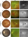
Case images from a series of patients where the application of fundus autofluorescence assisted in the differential diagnosis from AMD. A single case is displayed across the row. A–D, Medium-sized full-thickness macular hole without vitreomacular traction and central hyper-autofluorescence due to enhanced visibility of the RPE associated with the absence of overlying retinal tissue. E–H, Adult-onset vitelliform foveomacular dystrophy; the acquired vitelliform lesions appear as discrete lesions of intense hyper-autofluorescence. I–L, Macular telangiectasia type 2 that displayed a subtle increase in FAF at the temporal fovea. Abbreviations as in Fig. 1. IR, infrared reflectance image; OCT, optical coherence tomography.
FIGURE 6
FIGURE 6
Case series from patients that featured more extensive fundus changes involving the macula. A single case is displayed across the row. A–C, Fundus flavimaculatus: the characteristic flecks displayed prominent hyper-autofluorescence. D–F, Central serous chorioretinopathy distinguished by the typical appearance of a gravitational atrophic tract. G–I, Autosomal dominant drusen which appeared as discrete, radially distributed areas of hyper-autofluorescence. J–L, Retinitis pigmentosa characterized by patchy hypofluorescence in the periphery and a hyper-autofluorescent ring at the fovea. Abbreviations as in Fig. 1.

References

    1. Wong WL, Su X, Li X, et al. Global prevalence of age-related macular degeneration and disease burden projection for 2020 and 2040: a systematic review and meta-analysis. Lancet Glob Health 2014;2:e106–16. - PubMed
    1. Nivison-Smith L, Milston R, Madigan M, et al. Age-related macular degeneration: linking clinical presentation to pathology. Optom Vis Sci 2014;91:832–48. - PubMed
    1. von Ruckmann A, Fitzke FW, Bird AC. Distribution of fundus autofluorescence with a scanning laser ophthalmoscope. Br J Ophthalmol 1995;79:407–12. - PMC - PubMed
    1. Smith RT, Chan JK, Busuoic M, et al. Autofluorescence characteristics of early, atrophic, and high-risk fellow eyes in age-related macular degeneration. Invest Ophthalmol Vis Sci 2006;47:5495–504. - PMC - PubMed
    1. American Optometric Association Consensus Panel on Care of the Patient with Age-Related Macular Degeneration. Optometric Clinical Practice Guideline. Care of the Patient with Age-Related Macular Degeneration; 2004. Available at: http://www.aoa.org/documents/optometrists/CPG-6.pdf. Accessed: January 4, 2016.