. 2016 Nov;23(11):995-1002.
doi: 10.1038/nsmb.3296. Epub 2016 Sep 26.

E4 ligase-specific ubiquitination hubs coordinate DNA double-strand-break repair and apoptosis

Affiliations

E4 ligase-specific ubiquitination hubs coordinate DNA double-strand-break repair and apoptosis

Leena Ackermann et al. Nat Struct Mol Biol. 2016 Nov.

Abstract

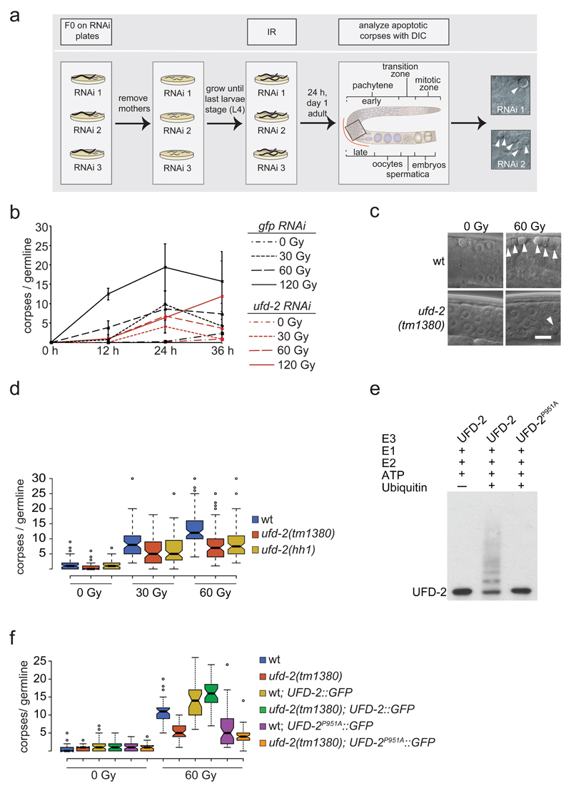
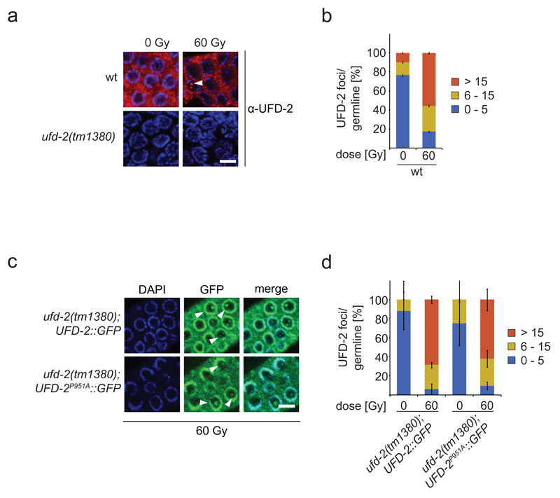
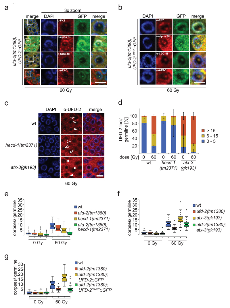
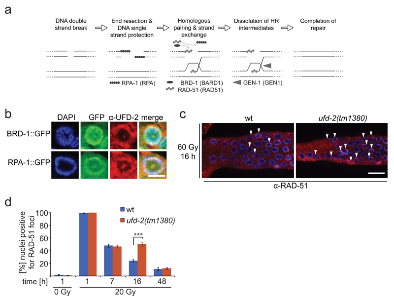
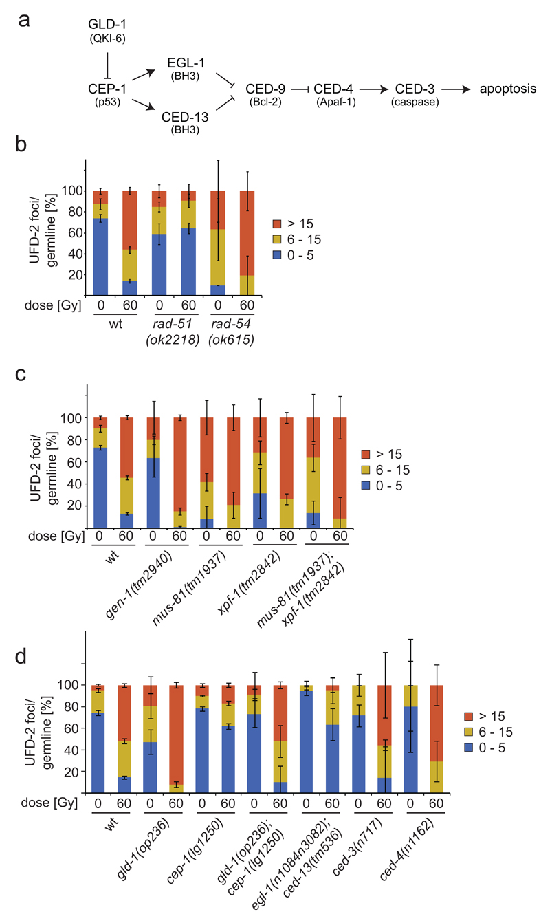
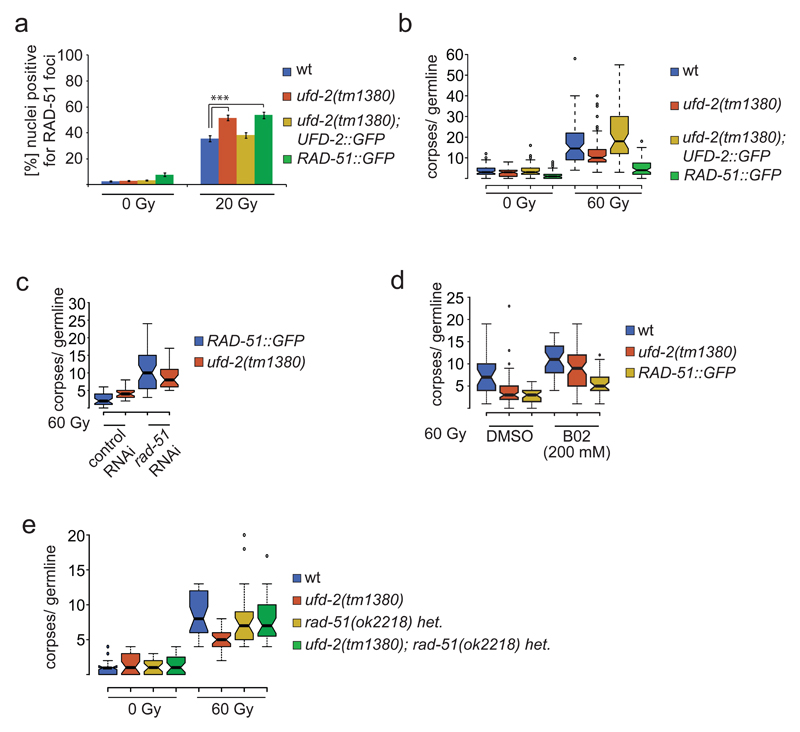
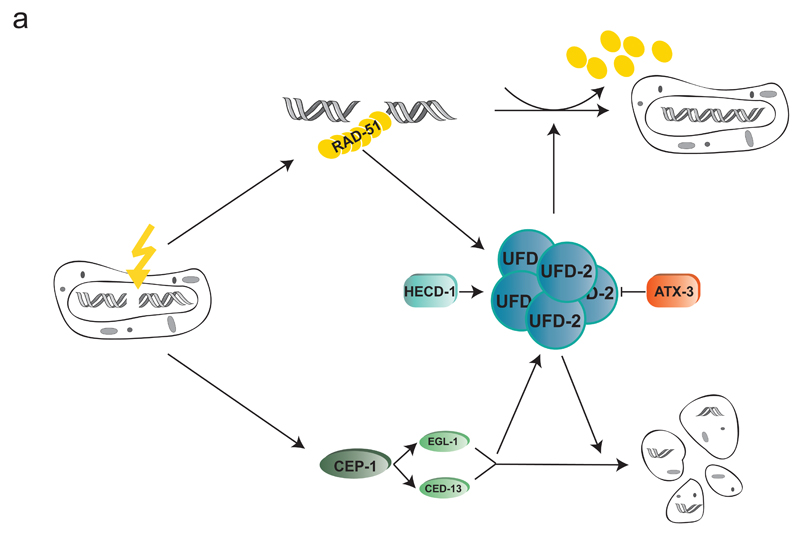
Multiple protein ubiquitination events at DNA double-strand breaks (DSBs) regulate damage recognition, signaling and repair. It has remained poorly understood how the repair process of DSBs is coordinated with the apoptotic response. Here, we identified the E4 ubiquitin ligase UFD-2 as a mediator of DNA-damage-induced apoptosis in a genetic screen in Caenorhabditis elegans. We found that, after initiation of homologous recombination by RAD-51, UFD-2 forms foci that contain substrate-processivity factors including the ubiquitin-selective segregase CDC-48 (p97), the deubiquitination enzyme ATX-3 (Ataxin-3) and the proteasome. In the absence of UFD-2, RAD-51 foci persist, and DNA damage-induced apoptosis is prevented. In contrast, UFD-2 foci are retained until recombination intermediates are removed by the Holliday-junction-processing enzymes GEN-1, MUS-81 or XPF-1. Formation of UFD-2 foci also requires proapoptotic CEP-1 (p53) signaling. Our findings establish a central role of UFD-2 in the coordination between the DNA-repair process and the apoptotic response.


Conflict of interest statement

The authors declare no competing financial interests.

Figures

Figure 1
Figure 1
Ubiquitin ligase activity of UFD-2 is required for apoptosis execution. (a) Schematic illustration of RNAi screen for identification of DNA damage-induced apoptosis mediators. After RNAi treatment worms were subjected to IR and scored for apoptotic corpses (indicated by filled arrowheads) 24 hrs later by differential interference contrast (DIC) microscopy. (b) Worms treated with indicated RNAi constructs were exposed to IR of increasing dose and scored for apoptotic corpses 24 hrs after treatment. Data represent mean ± s.e.m. of selected data of RNAi screen. (c) Representative images of late pachytene cells of C. elegans germline 24 hrs after IR treatment. Filled arrowheads indicate an apoptotic corpse. Scale bar 5 µm. (d) Indicated genotypes were scored for DNA damage induced apoptosis 24 hrs after IR. Center lines show the medians; box limits indicate the 25th and 75th percentiles as determined by R software; whiskers extend 1.5 times the interquartile range from the 25th and 75th percentiles, outliers are represented by dots. The notches are defined as +/- 1.58*IQR/sqrt(n) and represent the 95% confidence interval for each median. Non-overlapping notches give roughly 95% confidence that two medians differ. Sample points of 5 independent experiments. (e) Auto-ubiquitylation of UFD-2. Ubiquitylation reactions were carried out as indicated using UFD-2 (wild-type) and UFD-2P951A as ubiquitin ligases. Representative immunoblot of 3 independent experiments. (f) Indicated genotypes were scored for DNA damage induced apoptosis 24 hrs after IR. Sample points of 3 independent experiments. For n-values see Supplementary Table 1.
Figure 2
Figure 2
UFD-2 forms foci late after IR treatment. (a) Representative images of worm germlines of indicated genotypes irradiated with 60 Gy IR and stained with α-UFD-2 antibody and DAPI 24 hrs later. Filled arrowhead indicated nucleus with UFD-2 foci. Scale bar, 5 µm and (b) corresponding quantification of UFD-2 foci in pachytene region of germlines. Data show means ± s.e.m. of 12 independent experiments. (c) Representative images of worm germlines of indicated genotypes irradiated with 60 Gy IR. Germlines were isolated and stained with GFP-booster and DAPI 24 hrs after IR. Filled arrowheads indicate nuclei with UFD-2 foci. Scale bar, 5 µm and (d) corresponding quantification of UFD-2 foci in pachytene region of germlines. Data show means ± s.e.m. of 3 independent experiments. For n-values see Supplementary Table 1.
Figure 3
Figure 3
UPS factors accumulate in UFD-2 hubs and balance apoptotic signalling. Representative images of (a) ufd-2(tm1380); ufd-2::gfp and (b) ufd-2(tm1380); ufd-2P951A::gfp immunostained with indicated antibodies. Germlines were isolated 24 hrs after treatment with 60 Gy. DNA stained with DAPI. The boxed area is three times magnified (3x zoom). α-alpha SU, α-Proteasome 20S alpha subunits. Scale bars, 5 µm. Representative images of 3 independent experiments. (c) Representative images of worm germlines of indicated genotypes irradiated with 60 Gy IR and stained with α-UFD-2 antibody and DAPI 24 hrs later. Empty and filled arrowhead indicated nuclei positive or negative for UFD-2 foci, respectively. Scale bar, 5 µm and (d) corresponding quantification of UFD-2 foci in pachytene region of germlines. Data show means ± s.e.m. of 3 independent experiments. (e, f and g) Indicated genotypes were scored for apoptosis 24 hrs after 60 Gy IR. Sample points of 3 independent experiments. For n-values see Supplementary Table 1.
Figure 4
Figure 4
Loss of ufd-2 delays DSB repair processing. (a) Schematic illustration of DNA DSB repair by HR in C. elegans. Upon DSB induction RPA binds resected single stranded DNA, BRD-1 acts together with BRCA-1 at DSB site, RPA is exchanged for RAD-51, which mediates strand invasion, Gen-1 resolves HJ resulting in repaired DSB. Names in brackets indicate human homologues. (b) Representative images of brd-1::gfp and rpa-1::gfp germlines isolated and stained with α-UFD-2 and DAPI 24 hrs after treatment with 60 Gy of IR. Scale bar, 5 µm. Representative images of 3 independent experiments. (c) Representative images of germlines isolated from wild-type and ufd-2(tm1380) worms 16 hrs after IR treatment with 20 Gy. Germlines were stained with α-RAD-51 and DAPI. Filled arrowheads indicate nuclei positive for RAD-51 staining. Scale bar, 10 µm. (d) Quantification of germ cells that were positive for RAD-51 staining. Wild-type and ufd-2(tm1380) worms were treated with 0 or 20 Gy of IR and isolated 1, 7, 16, 48 hrs after treatment (7, 16, 48 hrs only for 60 Gy treated worms) and immunostained with α-RAD-51 and DAPI to stain DNA. The last 50 nuclei of pachytene germ cells prior entering diakinesis were evaluated. Data show means ± s.e.m. of 3 independent experiments. The triple asterisk indicates P value of ≤ 0.001 in Student’s t-test. For n-values see Supplementary Table 1.
Figure 5
Figure 5
UFD-2 foci in repair and apoptosis after DNA damage. (a) Schematic illustration of apoptosis pathway in C. elegans. Names in brackets indicate human homologues. (b, c and d) Quantification of UFD-2 foci in pachytene region of germlines of indicated genotypes isolated 24 hrs after irradiation with 60 Gy. Data show means ± s.e.m. of 3 independent experiments. For n-values see Supplementary Table 1.
Figure 6
Figure 6
UFD-2 coordinates communication between repair and apoptosis after DNA damage. (a) Quantification of germ cells positive for RAD-51 staining. Worms of indicated genotypes were treated with 0 or 20 Gy of IR. Germlines were isolated 24 hrs after treatment and stained with α-RAD-51 and DAPI. The last 50 nuclei of pachytene germ cells prior entering diakinesis were evaluated. Data show means ± s.e.m. of 3 independent experiments. The triple asterisk indicates P value of ≤ 0.001 in Student’s t-test. (b) Indicated genotypes were scored for apoptosis 24 hrs after 60 Gy IR. Sample points of 3 independent experiments. (c) ufd-2 and RAD-51::GFP worms were treated with rad-51 or control RNAi and scored for apoptosis 24 hrs after 60 Gy IR. Sample points of 3 independent experiments. (d) wild-type, ufd-2 and RAD-51::GFP worms were treated with RAD51 inhibitor B02 (200mM) from L1 larvae on and scored for apoptosis 24 hrs after 60 Gy IR. Sample points of 3 independent experiments. (e) Indicated genotypes were scored for apoptosis 24 hrs after 60 Gy IR. Sample points of 3 independent experiments. For n-values see Supplementary Table 1.
Figure 7
Figure 7
Model of how UFD-2 integrates HR repair and apoptotic signalling. UFD-2 forms hubs late after IR (additionally containing proteolytic factors as CDC-48 and the proteasome (not shown)) that are dependent on active repair and apoptotic CEP-1 signalling. UFD-2 hub formation is balanced by the E3 ligase HECD-1 and the DUB ATX-3. In accordance with hub formation at later stages after DSB induction, UFD-2 supports RAD-51 dissociation from DSB site at advanced time points and mediates signal to apoptosis pathway.

References

    1. Hoeijmakers JH. Genome maintenance mechanisms for preventing cancer. Nature. 2001;411:366–374. doi: 10.1038/35077232. - DOI - PubMed
    1. Clejan I, Boerckel J, Ahmed S. Developmental modulation of nonhomologous end joining in Caenorhabditis elegans. Genetics. 2006;173:1301–1317. doi: 10.1534/genetics.106.058628. - DOI - PMC - PubMed
    1. Lemmens BB, Tijsterman M. DNA double-strand break repair in Caenorhabditis elegans. Chromosoma. 2011;120:1–21. doi: 10.1007/s00412-010-0296-3. - DOI - PMC - PubMed
    1. Bizard AH, Hickson ID. The dissolution of double Holliday junctions. Cold Spring Harb Perspect Biol. 2014;6:a016477. doi: 10.1101/cshperspect.a016477. - DOI - PMC - PubMed
    1. Matos J, West SC. Holliday junction resolution: regulation in space and time. DNA Repair (Amst) 2014;19:176–181. doi: 10.1016/j.dnarep.2014.03.013. - DOI - PMC - PubMed

MeSH terms

Substances