. 2017 Mar 30;36(13):1770-1778.
doi: 10.1038/onc.2016.353. Epub 2016 Sep 26.

Cancer-associated fibroblast exosomes regulate survival and proliferation of pancreatic cancer cells

Affiliations

Cancer-associated fibroblast exosomes regulate survival and proliferation of pancreatic cancer cells

K E Richards et al. Oncogene. .

Abstract

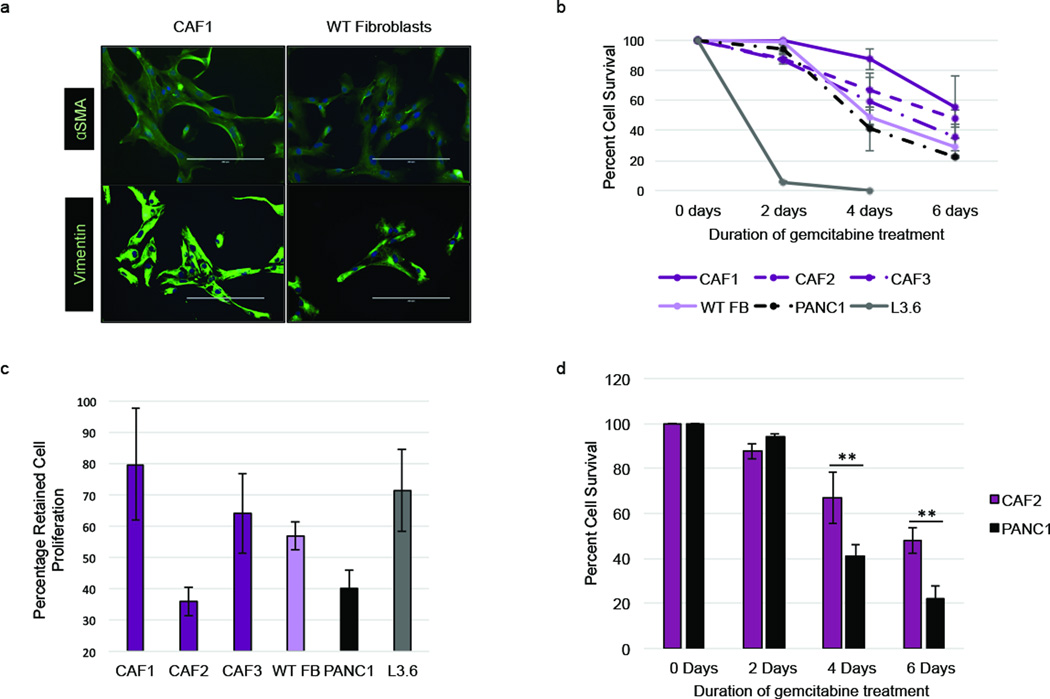
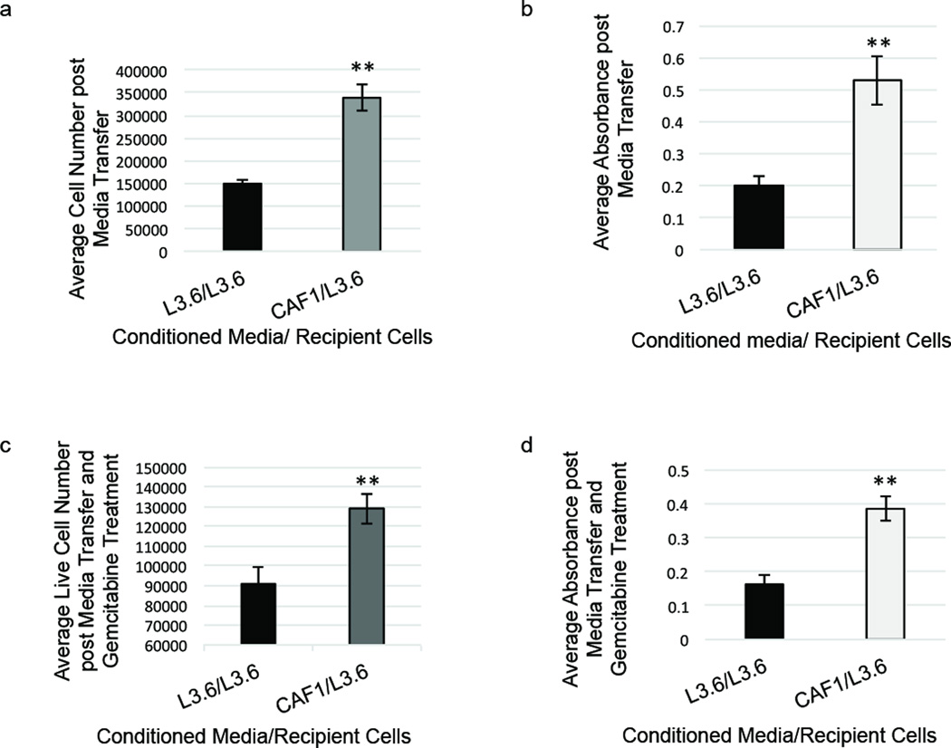
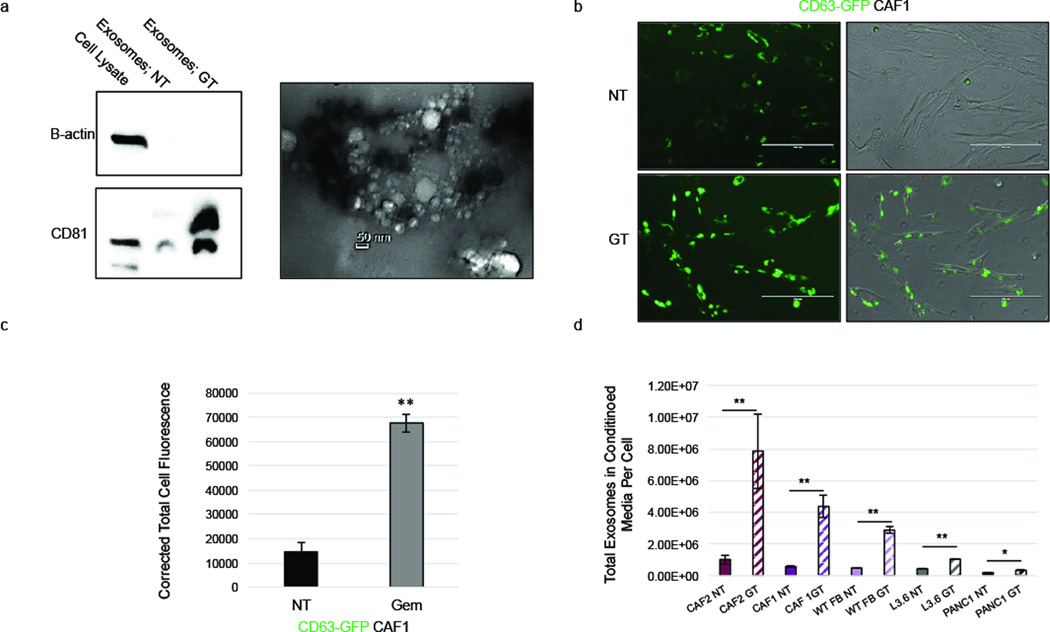
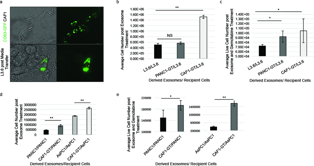
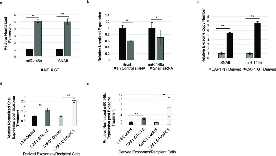
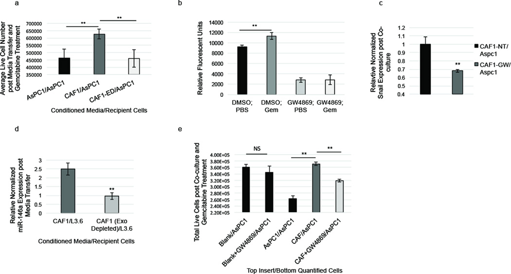
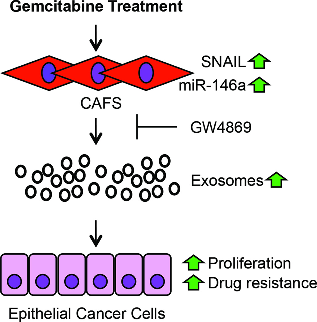
Cancer-associated fibroblasts (CAFs) comprise the majority of the tumor bulk of pancreatic ductal adenocarcinomas (PDACs). Current efforts to eradicate these tumors focus predominantly on targeting the proliferation of rapidly growing cancer epithelial cells. We know that this is largely ineffective with resistance arising in most tumors following exposure to chemotherapy. Despite the long-standing recognition of the prominence of CAFs in PDAC, the effect of chemotherapy on CAFs and how they may contribute to drug resistance in neighboring cancer cells is not well characterized. Here, we show that CAFs exposed to chemotherapy have an active role in regulating the survival and proliferation of cancer cells. We found that CAFs are intrinsically resistant to gemcitabine, the chemotherapeutic standard of care for PDAC. Further, CAFs exposed to gemcitabine significantly increase the release of extracellular vesicles called exosomes. These exosomes increased chemoresistance-inducing factor, Snail, in recipient epithelial cells and promote proliferation and drug resistance. Finally, treatment of gemcitabine-exposed CAFs with an inhibitor of exosome release, GW4869, significantly reduces survival in co-cultured epithelial cells, signifying an important role of CAF exosomes in chemotherapeutic drug resistance. Collectively, these findings show the potential for exosome inhibitors as treatment options alongside chemotherapy for overcoming PDAC chemoresistance.


Conflict of interest statement

CONFLICT OF INTEREST

The authors declare no conflict of interest.

Figures

Figure 1
Figure 1
Pancreatic fibroblasts are innately chemoresistant. (a) Immunofluorescence stain for αSMA and vimentin of cancer-associated fibroblasts (CAF1) and wild-type (WT) fibroblasts. (b) Cells were treated with 1µM gemcitabine for 2–6 days and live and dead cells were counted to obtain percent cell survival. (c) Cells were treated with 1µM gemcitabine for 2 days or left untreated and total cells were counted to obtain percentage of proliferation retention during GEM treatment. (d) Percent cell survival of CAFs (CAF2) and epithelial cells (PANC1) with similar proliferation retention rate over 6 days 1µM gemcitabine treatment. **p-value<0.01
Figure 2
Figure 2
Pancreatic CAF1-conditioned media increases proliferation and survival of epithelial cells. (a) L3.6 cells were grown in CAF1-conditioned media or L3.6-conditioned media for 8 days and total cells were counted. (b) Cell proliferation assay (MTT assay) was performed after 8 days L3.6 cell growth in conditioned media. (c) L3.6 cells were grown in cell-conditioned media for 6 days then treated with 100nM gemcitabine for 3 days, and live cells were counted. (d) Cell proliferation assay (MTT) was performed after 6 days L3.6 cell growth in conditioned media and 3 days of 100nM gemcitabine treatment. **p-value<0.01
Figure 3
Figure 3
Gemcitabine increases CAF exosome secretion. (a) CAF1s were left untreated (NT) or treated with 1µM gem (GT) for 4 days. Exosomes were isolated from conditioned cell media, and protein lysates were used to perform a western blot for CD81 and beta-actin (left). Isolated exosomes were examined for size and structure via transmission electron microscopy (right). (b) CAF1 cells transduced with a GFP-CD63 lentivirus (GFP-CD63-CAF1s) were treated with 1uM gemcitabine (GT) or left untreated (NT) and fluorescence was analyzed via microscopy. (c) Total corrected cell fluorescence of GFP-CD63-CAF1 cells was quantified using ImageJ. (d) Cells were treated with 1µM gemcitabine (Fibroblasts and PANC1), 10nM gemcitabine (L3.6), or left untreated for 4 days (NT), and exosomes were collected and quantified. Scale bar, 200 µm. *p-value<0.05; **p-value<0.01
Figure 4
Figure 4
GT-CAF exosomes increase cell number and survival of epithelial cells. (a) L3.6 cells were treated with GFP-CD63-CAF1 conditioned media for 48 hours and exosome uptake was visualized. (b) L3.6 cells were treated with L3.6, GT-PANC1, or GT-CAF1 exosomes for 6 days and total cells were counted. (c) L3.6 cells were treated with L3.6, GT-PANC1 or GT-CAF1 exosomes for 6 days and 1µM GEM for 3 days, and live cells were counted. (d) PANC1 cells were treated with PANC1 or GT-CAF1 exosomes for 6 days. AsPC1 cells were treated with AsPC1 or GT-CAF1 exosomes for 6 days. Total cells were counted. (e) PANC1 cells were treated with PANC1 or GT-CAF1 exosomes for 6 days, and AsPC1 cells were treated with AsPC1 or GT-CAF1 exosomes for 6 days. All cells were then treated with 1µM GEM for 3 days, and live cells were counted. *p-value<0.05; **p-value<0.01
Figure 5
Figure 5
Pancreatic fibroblasts upregulate and secrete miR-146a and Snail during gemcitabine treatment. (a) RT-PCR. miR-146a and Snail levels were altered in CAF1s during 1 µM GEM treatment (GT) (3 days) compared to untreated control (NT). (b) RT-PCR. CAF1s were treated with Snail-siRNA, and Snail and miR-146a expression was measured compared to negative siRNA control treated CAFs. (c) Exosomes from untreated and 1µM GEM-treated CAF1s were isolated and Snail mRNA and miR-146a within CAF1 exosomes was quantified via RT-PCR using relative Ct values. (d–e) L3.6 cells were treated with GT-CAF1 exosomes for 6 days (GT-CAF1/L3.6) or left untreated (L3.6 control). AsPC1 cells were treated with GT-CAF1 exosomes for 6 days (GT-CAF1/AsPC1) or left untreated (AsPC1 control). Snail (d) and miR-146a (e) levels were quantified in recipient cells via RT-PCR. *p-value<0.05; **p-value<0.01
Figure 6
Figure 6
Inhibition of CAF exosome signaling suppresses chemoresistance. (a) AsPC1 cells were grown for 5 days in AsPC1-conditioned media (AsPC1/AsPC1), CAF1-conditioned media (CAF1/AsPC1) or CAF1-conditioned media depleted of exosomes (CAF1-ED/AsPC1) and then treated with 1µM GEM for 3 days and live cells were counted. (b) CAF1s were treated with 20µm GW4869 or DMSO along with 1µM gemcitabine or PBS for 3 days. Exosomes in media were collected, dyed with CFSE, and quantified. (c) AsPC1 cells were co-cultured with AsPC1 cells, CAFs, GW4869-treated CAF1s, DMEM alone (Blank/AsPC1), or GW4869 in DMEM (Blank+GW4869) for 6 days then treated during co-culture with 1µM gemcitabine for 3 days. Live co-cultured AsPC1 cells at the bottom of the plate were counted. (d) Snail expression was measured via RT-PCR in AsPC1 cells co-cultured with untreated CAFs (CAF-NT/AsPC1) or GW4869-treated CAFs (CAF-GW/AsPC1). (e) L3.6 cells were cultured in CAF1-conditioned (CAF1/L3.6) media or CAF1-conditioned media depleted of exosomes (CAF1-ED/L3.6). miR-146a expression was measured via RT-PCR. **p-value<0.01
Figure 7
Figure 7
Schematic overview of CAF exosome signaling during gemcitabine treatment. Gemcitabine treatment leads to upregulation of Snail and miR-146a as well as exosome secretion in CAFs that could lead to increased cell proliferation, tumor growth, and chemoresistance of adjacent cancer epithelial cells. GW4869 suppresses exosome release and therefore exosomal transfer of Snail and miR-146a.

Comment in

References

    1. Stathis A, Moore MJ. Advanced pancreatic carcinoma: current treatment and future challenges. Nature reviews. 2010;7(3):163–172. - PubMed
    1. Rahib L, Smith BD, Aizenberg R, Rosenzweig AB, Fleshman JM, Matrisian LM. Projecting cancer incidence and deaths to 2030: the unexpected burden of thyroid, liver, and pancreas cancers in the United States. Cancer Res. 2014;74(11):2913–2921. - PubMed
    1. Oettle H, Post S, Neuhaus P, Gellert K, Langrehr J, Ridwelski K, et al. Adjuvant chemotherapy with gemcitabine vs observation in patients undergoing curative-intent resection of pancreatic cancer: a randomized controlled trial. JAMA : the journal of the American Medical Association. 2007;297(3):267–277. - PubMed
    1. Kleeff J, Beckhove P, Esposito I, Herzig S, Huber PE, Lohr JM, et al. Pancreatic cancer microenvironment. Int J Cancer. 2007;121(4):699–705. - PubMed
    1. Chu GC, Kimmelman AC, Hezel AF, DePinho RA. Stromal biology of pancreatic cancer. Journal of cellular biochemistry. 2007;101(4):887–907. - PubMed

Publication types

MeSH terms