. 2016 Nov 1;126(11):4088-4102.
doi: 10.1172/JCI87148. Epub 2016 Sep 26.

Targeting cancer metabolism by simultaneously disrupting parallel nutrient access pathways

Targeting cancer metabolism by simultaneously disrupting parallel nutrient access pathways

Seong M Kim et al. J Clin Invest. .

Abstract

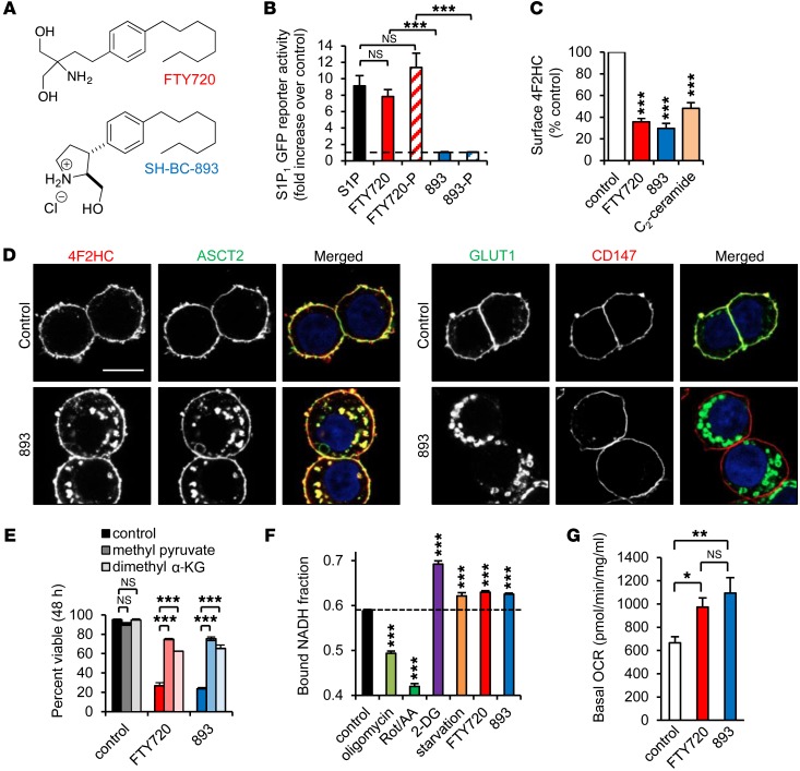
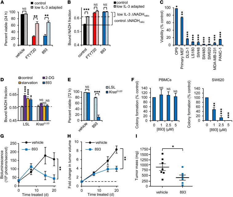
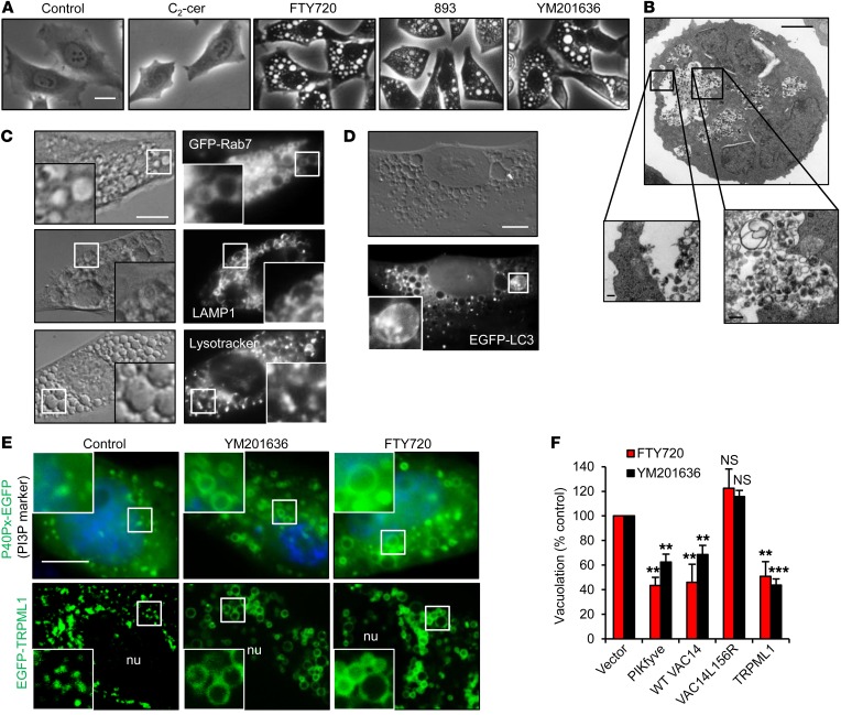
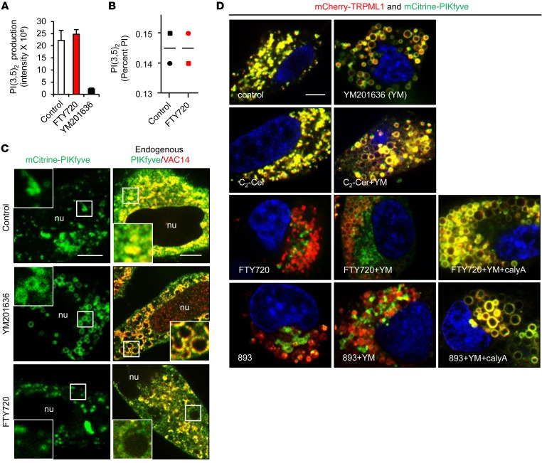
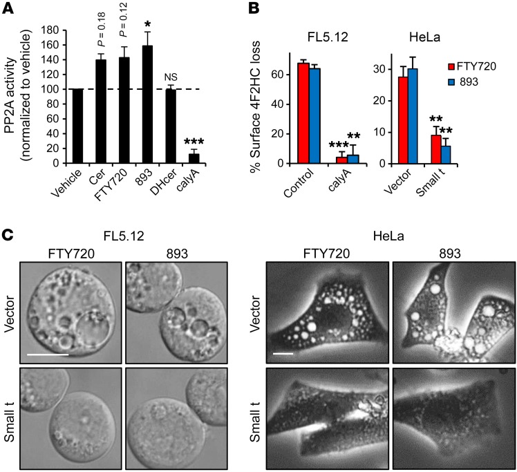
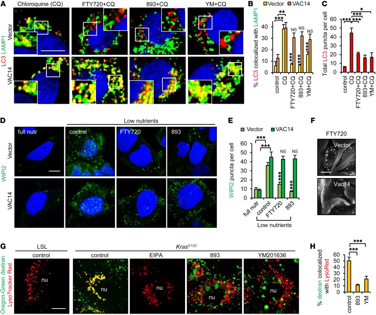
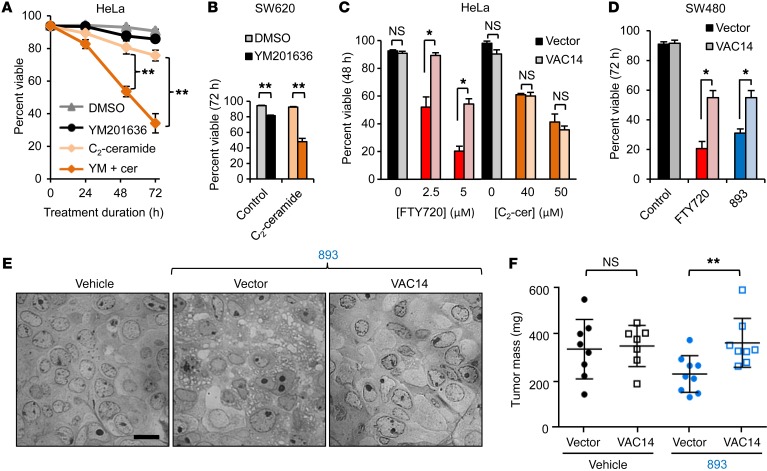
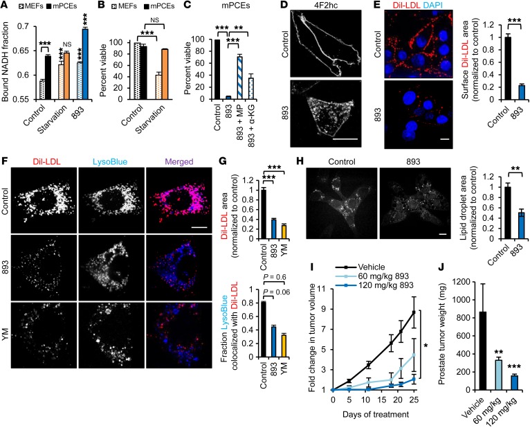
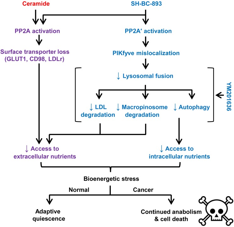
Oncogenic mutations drive anabolic metabolism, creating a dependency on nutrient influx through transporters, receptors, and macropinocytosis. While sphingolipids suppress tumor growth by downregulating nutrient transporters, macropinocytosis and autophagy still provide cancer cells with fuel. Therapeutics that simultaneously disrupt these parallel nutrient access pathways have potential as powerful starvation agents. Here, we describe a water-soluble, orally bioavailable synthetic sphingolipid, SH-BC-893, that triggers nutrient transporter internalization and also blocks lysosome-dependent nutrient generation pathways. SH-BC-893 activated protein phosphatase 2A (PP2A), leading to mislocalization of the lipid kinase PIKfyve. The concomitant mislocalization of the PIKfyve product PI(3,5)P2 triggered cytosolic vacuolation and blocked lysosomal fusion reactions essential for LDL, autophagosome, and macropinosome degradation. By simultaneously limiting access to both extracellular and intracellular nutrients, SH-BC-893 selectively killed cells expressing an activated form of the anabolic oncogene Ras in vitro and in vivo. However, slower-growing, autochthonous PTEN-deficient prostate tumors that did not exhibit a classic Warburg phenotype were equally sensitive. Remarkably, normal proliferative tissues were unaffected by doses of SH-BC-893 that profoundly inhibited tumor growth. These studies demonstrate that simultaneously blocking parallel nutrient access pathways with sphingolipid-based drugs is broadly effective and cancer selective, suggesting a potential strategy for overcoming the resistance conferred by tumor heterogeneity.


Figures

Figure 1
Figure 1. SH-BC-893 triggers nutrient transporter internalization mimicking starvation.
(A) Structures of FTY720 and SH-BC-893. (B) S1P1 receptor–driven GFP expression measured by flow cytometry in reporter MEFs after a 24-hour incubation with the indicated compounds at 2.5 μM. 893, SH-BC-893; 893-P, (SH-BC-893-phosphate); FTY720-P, (FTY720-phosphate). (C) Surface 4F2HC measured by flow cytometry in FL5.12 cells treated with 5 μM FTY720 or SH-BC-893 or 10 μM C2-ceramide. (D) SH-BC-893–treated SW620 cells stained as indicated. Scale bar: 10 μm. (E) Viability of BCLXL-overexpressing FL5.12 cells treated as indicated, with or without 5.5 mM methyl pyruvate or 2 mM dimethyl α-ketoglutarate (α-KG). (F) Bound NADH fraction in MEFs treated with mitochondrial inhibitors (1 μM oligomycin or 1 μM rotenone and antimycin A [Rot/AA]), 1 mM 2-DG, 5 μM FTY720, or 7.5 μM SH-BC-893 for 16 hours. For starvation, growth media were replaced with DMEM lacking glucose and amino acids, supplemented with 10% dialyzed FCS. Statistical significance was compared with respective controls. (G) Oxygen consumption rate (OCR) in MEFs treated with FTY720 or SH-BC-893 for 16 hours measured by an XF24 Extracellular Flux Analyzer (Seahorse Bioscience). Error bars indicate the mean ± SEM. *P <0.05, **P < 0.01, and ***P <0.001, by unpaired, 2-tailed Student’s t test. P values were determined using Tukey’s method when correcting for multiple comparisons. Data are representative of at least 3 independent experiments.
Figure 2
Figure 2. SH-BC-893 selectively kills cancer cells.
(A) Viability of FL5.12 cells cultured in control medium (500 pg/ml IL-3) or adapted to low IL-3 (25 pg/ml) treated with 5 μM FTY720 or SH-BC-893. (B) Bound NADH fraction in FL5.12 cells cultured as in A and treated with FTY720 or SH-BC-893. (C) Viability of the indicated cells treated with 5 μM SH-BC-893 for 72 hours. Statistical significance was compared with vehicle. OP9 (murine bone marrow stromal cells), and primary MEF were nontransformed cells. DLD-1, LS180, SW480, SW480, and SW620 were colon cancer cell lines. MDA-MB-231 and PANC-1 were breast cancer and pancreatic cancer cell lines, respectively. (D) Bound NADH fraction in control or KrasG12D-expressing MEFs treated with 1 mM 2-DG, starvation media (glucose and amino acids supplemented with 10% dialyzed FCS), or 7.5 μM SH-BC-893 for 16 hours. Statistical significance was compared with respective controls. (E) Viability of p53–/– MEFs, with or without KrasG12D expression, that were treated with 6 μM SH-BC-893. (F) Colony formation by normal human PBMCs or SW620 CRC cells, with or without SH-BC-893 at the indicated doses. Error bars indicate the mean ± SEM. (GI) Subcutaneous SW620 tumor growth as measured by bioluminescence imaging (BLI) (G), calipers (H), or tumor weight at sacrifice (I). Because FTY720 and SH-BC-893 were similarly potent in vitro (Figure 1, C and E) (26), SH-BC-893 was given i.p. at a dose of 10 mg/kg for 7 days. On the basis of BLI (G), the dose was increased to 20 mg/kg on day 8. Error bars indicate the mean ± SD. n = 7 mice per group. *P <0.05, **P <0.01, and ***P <0.001, by unpaired, 2-tailed Student’s t test. P values were determined using Tukey’s method when correcting for multiple comparisons. In vitro data are representative of at least 3 independent experiments.
Figure 3
Figure 3. Sphingolipid-induced vacuolation resembles PIKfyve inhibition.
(A) HeLa cells treated as indicated with 25 μM C2-ceramide, 5 μM FTY720 or SH-BC-893, or 800 nM YM201636 for 6 hours. (B) Electron micrograph of an FL5.12 cell treated with FTY720 for 24 hours. Scale bars: 2 μm, 0.2 μm in zoom (insets). (C) GFP-RAB7–expressing HeLa cells (top) or MEFs treated with FTY720 and stained as indicated. Images of 3 separate cells are shown. Scale bar: 10 μm; ×2.5 magnification. (D) EGFP-LC3–expressing MEFs treated with FTY720. Scale bar: 10 μm; ×2.75 magnification. (E) P40Px-EGFP– or EGFP-TRPML1–expressing HeLa cells treated with YM201636 or FTY720 for 6 hours. Scale bar: 10 μm; ×2.5 magnification. nu, nucleus. (F) HeLa cells overexpressing PIKfyve, WT VAC14, mutant (L156R) VAC14, or TRPML1 treated with YM201636 or FTY720. Error bars indicate the mean ± SEM. n ≥3 independent experiments; ≥30 cells per condition were analyzed per experiment. Statistical significance was compared with respective controls. **P < 0.01 and ***P < 0.001, by unpaired, 2-tailed Student’s t test. P values were determined using Tukey’s method to correct for multiple comparisons.
Figure 4
Figure 4. SH-BC-893 and FTY720 disrupt PIKfyve localization but not its activity.
(A) In vitro kinase assay with purified FLAG-PIKfyve, with or without 5 μM FTY720 or 800 nM YM201636. Error bars indicate the mean ± SD from 3 technical replicates. (B) PI(3,5)P2 levels in HeLa cells treated with FTY720 expressed as a percentage of total phosphatidylinositol (PI). Bars indicate the average from 2 independent experiments. (C) mCitrine-PIKfyve or endogenous PIKfyve/VAC14 in HeLa cells treated with 800 nM YM201636 or 5 μM FTY720 for 6 hours. Scale bars: 15 μM; ×2.5 magnification (C and D). (D) HeLa cells expressing low levels of mCitrine-PIKfyve and mCherry-TRPML1 treated as indicated: 800 nM YM201636, 5 μM FTY720 or SH-BC-893, 25 μM C2-ceramide (C2-Cer), or 5 nM calyculin A (calyA). Scale bar: 10 μm.
Figure 5
Figure 5. SH-BC-893 and FTY720 activate PP2A to induce vacuolation.
(A) PP2A phosphatase activity was measured in FL5.12 cell lysates in the presence or absence of 50 μM C2-ceramide (cer) or dihydro-C2-ceramide (DHcer), 5 μM FTY720 or SH-BC-893 (893), or 5 nM calyculin A. (B and C) Surface 4F2HC (B) or vacuolation (C) in FL5.12 cells pretreated with vehicle or 5 nM calyculin A or HeLa cells expressing vector or SV40 small t antigen treated as indicated. Error bars indicate the mean ± SEM. *P < 0.05, **P < 0.01, and ***P < 0.001, by unpaired, 2-tailed Student’s t test. P values were determined using Tukey’s method when correcting for multiple comparisons. Data are representative of at least 3 independent experiments. Scale bars: 10 μm.
Figure 6
Figure 6. SH-BC-893 reduces autophagic flux and macropinosome degradation.
(A) Control or VAC14-overexpressing HeLa cells treated with 25 μM chloroquine (CQ), with or without 5 μM FTY720, were stained for LC3 and LAMP1 and evaluated by confocal microscopy. (B and C) Quantification of LC3-LAMP1 colocalization (B) and total LC3 puncta (C) in cells treated with chloroquine, with or without FTY720, SH-BC-893, or 800 nM YM201636 for 6 hours. Error bars indicate the mean ± SEM. n ≥35 cells evaluated per condition. (D) Vector or VAC14-overexpressing HeLa cells were nutrient stressed in DMEM lacking amino acids and glucose in the presence or absence of 5 μM FTY720 or SH-BC-893 and stained for WIPI2. (E) Quantification of WIPI2 puncta. Error bars indicate the mean ± SEM. n ≥50 cells evaluated per condition. Statistical significance was compared with the respective low-nutrient control unless otherwise indicated. (F) HeLa cells overexpressing VAC14 were treated with 5 μM FTY720 for 6 hours. (G and H) Dextran uptake in p53–/– LSL-KrasG12D MEFs before (LSL) or after (KrasG12D) introduction of Cre, with or without the macropinocytosis inhibitor EIPA, SH-BC-893, or YM201636 (G). Colocalization of dextran and LysoTracker Red (H) was determined using ImageJ. Error bars indicate the mean ± SEM shown. n ≥15 cells evaluated per condition. *P < 0.05, **P < 0.01, and ***P < 0.001, by unpaired, 2-tailed Student’s t test. P values were determined using Tukey’s method when correcting for multiple comparisons. Scale bars: 20 μm; magnification in (A) ×1.8. nutr, nutrient; YM, YM201636.
Figure 7
Figure 7. Vacuolation enhances the antineoplastic effects of SH-BC-893 in vitro and in vivo.
(A and B) Viability of HeLa (A) or SW620 (B) cells treated with DMSO, 800 nM YM201636, 25 μM C2-ceramide, or YM201636 plus C2-ceramide (YM + cer) for the indicated durations. (C and D) Viability of control or VAC14-overexpressing cells treated with FTY720 or C2-ceramide. Error bars indicate the mean ± SEM. (E) Vacuolation evaluated by bright-field microscopy in SW480 tumors from mice treated with vehicle (water) or 60 mg/kg SH-BC-893 by gavage daily for 18 days. Scale bar: 10 μm. (F) Tumor weight at sacrifice. P = 0.06 between vehicle and SH-BC-893–treated vector groups (2-tailed Student’s t test). Error bars indicate the mean ± SD. n ≥7 mice per group. *P < 0.05 and **P < 0.01, by unpaired, 2-tailed Student’s t test. In vitro data are representative of at least 3 independent experiments.
Figure 8
Figure 8. SH-BC-893 starves Pten–/– prostate cancer cells.
(A) Bound NADH fraction in MEFs or mPCEs. Error bars indicate the mean ± SEM. n ≥50 cells evaluated per condition. (B) Viability of MEFs or mPCEs in starvation media (DMEM lacking glucose and amino acids supplemented with 10% FBS). (C) Viability of mPCEs treated with 5 μM SH-BC-893, with or without 11 mM methyl pyruvate or 7 mM dimethyl α-ketoglutarate. Error bars indicate the mean ± SEM. (D) mPCEs treated with 5 μM SH-BC-893 stained for 4F2HC. (E) Dil-LDL bound to surface LDL receptors of mPCEs maintained at 4°C. Error bars indicate the mean ± SEM. n ≥80 cells per condition. (F and G) Dil-LDL uptake and lysosomes in mPCEs treated with SH-BC-893 or YM201636. Quantification of total Dil-LDL area (F) and colocalization of Dil-LDL with LysoTracker Blue (G). Error bars indicate the mean ± SEM. n ≥30 cells per condition. (H) mPCEs treated with SH-BC-893 for 3 hours were imaged using CARS to detect lipid droplets. Error bars indicate the mean ± SEM. n = 5 fields. (I) Growth of mPCE s.c. isografts measured by calipers. n = 7 per group. (J) Mean weight of autochthonous prostate tumors at sacrifice. n = 9 for the vehicle-treated and the 60 mg/kg SH-BC-893 groups; n = 8 for the 120 mg/kg group. Statistical significance was compared with respective controls unless otherwise indicated. *P < 0.05, **P < 0.01, and ***P < 0.001, by unpaired, 2-tailed Student’s t test (AC, E, and GI) and Mann-Whitney U test for comparison of treated mice with controls (J). P values were determined using Tukey’s method when correcting for multiple comparisons. Scale bars: 20 μm. LysoBlue, LysoTracker Blue.
Figure 9
Figure 9. Vacuolating sphingolipid SH-BC-893 targets primary and adaptive pathways for nutrient acquisition.
Like ceramide, the synthetic sphingolipid SH-BC-893 activates PP2A to downregulate nutrient transporters. In addition, SH-BC-893 activates a second PP2A complex, PP2A′ that is not affected by ceramide. Activation of PP2A′ leads to mislocalization of PIKfyve and PI(3,5)P2, reducing lysosomal fusion reactions. Because PI(3,5)P2 is membrane anchored and cannot diffuse to its target, loss of PI(3,5)P2 (YM201636 treatment) and PI(3,5)P2 mislocalization (SH-BC-893 treatment) produce similar phenotypes. While ceramide limits access to extracellular nutrients, SH-BC-893 blocks access to both extracellular and intracellular nutrients. Substrate limitation in the context of oncogene-driven anabolism is lethal, while nontransformed cells can make adaptive metabolic changes that allow them to survive nutrient stress.

References

    1. McCracken AN, Edinger AL. Nutrient transporters: the Achilles’ heel of anabolism. Trends Endocrinol Metab. 2013;24(4):200–208. doi: 10.1016/j.tem.2013.01.002. - DOI - PMC - PubMed
    1. Garcia-Cao I, et al. Systemic elevation of PTEN induces a tumor-suppressive metabolic state. Cell. 2012;149(1):49–62. doi: 10.1016/j.cell.2012.02.030. - DOI - PMC - PubMed
    1. Shroff EH, et al. MYC oncogene overexpression drives renal cell carcinoma in a mouse model through glutamine metabolism. Proc Natl Acad Sci U S A. 2015;112(21):6539–6544. doi: 10.1073/pnas.1507228112. - DOI - PMC - PubMed
    1. Ying H, et al. Oncogenic Kras maintains pancreatic tumors through regulation of anabolic glucose metabolism. Cell. 2012;149(3):656–670. doi: 10.1016/j.cell.2012.01.058. - DOI - PMC - PubMed
    1. Yue S, et al. Cholesteryl ester accumulation induced by PTEN loss and PI3K/AKT activation underlies human prostate cancer aggressiveness. Cell Metab. 2014;19(3):393–406. doi: 10.1016/j.cmet.2014.01.019. - DOI - PMC - PubMed

Publication types

MeSH terms