. 2016 Nov 28;36(24):3010-3018.
doi: 10.1128/MCB.00474-16. Print 2016 Dec 15.

Regulation of Linear Ubiquitin Chain Assembly Complex by Caspase-Mediated Cleavage of RNF31

Affiliations

Regulation of Linear Ubiquitin Chain Assembly Complex by Caspase-Mediated Cleavage of RNF31

Donghyun Joo et al. Mol Cell Biol. .

Abstract

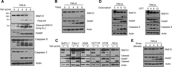
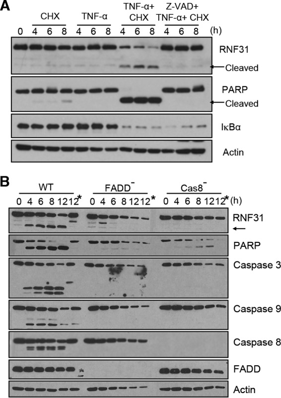
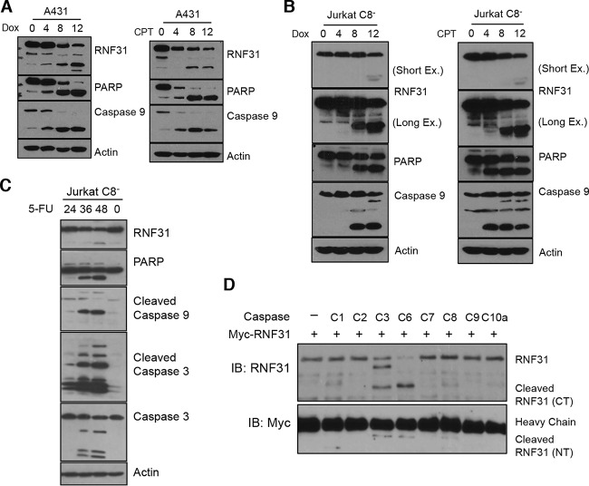
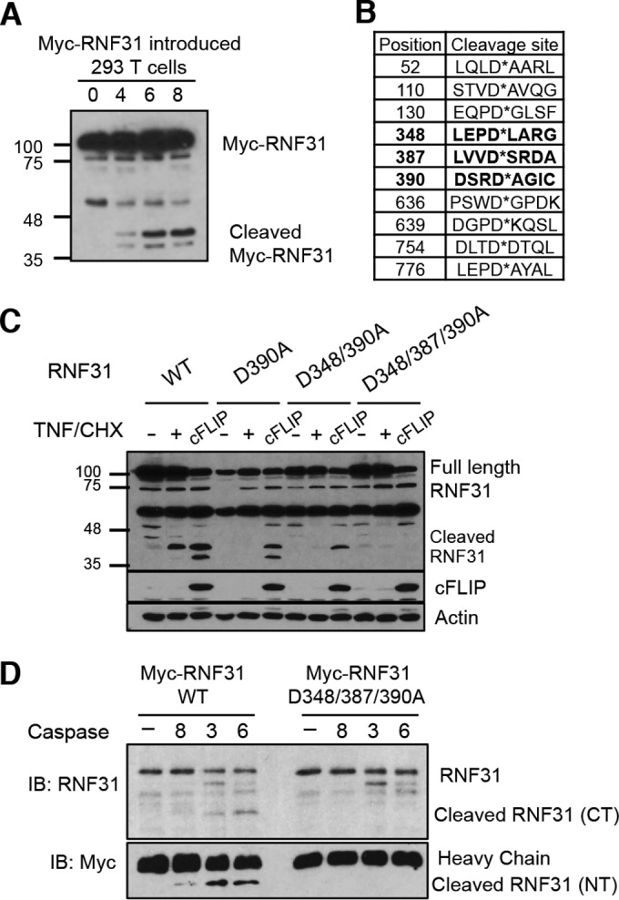
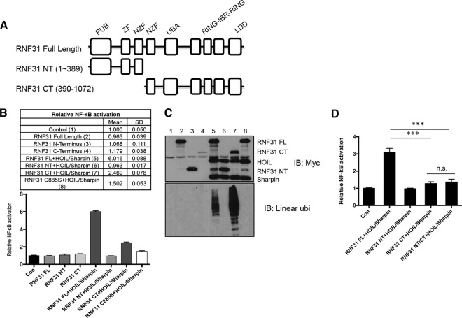
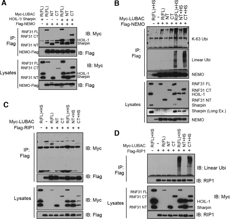
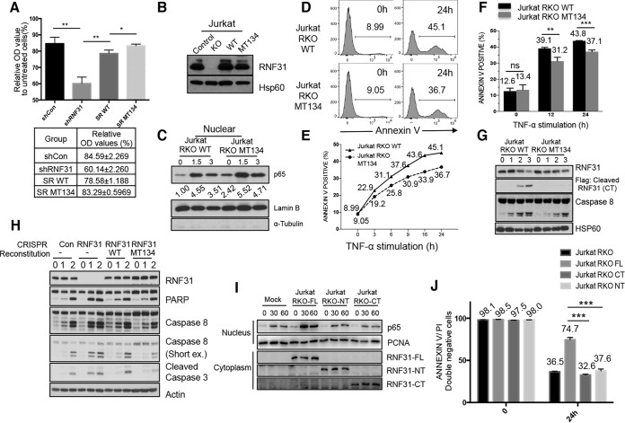
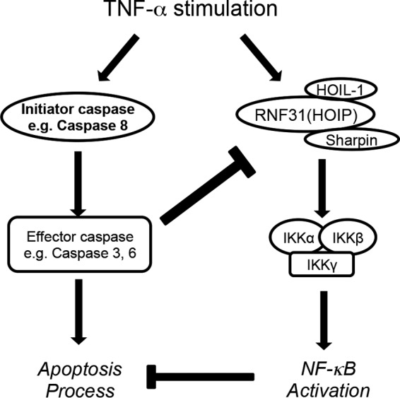
Cell death and survival signaling pathways have opposed but fundamental functions for various cellular processes and maintain cell homeostasis through cross talk. Here we report a novel mechanism of interaction between these two pathways through the cleavage of RNF31 by caspases. RNF31, a component of the linear ubiquitin chain assembly complex (LUBAC), regulates cell survival by inducing linear ubiquitination of NF-κB signaling components. We found that RNF31 is cleaved under apoptosis conditions through various stimulations. The effector caspases caspase 3 and caspase 6 are responsible for this event, and aspartates 348, 387, and 390 were identified as target sites for this cleavage. Cleavage of RNF31 suppressed its ability to activate NF-κB signaling; thus, mutation of cleavage sites inhibited the induction of apoptosis by treatment with tumor necrosis factor alpha (TNF-α). Our findings elucidate a novel regulatory loop between cell death and the survival signal and may provide guidance for the development of therapeutic strategies for cancers through the sensitization of tumor cells to death-inducing drugs.


Figures

FIG 1
FIG 1
RNF31 is cleaved during the process of apoptosis. (A and B) WB analysis of the indicated proteins in HeLa cells treated either with TNF-α (40 ng/ml) and CHX (10 μg/ml) (A) or with TRAIL (100 ng/ml) (B). Ex., exposure. (C) WB analysis of lysates of BxPC-1, Panc-1, A549, HCT116, HT29, and HeLa cells stimulated with TNF-α (20 ng/ml) and CHX (10 μg/ml) for 6 h. (D) WB analysis of HeLa cells exposed to Dox (3 μg/ml) or CPT (20 μM). (E) WB analysis of HeLa cells treated with a Smac mimetic (20 μM).
FIG 2
FIG 2
RNF31 is cleaved by caspases during apoptosis but not during necroptosis. (A) Nonpretreated HeLa cells or HeLa cells pretreated with Z-VAD-FMK (20 μM) were treated with TNF-α (20 ng/ml) alone, CHX (10 μg/ml) alone, or both TNF-α and CHX and were then subjected to WB analysis. (B) WB analysis of WT, FADD-deficient, and caspase 8-deficient Jurkat cells stimulated with TNF-α (20 ng/ml) and CHX (10 μg/ml). Asterisks indicate pretreatment with the pancaspase inhibitor Z-VAD-FMK.
FIG 3
FIG 3
RNF31 is cleaved by the effector caspases caspase 3 and caspase 6. (A) WB analysis of A431 cells treated with Dox (3 μg/ml) or CPT (20 μM). (B and C) WB analysis of caspase 8-deficient Jurkat cells stimulated with Dox (3 μg/ml), CPT (20 μM), or 5-FU (20 μg/ml). (D) Results of an in vitro cleavage assay in which Myc-tagged RNF31 proteins were incubated with or without the indicated recombinant caspases for 2 h. C, caspase; IB, immunoblot.
FIG 4
FIG 4
RNF31 is cleaved at aspartates 348, 387, and 390. (A) WB analysis of 293T cells transfected with RNF31 tagged with Myc at the N terminus, followed by treatment with TNF-α (40 ng/ml) and CHX (10 μg/ml). (B) Estimated sites of RNF31 cleavage by caspases. (C) WB analysis of 293T cells transfected with a plasmid encoding Myc-conjugated WT, D390A, D348/390A, or D348/387/390A RNF31. (D) Results of an in vitro cleavage assay in which recombinant WT or D348/387/390A mutant RNF31 was incubated with or without caspase 8, caspase 3, or caspase 6 for 1 h.
FIG 5
FIG 5
Activation of the NF-κB pathway is suppressed by RNF31 cleavage. (A) Schematic diagram of RNF31 domains. PUB, PNGase/UBA- or UBX-containing proteins; UBA, ubiquitin associated; IBR, in between Ring fingers; LDD, linear ubiquitin chain-determining domain. (B) Luciferase analysis of lysates from 293T cells transfected with NF-κB luciferase reporter genes and the indicated RNF31 mutants with or without HOIL-1 and Sharpin. FL, full length. (C) WB analysis of lysates from the cells analyzed in panel B. ubi, ubiquitination. (D) Luciferase analysis of lysates from 293T cells transfected with NF-κB luciferase reporter genes and the indicated RNF31 mutants with HOIL-1 and Sharpin. Con, control; n.s., not significant. Triple asterisks indicate significant differences (***, P < 0.001.).
FIG 6
FIG 6
NEMO and RIP1 are conjugated with linear ubiquitination chains by the C-terminal RNF31 fragment. (A and C) WB analysis of immunoprecipitates (IP) from 293T cells transfected with FLAG-NEMO (A) or FLAG-RIP1 (C) and the indicated LUBAC components using anti-FLAG beads. (B and D) WB analysis of immunoprecipitates from lysates (prepared with 2% SDS lysis buffer and boiling) of 293T cells transiently transfected with the indicated plasmids using anti-FLAG beads. HS, HOIL-1 and Sharpin.
FIG 7
FIG 7
A cleavage-resistant RNF31 mutant enhances resistance to apoptosis. (A) MTT analysis of lysates from control KD (shCon), RNF31 KD (shRNF31), WT RNF31-reconstituted RNF31 KD (SR WT), and cleavage-resistant RNF31 mutant-reconstituted RNF31 KD (SR MT134) HeLa cells treated with TNF-α (100 ng/ml). Single and double asterisks indicate significant differences at P values of <0.05 and <0.01, respectively. OD, optical density. (B) WB analysis of control, RNF31-deficient, RKO-WT, and RKO-MT134 Jurkat cells. (C) WB analysis of nuclear lysates from RKO-WT and RKO-MT134 Jurkat cells treated with TNF-α (20 ng/ml). (D) Flow cytometry analysis of Jurkat RKO-WT and Jurkat RKO-MT134 cells treated with TNF-α (200 ng/ml) for 24 h. The percentage of annexin V-stained cells is given in each panel. (E) Sensitivities to apoptosis of Jurkat RKO-WT and Jurkat RKO-MT134 cells treated with TNF-α (200 ng/ml) for the indicated periods. (F) Flow cytometry analysis of Jurkat RKO-WT and Jurkat RKO-MT134 cells treated with TNF-α (200 ng/ml) plus cycloheximide for the times shown. ns, not significant. Double and triple asterisks indicate significant differences at P values of <0.01 and <0.001, respectively. (G) WB analysis of RKO-WT and RKO-MT134 Jurkat cells stimulated with TNF-α (50 ng/ml). (H) WB analysis of control, RNF31-deficient, RKO-WT, and RKO-MT134 Jurkat cells treated with TNF-α (10 ng/ml) and CHX (10 μg/ml). (I) WB analysis of Jurkat RKO-Mock, Jurkat RKO-FL, Jurkat RKO-NT, and Jurkat RKO-CT cells stimulated with TNF-α (10 ng/ml). (J) Flow cytometry analysis of Jurkat RKO, Jurkat RKO-FL, Jurkat RKO-CT, and Jurkat RKO-NT cells treated with TNF-α (100 ng/ml) for 24 h.
FIG 8
FIG 8
Proposed model of the current study. Upon TNF-α stimulation, activation of the caspase cascade leads to the initiation of apoptosis. Simultaneously, NF-κB signaling is activated through the activation of the LUBAC/IKKs, which promotes cell survival by regulating gene expression. Here we report the negative regulatory loop from apoptosis to NF-κB signaling. Activated effector caspases cleave RNF31, suppressing NF-κB activation and accelerating the apoptosis process.

References

    1. Oeckinghaus A, Ghosh S. 2009. The NF-κB family of transcription factors and its regulation. Cold Spring Harb Perspect Biol 1:a000034. doi:10.1101/cshperspect.a000034. - DOI - PMC - PubMed
    1. Wu CJ, Conze DB, Li T, Srinivasula SM, Ashwell JD. 2006. Sensing of Lys 63-linked polyubiquitination by NEMO is a key event in NF-κB activation [corrected]. Nat Cell Biol 8:398–406. doi:10.1038/ncb1384. - DOI - PubMed
    1. Ikeda F, Deribe YL, Skanland SS, Stieglitz B, Grabbe C, Franz-Wachtel M, van Wijk SJ, Goswami P, Nagy V, Terzic J, Tokunaga F, Androulidaki A, Nakagawa T, Pasparakis M, Iwai K, Sundberg JP, Schaefer L, Rittinger K, Macek B, Dikic I. 2011. SHARPIN forms a linear ubiquitin ligase complex regulating NF-κB activity and apoptosis. Nature 471:637–641. doi:10.1038/nature09814. - DOI - PMC - PubMed
    1. Tokunaga F, Nakagawa T, Nakahara M, Saeki Y, Taniguchi M, Sakata S, Tanaka K, Nakano H, Iwai K. 2011. SHARPIN is a component of the NF-κB-activating linear ubiquitin chain assembly complex. Nature 471:633–636. doi:10.1038/nature09815. - DOI - PubMed
    1. Gerlach B, Cordier SM, Schmukle AC, Emmerich CH, Rieser E, Haas TL, Webb AI, Rickard JA, Anderton H, Wong WW, Nachbur U, Gangoda L, Warnken U, Purcell AW, Silke J, Walczak H. 2011. Linear ubiquitination prevents inflammation and regulates immune signalling. Nature 471:591–596. doi:10.1038/nature09816. - DOI - PubMed

Publication types

MeSH terms

LinkOut - more resources