Review
. 2016 Oct;35(4):267-80.
doi: 10.14366/usg.16035. Epub 2016 Aug 30.

Clinical photoacoustic imaging of cancer

Affiliations
Review

Clinical photoacoustic imaging of cancer

Keerthi S Valluru et al. Ultrasonography. 2016 Oct.

Abstract

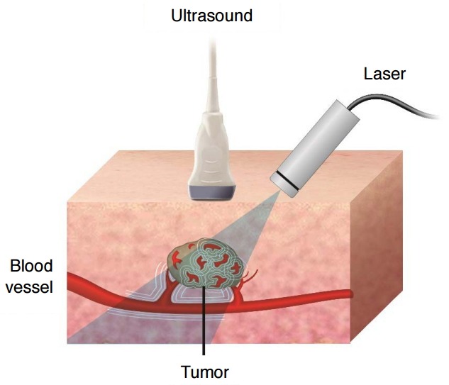
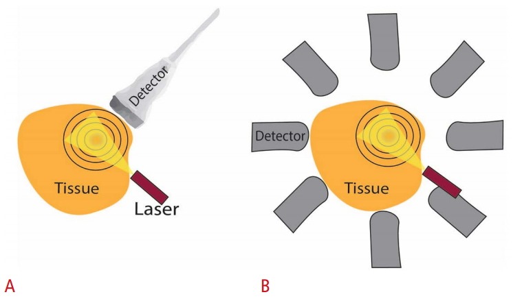
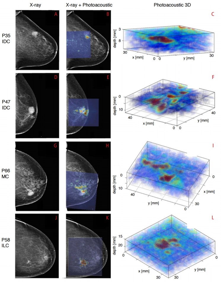
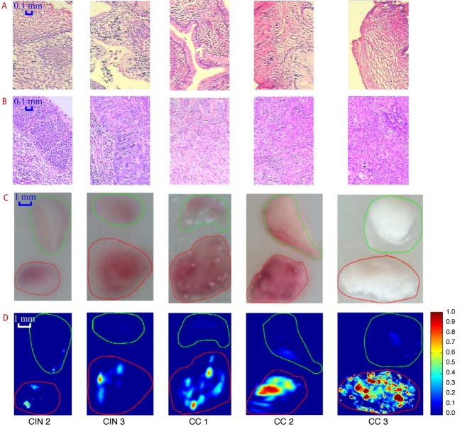
Photoacoustic imaging is a hybrid technique that shines laser light on tissue and measures optically induced ultrasound signal. There is growing interest in the clinical community over this new technique and its possible clinical applications. One of the most prominent features of photoacoustic imaging is its ability to characterize tissue, leveraging differences in the optical absorption of underlying tissue components such as hemoglobin, lipids, melanin, collagen and water among many others. In this review, the state-of-the-art photoacoustic imaging techniques and some of the key outcomes pertaining to different cancer applications in the clinic are presented.

Keywords: Neoplasms; Oncology; Photoacoustic techniques; Spectroscopy, near-infrared.


Conflict of interest statement

No potential conflict of interest relevant to this article was reported.

Figures

Fig. 1.
Fig. 1.. Principle of photoacoustic effect and imaging.
When a tissue is exposed to pulsed near infrared laser, the tissue constituents such as hemoglobin, lipid, water, collagen, etc. absorb light and undergo thermoelastic expansion, thereby emanating ultrasound signals (photoacoustic effect). An ultrasound transducer can thus detect these laser-induced ultrasound signals (photoacoustic signals) and facilitate photoacoustic (or optoacoustic) imaging. Each of these bio-absorbers can be targeted by irradiating tissue at its corresponding dominant absorption wavelength. Hence, by using a tunable laser operating at the relevant wavelengths of interest, one can obtain multiple photoacoustic images that can be spectrally resolved to allow assessment of the tissue composition based on endogenous contrast. For example, by assessing blood oxygenation in the tissue, contrast obtained from photoacoustic imaging can potentially be helpful in detection, characterization, and monitoring of cancer.
Fig. 2.
Fig. 2.. Photoacoustic imaging configurations.
Laser source and the detectors may be on the same side or at an angle to each other. However, an overlap must exist between the laser beam and the detector’s directivity pattern to ensure that laser-induced ultrasound signals are detected and visualized. A. Photoacoustic imaging performed with a conventional ultrasound transducer where only a portion of the spherical wavefront (black rings) originated from the target (red absorber at the center of the rings) is seen by the transducer. B. Photoacoustic tomography approach showing X-ray computed tomography–like reconstruction where a single detector can be rotated around the target or an array of multiple stationary detector elements around the target can be employed. The signal arriving at each detector is filtered back-projected along circular arcs in the spatial domain, and then all the back-projections are added together to obtain the final photoacoustic image which represents the spatial distribution of optical absorption within the target.
Fig. 3.
Fig. 3.. Breast imaging with photoacoustic mammoscope.
Photoacoustic images were overlaid on X-ray mammograms to show lesions detected on both modalities. Reconstructed 3D photoacoustic volume encompassing each lesion of interest is also shown here. Infiltrating ductal carcinoma (IDC) lesion was seen on X-ray and photoacoustic imaging in a 79-year-old patient (A-C) and a 69-year-old patient (D-F), respectively. Mucinous carcinoma (MC) was detected in an 83-year-old patient (G-I) while infiltrating lobular carcinoma (ILC) was seen in a 65-year-old patient (J-L). The lesions were co-localized on photoacoustic images with respect to X-ray mammograms and were visualized at depths of more than 20 mm with good contrast on photoacoustic images. PXX indicates patient-identifier in the study. Reproduced from Heijblom M et al. Eur Radiol 2016, http://dx.doi.org/10.1007/s00330-016-4240-7 [55], according to the Creative Commons license.
Fig. 4.
Fig. 4.. Photoacoustic imaging of human cervix ex vivo .
Each frozen cervical tissue sample consisting of normal tissue and lesion was embedded in a cylindrical agar phantom. Histopathology images showing normal cervical tissue (A) and cervical lesion (B). Photographs of gross tissue specimens embedded in agar showing normal tissue on the top and lesion on the bottom (C). Corresponding photoacoustic images (D) showing signal intensity proportional to the lesion staging. Normal tissue is marked in green and cervical lesion is marked in red. The scale bar for the histological images represents 0.1 mm, and that for the photographs and photoacoustic images is 1 mm. CIN, cervical intraepithelial neoplasia; CC, cervical cancer. Reprinted from Peng K et al. Biomed Opt Express 2015;6:135-143 [67], with permission of Optical Society of America.
Fig. 5.
Fig. 5.. Identification of sentinel lymph node (SLN) in breast cancer patients using photoacoustic imaging.
A. Sonogram shows lymph node in a breast cancer patient. B. Photoacoustic image shows a strong signal from the lymph node due to methylene blue accumulation confirming the ultrasound detected node as a SLN. C. Co-registered photoacoustic image of the SLN overlaid on sonogram. D. Radiograph of surgically removed SLN shows the presence of tissue-marking titanium clip which was implanted under the guidance of photoacoustic imaging, validating the feasibility of photoacoustic imaging to identify SLNs noninvasively. Reproduced from Garcia-Uribe A et al. Sci Rep 2015;5:15748 [75], according to the Creative Commons license.
Fig. 6.
Fig. 6.. Sentinel lymph node (SLN) visualization using a handheld multispectral optoacoustic tomography (MSOT) transducer in patients with melanoma.
Indocyanine green (ICG) dye was injected peritumorally to visualize the lymphatic drainage using photoacoustic imaging. While ICG indicates the location of SLNs, melanin inside the SLN suggests melanoma metastasis. A. Absence of melanin signal in ICG-positive right axillary node suggests that the patient has no detectable metastasis, confirmed as such by histology. B. Photoacoustic imaging of left axillary node (ICG-positive) in a patient suggests metastasis as indicated by strong melanin signal. Immunohistochemistry revealed no evidence of metastasis when stained for Melan A indicating false-positive photoacoustic imaging diagnosis. Left axillary lymph node in a patient showing high melanin signal on photoacoustic image (C) which was confirmed to be metastatic on histology (true positive finding). Adapted from Stoffels I et al. Sci Transl Med 2015;7:317ra199 [71], with permission of The American Association for the Advancement of Science.

References

    1. Willmann JK, van Bruggen N, Dinkelborg LM, Gambhir SS. Molecular imaging in drug development. Nat Rev Drug Discov. 2008;7:591–607. - PubMed
    1. Pysz MA, Willmann JK. Targeted contrast-enhanced ultrasound: an emerging technology in abdominal and pelvic imaging. Gastroenterology. 2011;140:785–790. - PMC - PubMed
    1. Kircher MF, Hricak H, Larson SM. Molecular imaging for personalized cancer care. Mol Oncol. 2012;6:182–195. - PMC - PubMed
    1. Hussain T, Nguyen QT. Molecular imaging for cancer diagnosis and surgery. Adv Drug Deliv Rev. 2014;66:90–100. - PMC - PubMed
    1. Seaman ME, Contino G, Bardeesy N, Kelly KA. Molecular imaging agents: impact on diagnosis and therapeutics in oncology. Expert Rev Mol Med. 2010;12:e20. - PMC - PubMed