Clinical Trial
. 2016 Jul 18;1(10):16113.
doi: 10.1038/nmicrobiol.2016.113.

Enrichment of the lung microbiome with gut bacteria in sepsis and the acute respiratory distress syndrome

Affiliations
Clinical Trial

Enrichment of the lung microbiome with gut bacteria in sepsis and the acute respiratory distress syndrome

Robert P Dickson et al. Nat Microbiol. .

Abstract

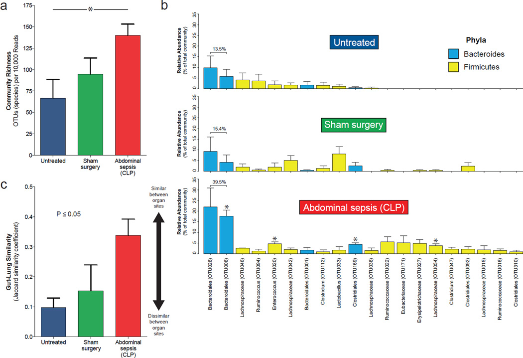
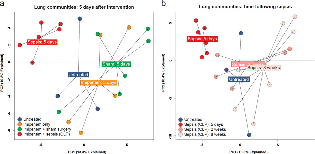
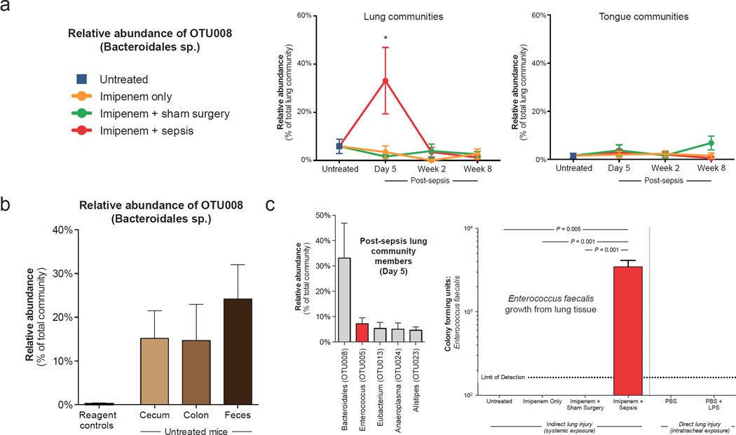
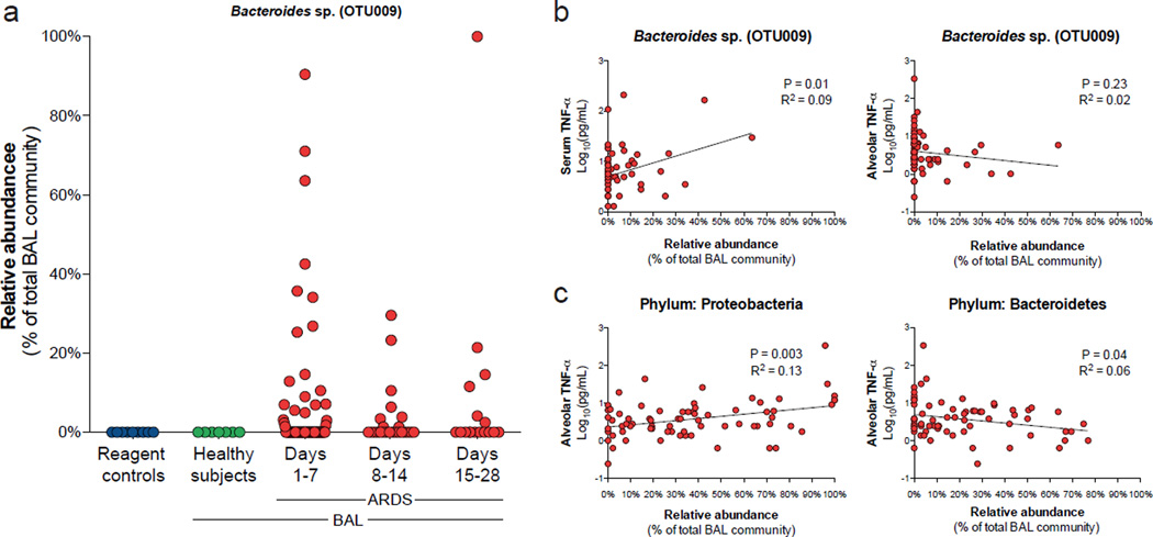
Sepsis and the acute respiratory distress syndrome (ARDS) are major causes of mortality without targeted therapies. Although many experimental and clinical observations have implicated gut microbiota in the pathogenesis of these diseases, culture-based studies have failed to demonstrate translocation of bacteria to the lungs in critically ill patients. Here, we report culture-independent evidence that the lung microbiome is enriched with gut bacteria both in a murine model of sepsis and in humans with established ARDS. Following experimental sepsis, lung communities were dominated by viable gut-associated bacteria. Ecological analysis identified the lower gastrointestinal tract, rather than the upper respiratory tract, as the likely source community of post-sepsis lung bacteria. In bronchoalveolar lavage fluid from humans with ARDS, gut-specific bacteria (Bacteroides spp.) were common and abundant, undetected by culture and correlated with the intensity of systemic inflammation. Alveolar TNF-α, a key mediator of alveolar inflammation in ARDS, was significantly correlated with altered lung microbiota. Our results demonstrate that the lung microbiome is enriched with gut-associated bacteria in sepsis and ARDS, potentially representing a shared mechanism of pathogenesis in these common and lethal diseases.


Figures

Figure 1
Figure 1
Altered lung microbiota 24 hours after experimental sepsis. Abdominal sepsis was induced in mice via cecal ligation and puncture, and lung bacterial communities were sequenced and analyzed after 24 hours. (a) The lungs of post-sepsis mice contained increased species richness compared to untreated mice, consistent with immigration of new species. (b) Relative abundance of bacteria in the lung microbiome following sepsis. The 20 most abundant operational taxonomic units detected in post-sepsis lungs are shown across experimental arms. Asterisks indicate OTUs significantly enriched in post-sepsis lungs compared to control groups. (c) Gut-lung community similarity after sepsis. For each mouse, the community similarity was calculated for paired lung and colon communities. Four mice were used in each intervention group. Group means and standard errors of the mean are depicted. Statistical significance was determined with two-way analysis of variation (ANOVA) with Tukey’s multiple comparisons test. * P ≤ 0.05. Values presented as means ± standard error of the mean. Significance in 1c reflects overall effect of intervention on community richness; between-group differences were not significant after controlling for multiple comparisons.
Figure 2
Figure 2
Transient alteration of the lung microbiome after experimental sepsis. Mice were exposed to abdominal sepsis (cecal ligation and puncture and imipenem) and compared at multiple timepoints to three experimental control groups: untreated, imipenem only, and imipenem with sham surgery. (a) Five days after exposure, bacterial communities in the lungs of post-sepsis mice were distinct from those of all control groups. (b) Lung communities of post-sepsis mice were distinct from untreated mice after five days but indistinguishable at 2 and 8 weeks following injury.
Figure 3
Figure 3
Evidence suggesting gut-lung translocation after experimental sepsis. (a) The bacterial communities of post-sepsis lungs were dominated by an uncultured bacterium, OTU008 (Bacteroidales sp.) that normalized after 2 weeks. This bacterium did not dominate the bacterial communities detected in simultaneously collected tongue specimens. (b) This Bacteroidales sp. was of minimal abundance in reagent control specimens, but was the most abundant community member in all lower gastrointestinal sites of mice before injury. (c) This Bacteroidales sp. was not detected by culture, but Enterococcus faecalis (the second most abundant species detected in the lungs of mice 5 days after sepsis) was isolated via aerobic culture from the lungs of all post-sepsis mice 5 days after injury. It was not isolated from the lungs of any control group, including mice exposed to direct lung injury via intratracheal LPS instillation. Statistical significance was determined by Kruskal–Wallis one-way analysis of variance with Dunn’s multiple comparisons test (a, c). Values presented as means ± standard error of the mean. Six mice were used in each intervention arm and timepoint. PBS: phosphate-buffered saline. LPS: lipopolysaccharide.
Figure 4
Figure 4
Evidence suggesting gut-lung translocation of bacteria in humans with the acute respiratory distress syndrome (ARDS). A Bacteroides sp. (OTU009), a representative of most abundant bacterial genus in the human gut microbiome, was absent from reagent control specimens and the lungs of healthy subjects, but common (33% of specimens) in the lungs of patients with ARDS (a). The relative abundance of this Bacteroides sp. was positively correlated with serum TNF-α but not associated with alveolar TNF-α (b). By contrast, alveolar TNF-α was positively correlated with relative abundance of the Proteobacteria phylum and negatively correlated with the Bacteroidetes phylum (c). 100 bronchoalveolar lavage specimens from 68 unique patients were studied. Statistical significance was determined by linear regression using logarithmically-transformed cytokine data (b, c).

Comment in

References

    1. Angus DC, et al. Epidemiology of severe sepsis in the United States: analysis of incidence, outcome, and associated costs of care. Crit. Care Med. 2001;29:1303–1310. - PubMed
    1. Rubenfeld GD, et al. Incidence and outcomes of acute lung injury. N. Engl. J. Med. 2005;353:1685–1693. - PubMed
    1. Hudson LD, Milberg JA, Anardi D, Maunder RJ. Clinical risks for development of the acute respiratory distress syndrome. Am. J. Respir. Crit. Care Med. 1995;151:293–301. - PubMed
    1. Sheu CC, et al. Clinical characteristics and outcomes of sepsis-related vs non-sepsis-related ARDS. Chest. 2010;138:559–567. - PMC - PubMed
    1. Dickson RP. The microbiome and critical illness. Lancet Respir. Med. 2016;4:59–72. - PMC - PubMed

MeSH terms

Substances