Comparative Study
. 2017 Jan;14(1):25-33.
doi: 10.1016/j.hrthm.2016.09.019. Epub 2016 Sep 23.

Simultaneous noninvasive recording of skin sympathetic nerve activity and electrocardiogram

Affiliations
Comparative Study

Simultaneous noninvasive recording of skin sympathetic nerve activity and electrocardiogram

Anisiia Doytchinova et al. Heart Rhythm. 2017 Jan.

Abstract

Background: Sympathetic nerve activity is important to cardiac arrhythmogenesis.

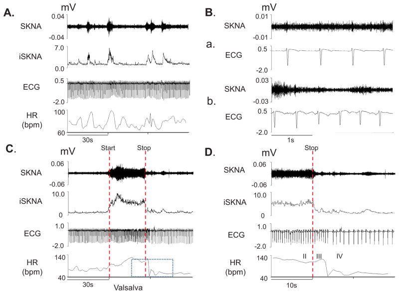
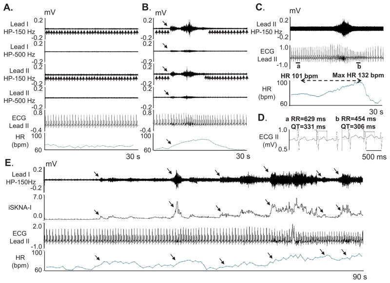
Objective: The purpose of this study was to develop a method for simultaneous noninvasive recording of skin sympathetic nerve activity (SKNA) and electrocardiogram (ECG) using conventional ECG electrodes. This method (neuECG) can be used to adequately estimate sympathetic tone.

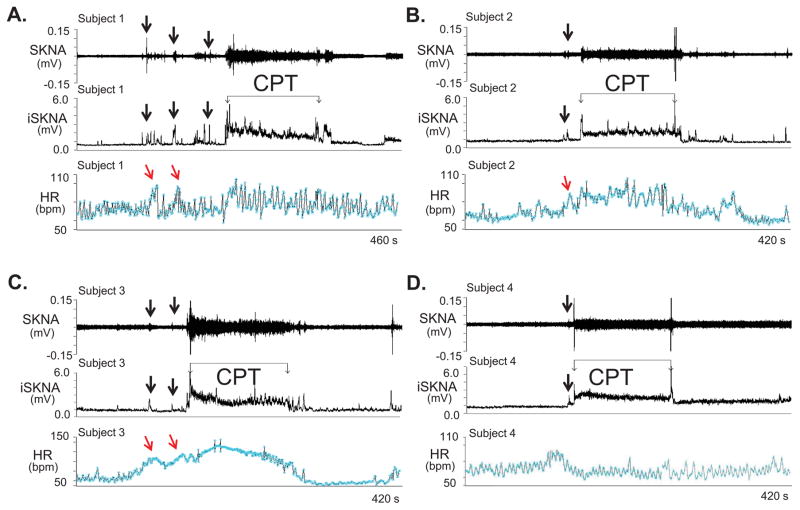
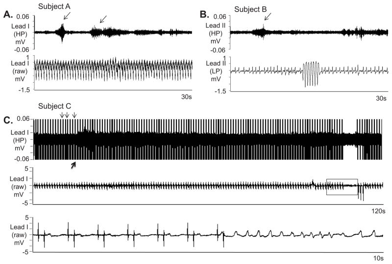
Methods: We recorded neuECG signals from the skin of 56 human subjects. The signals were low-pass filtered to show the ECG and high-pass filtered to show nerve activity. Protocol 1 included 12 healthy volunteers who underwent cold water pressor test and Valsalva maneuver. Protocol 2 included 19 inpatients with epilepsy but without known heart diseases monitored for 24 hours. Protocol 3 included 22 patients admitted with electrical storm and monitored for 39.0 ± 28.2 hours. Protocol 4 included 3 patients who underwent bilateral stellate ganglion blockade with lidocaine injection.

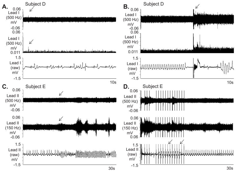
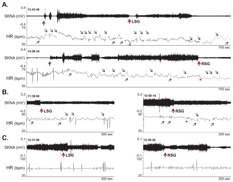
Results: In patients without heart diseases, spontaneous nerve discharges were frequently observed at baseline and were associated with heart rate acceleration. SKNA recorded from chest leads (V1-V6) during cold water pressor test and Valsalva maneuver (protocol 1) was invariably higher than during baseline and recovery periods (P < .001). In protocol 2, the average SKNA correlated with heart rate acceleration (r = 0.73 ± 0.14, P < .05) and shortening of QT interval (P < .001). Among 146 spontaneous ventricular tachycardia episodes recorded in 9 patients of protocol 3, 106 episodes (73%) were preceded by SKNA within 30 seconds of onset. Protocol 4 showed that bilateral stellate ganglia blockade by lidocaine inhibited SKNA.

Conclusion: SKNA is detectable using conventional ECG electrodes in humans and may be useful in estimating sympathetic tone.

Keywords: Cold water pressor test; Microneurography; Sympathetic nerve activity; Ventricular tachycardia.


Conflict of interest statement

Shien-Fong Lin and Peng-Sheng Chen have equity interest in Arrhythmotech, LLC. Medtronic, St Jude and Cyberonics Inc. donated research equipment to Dr Chen’s research laboratory.

Figures

Figure 1
Figure 1
neuECG recordings from subject 12 of Protocol 1. Signals from Lead V1 were bandpass filtered between 500 Hz and 1000 Hz to detect SKNA and bandpass filtered between 0.5 Hz and 150 Hz to detect ECG. Integrated SKNA (iSKNA) was calculated over 100 ms window. A: Increased SKNA was associated with heart rate (HR) acceleration. B: Higher magnification of SKNA showing baseline spontaneous nerve activities (a) and large variations of nerve discharges associated with tachycardia (b). C: increased SKNA and HR were evident during Valsalva maneuver (VM); dotted red lines mark the start and the stop of the maneuver. D shows the magnified boxed segment from C, showing phases II–IV of the VM and demonstrating that the SKNA is not synchronous with the QRS complex.
Figure 2
Figure 2
neuECG recordings during the cold water pressor test (CPT) in Protocol 1. The electrode location was on the right and left arm for ECG Lead I recording. AD: Increased skin sympathetic nerve activity (SKNA) was detected in subjects 1–4, respectively, during the CPT. Black downward arrows point to increased SKNA prior to CPT, likely due to the anticipation of the impending cold water immersion. The increased SKNA was associated with heart rate acceleration in patients 1–3, but not in patient 4. Integrated SKNA (iSKNA) shows the total SKNA over 100 ms windows after applying 500 Hz high pass filter. HR= heart rate, bmp=beats per minute.
Figure 3
Figure 3
neuECG recording in patients without known heart diseases in Protocol 2. The neuECG electrodes were placed on the chest to form Lead I and Lead II. A shows baseline recording in leads I and II filtered at either 150 Hz or 500 Hz high pass to display SKNA and low pass filtered at 10 Hz to display the ECG. B shows an episode of SKNA associated with heart rate (HR) acceleration (downward arrows). The 150 Hz high pass filter resulted in better signal to noise ratio and higher amplitude of SKNA, but some ECG signals remained (upward arrows). High pass filter at 500 Hz largely eliminated the ECG signals, but also reduced nerve amplitude and the signal to noise ratio. The baseline artifact on the surface ECG occurred after the onset of SKNA, suggesting motion artifacts induced by muscle movement. C shows SKNA (500 Hz high pass, Lead II) and ECG tracings (125 Hz low pass) from a different patient. There was abrupt increase of HR from 101 beats per minute (bpm) to a maximum (max) of 132 bpm after SKNA activation, along with QT interval shortening. D shows the enlarged ECG from line segments a and b in C. Both the RR and the QT interval shortened after SKNA. E shows a 90 s recording at baseline, illustrating spontaneous SKNA episodes and their relationship with HR. HP=high pass, LP=low pass, bpm=beats per minute.
Figure 4
Figure 4
SKNA during sustained VT and before nonsustained VT. The neuECG electrodes were placed on the chest to form Lead I and Lead II. A: Nerve discharges (arrows) are noted throughout monomorphic ventricular tachycardia (VT). Signals simultaneously obtained from ECG lead I with the top panel representing the signal after 500 Hz high pass (HP) filter and the bottom panel displaying the raw signal. B: Similar discharges are observed in another patient preceding non-sustained VT. Signal simultaneously obtained from ECG lead II, the top panel is filtered at 500 Hz high pass and the bottom ECG is filtered at 10 Hz low pass. C: Pacing artifacts (downward arrows) are observed despite 500 Hz high pass filtering. Increased high frequency SKNA is still evident (upward arrow) beginning 90 s prior to VT. The bottom panel shows the boxed segment from the middle panel and the onset of VT. VT=ventricular tachycardia, ECG=electrocardiogram, HP=500 Hz high pass filter, LP=10 Hz low pass filter, SKNA=skin sympathetic nerve activity.
Figure 5
Figure 5
Further examples of SKNA and VT. A: Increased nerve activity, including a large spike (arrow) is noted in lead I in a intubated and sedated patient with electrical storm after 500 Hz high pass filtering. The signal is better appreciated in the middle panel after rectification and baseline adjustment. The simultaneous raw electrocardiogram (ECG) is displayed in the bottom panel and shows both premature ventircular contractions and a brief run of polymorphic VT. B displays similar simultaneous recordings in the same patient during sustained VT requiring external defibrillation (arrow, top panel). The defibrillation was followed immediately by large SKNA. The shock was only transiently successful, followed by recurrences of VT. Recordings from C and D are from another patient with VT and are continuous. In this recording after high pass filtering, the signal to noise ratio of SKNA prior to VT in C, top and middle panel does not reach 2:1. High pass filtering at 150 Hz again improves the signal to noise ratio (downward arrows, C). However as the arrhythmia continues in D and the patient’s device delivers successful burst of anti-tachycardia pacing (downward arrows, bottom panel), incraesed SKNA is detected both with 500 Hz high pass filter and 150 Hz high pass filter. VT=ventricular tachycardia, ECG=electrocardiogram, SKNA=skin sympathetic nerve activity.
Figure 6
Figure 6
Effects of lidocaine (10 ml, 2 %) stellate ganglion block on SKNA continuously recorded from the right arm. A shows patient 1. Needle insertion (black arrows) was followed by activation of SKNA. Lidocaine injection (red arrows) into the LSG transiently reduced SKNA. However, RSG injection was followed by a significant reduction of SKNA. Panels B and C show responses to lidocaine injection in the remaining 2 patients. The gaps in tachogram (small black upward arrows) occurred because artifacts prevented automated selections of the R waves.

References

    1. Kligfield P, Gettes LS, Bailey JJ, et al. Recommendations for the standardization and interpretation of the electrocardiogram. Part I: The electrocardiogram and its technology.A scientific statement from the American Heart Association Electrocardiography and Arrhythmias Committee, Council on Clinical Cardiology; the American College of Cardiology Foundation; and the Heart Rhythm Society. Heart Rhythm. 2007;4:394–412. - PubMed
    1. Chakravarthy Marx S, Kumar P, Dhalapathy S, Anitha Marx C. Distribution of sympathetic fiber areas in the sensory nerves of forearm: an immunohistochemical study in cadavers. Rom J Morphol Embryol. 2011;52:605–611. - PubMed
    1. Donadio V, Nolano M, Provitera V, Stancanelli A, Lullo F, Liguori R, Santoro L. Skin sympathetic adrenergic innervation: an immunofluorescence confocal study. Ann Neurol. 2006;59:376–381. - PubMed
    1. Baron R, Janig W, With H. Sympathetic and afferent neurones projecting into forelimb and trunk nerves and the anatomical organization of the thoracic sympathetic outflow of the rat. J Auton Nerv Syst. 1995;53:205–214. - PubMed
    1. Taniguchi T, Morimoto M, Taniguchi Y, Takasaki M, Totoki T. Cutaneous distribution of sympathetic postganglionic fibers from stellate ganglion: A retrograde axonal tracing study using wheat germ agglutinin conjugated with horseradish peroxidase. J Anesth. 1994;8:441–449. - PubMed

Publication types