Review
. 2016 Nov 2;35(21):2270-2284.
doi: 10.15252/embj.201694613. Epub 2016 Sep 26.

Membrane fission by dynamin: what we know and what we need to know

Affiliations
Review

Membrane fission by dynamin: what we know and what we need to know

Bruno Antonny et al. EMBO J. .

Abstract

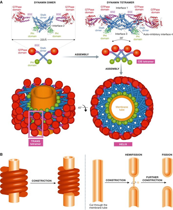
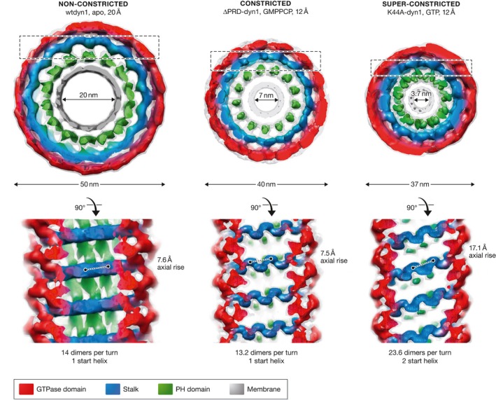
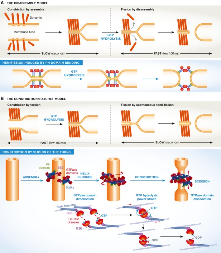
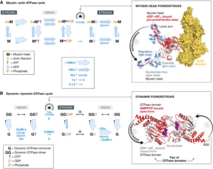
The large GTPase dynamin is the first protein shown to catalyze membrane fission. Dynamin and its related proteins are essential to many cell functions, from endocytosis to organelle division and fusion, and it plays a critical role in many physiological functions such as synaptic transmission and muscle contraction. Research of the past three decades has focused on understanding how dynamin works. In this review, we present the basis for an emerging consensus on how dynamin functions. Three properties of dynamin are strongly supported by experimental data: first, dynamin oligomerizes into a helical polymer; second, dynamin oligomer constricts in the presence of GTP; and third, dynamin catalyzes membrane fission upon GTP hydrolysis. We present the two current models for fission, essentially diverging in how GTP energy is spent. We further discuss how future research might solve the remaining open questions presently under discussion.

Keywords: GTPase; dynamin; endocytosis; membrane fission; molecular motor.


Figures

Figure 1
Figure 1. Structure and assembly of dynamin
(A) Crystal structure of the dimer and of the tetramer, showing the interfaces required for assembly. A schematic representation shows how the tetramers further assemble into a helix, showing the basic CIS‐tetramer and TRANS‐tetramers. (B) The original constriction model for dynamin‐mediated membrane fission, as suggested by the helical structure of dynamin.
Figure 2
Figure 2. The three states of the dynamin helix observed by cryo‐EM, with dimensions and angles.
Figure 3
Figure 3. The two models of dynamin‐mediated membrane fission
(A) The two‐stage model, where constriction is mediated by assembly, and fission by disassembly. (B) The constriction/ratchet model in which constriction is realized by active sliding of the helical turns and fission by spontaneous fusion of the membrane. The one ring state presented here is proposed to be the most common in vivo (see text).
Figure 4
Figure 4. Comparison of the skeletal muscle myosin ATPase (A) with the dynamin GTPase (B) cycles
Both reaction pathways are populated by chemical intermediates defined in the figure. High energy states are indicated with an asterisk. Arrows indicate the reactions between each pair of intermediates. The sizes of the blue arrows are proportional to the rates under physiological conditions (taking into account the concentrations for bimolecular reactions) as defined at the bottom right. The black arrows in (B) indicate unknown rates. The bottom rows in (A) and (B) are reactions of myosin (M) and dynamin (G) monomers. The top rows are reactions of myosin bound to an actin filament (AM) or dynamin dimers (GG). The vertical arrows indicate the rates of myosin binding actin filaments and dynamin forming dimers. In (A), the right panel represents a superposition of myosin in the nucleotide‐free, pre‐power stroke state (pdb 2mys, white) and the ADPAlF4‐bound rigor state (pdb 1br1, red). ADPAlF4 is shown in magenta, and the two myosin light chains bound to the lever arm are shown in blue and dark blue. The positions of the second light chain and the distal end of the lever in pdb 1br1 were modeled based on pdb 2mys. Five actin molecules (yellow) are indicated (from pdb 5jlh). In (B), the right panel represents a superposition of the G domains in the dynamin GG construct in the GMPPCP‐bound open (pdb 3zyk in red) and the GDPAlF4‐bound closed form (pdb 2x2e in white). Nucleotides are shown in magenta. Note the 70° rotation of the BSE relative to the G domain.

References

    1. Abutbul‐Ionita I, Rujiviphat J, Nir I, McQuibban GA, Danino D (2012) Membrane tethering and nucleotide‐dependent conformational changes drive mitochondrial genome maintenance (Mgm1) protein‐mediated membrane fusion. J Biol Chem 287: 36634–36638 - PMC - PubMed
    1. Arimura S, Tsutsumi N (2002) A dynamin‐like protein (ADL2b), rather than FtsZ, is involved in Arabidopsis mitochondrial division. Proc Natl Acad Sci USA 99: 5727–5731 - PMC - PubMed
    1. Ban T, Heymann JA, Song Z, Hinshaw JE, Chan DC (2010) OPA1 disease alleles causing dominant optic atrophy have defects in cardiolipin‐stimulated GTP hydrolysis and membrane tubulation. Hum Mol Genet 19: 2113–2122 - PMC - PubMed
    1. Bashkirov PV, Akimov SA, Evseev AI, Schmid SL, Zimmerberg J, Frolov VA (2008) GTPase cycle of dynamin is coupled to membrane squeeze and release, leading to spontaneous fission. Cell 135: 1276–1286 - PMC - PubMed
    1. Binns DD, Helms MK, Barylko B, Davis CT, Jameson DM, Albanesi JP, Eccleston JF (2000) The mechanism of GTP hydrolysis by dynamin II: a transient kinetic study. Biochemistry 39: 7188–7196 - PubMed