Review
. 2017 Jan;26(1):52-68.
doi: 10.1002/pro.3052. Epub 2016 Oct 13.

A guide to the 3D structure of the ryanodine receptor type 1 by cryoEM

Affiliations
Review

A guide to the 3D structure of the ryanodine receptor type 1 by cryoEM

Montserrat Samsó. Protein Sci. 2017 Jan.

Abstract

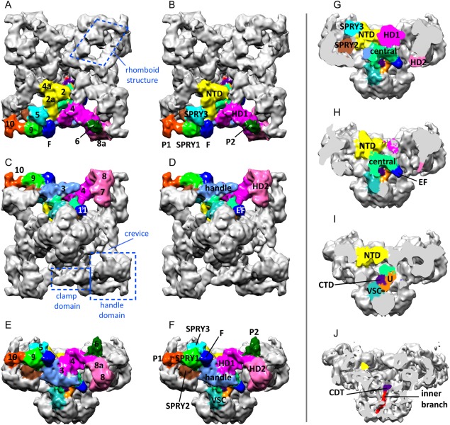
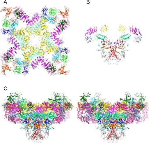
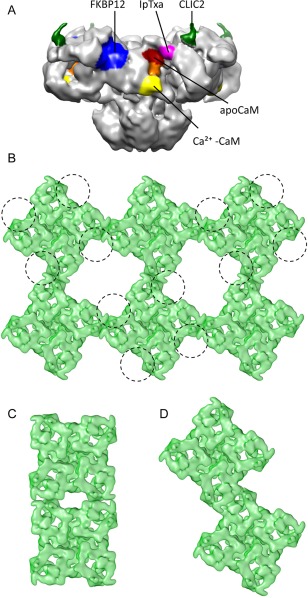
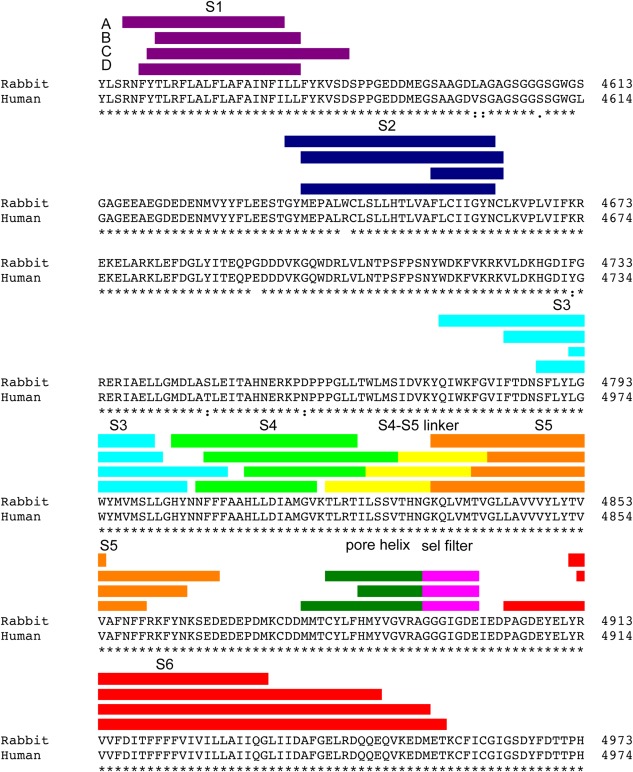
Signal transduction by the ryanodine receptor (RyR) is essential in many excitable cells including all striated contractile cells and some types of neurons. While its transmembrane domain is a classic tetrameric, six-transmembrane cation channel, the cytoplasmic domain is uniquely large and complex, hosting a multiplicity of specialized domains. The overall outline and substructure readily recognizable by electron microscopy make RyR a geometrically well-behaved specimen. Hence, for the last two decades, the 3D structural study of the RyR has tracked closely the technological advances in electron microscopy, cryo-electron microscopy (cryoEM), and computerized 3D reconstruction. This review summarizes the progress in the structural determination of RyR by cryoEM and, bearing in mind the leap in resolution provided by the recent implementation of direct electron detection, analyzes the first near-atomic structures of RyR. These reveal a complex orchestration of domains controlling the channel's function, and help to understand how this could break down as a consequence of disease-causing mutations.

Keywords: 3D reconstruction; allosterism; calcium; cryo electron microscopy; excitation-contraction coupling; ryanodine receptor.


Figures

Figure 1
Figure 1
Multi‐domain structure of the cytoplasmic domain of RyR1. Cytoplasmic domain in three orthogonal views. The domains in one subunit are color‐coded. (A, B) Cytoplasmic view. (C, D) Endo/sarcoplasmic reticulum luminal view. (E, F) Side view. In (A‐F) the panels on the left show the traditional numerical designation and the panels on the right show the new names of the domains. (G–J) Successive slices towards the fourfold axis showing the internal domains. Based on the cryoEM map EMDB‐1606.
Figure 2
Figure 2
Near‐atomic structure of RyR1. (A) Cytoplasmic view; the transmembrane domain is omitted for clarity. (B) Central slice of the side view. (C) Stereo view. Subunits are color‐coded as in Figure 1 and S6 is colored in red. Based on the atomic model 3J8H.pdb.
Figure 3
Figure 3
Quaternary interactions of RyR. (A) Side view showing the known binding sites for different RyR1 ligands: FKPB12 (blue), apoCaM (red), Ca2+‐CaM (yellow), approximate region of apo‐ and Ca2+‐CaM overlap (orange), CLIC2 (green) and IpTxa (magenta). (B) Cytoplasmic view showing an array of RyR1s and a tentative footprint of the DHPR tetrads (each DHPR represented with a circumference) interacting with every other RyR1. (C) Side‐by‐side interaction of RyR2. (D) Oblique interaction of RyR2.
Figure 4
Figure 4
Transmembrane domain sequence assignment. Predictions of the boundaries of the transmembrane segments of RyR1 based on (A) hydropathy plots,114 (B) refined secondary structure prediction,115 (C) modeling based on a 4.8 Å cryoEM map,46 and (D) modeling based on a 3.8 Å resolution cryoEM map.47
Figure 5
Figure 5
Structure of the transmembrane domain of RyR1. (A) Transmembrane domain seen from the cytoplasm; α‐helices are color‐coded as indicated in Figure 4. (B) Side view of the transmembrane domain for one subunit; cytoplasmic side is above and SR luminal side is below. The atomic model corresponds to 3J8H.pdb.
Figure 6
Figure 6
The closed, open and inactivated conformations of RyR1. (A) RyR1 closed at 3.8 Å resolution (cryoEM map EMDB‐2807); map filtered to 7 Å for easier comparison. The extra mass surrounding the transmembrane domain corresponds to the detergent micelle. (B) RyR1 in open conformation at 10‐Å resolution (cryoEM map EMDB‐1607). Green arrows indicate the swivel movement of the rhomboid structure in going form the closed to the open conformation, green dot shows the approximate center of rotation. (C) RyR1 inactivated at 8.5‐Å resolution (cryoEM map EMDB‐2752). The side view is cut along the fourfold axis, the panels below show a magnified image of the regions surrounding the ion gate (ig). The C‐terminal domain is highlighted in blue and S6, formed by the inner helices (ih) and inner branches (ib), is highlighted in red in all four subunits. The CTD and the region of the U‐motif contacting the inner branches are also highlighted.
Figure 7
Figure 7
A change in trajectory of S6 opens the channel passage. (A–F) Cross‐sections of S6 in the closed (green, cryoEM map EMDB‐1606) and open (magenta, cryoEM map EMDB‐1607) conformations, in going from the cytoplasm to the SR lumen, perpendicular to the fourfold axis. (A–B) Inner branches (C, D) Ion gate; the flexibility of the inner helices in the open state results in loss of continuity in section (D). (E, F) Inner helices. (G, H) Cross‐section of RyR1 in the closed and open conformations displayed at a lower threshold to reveal the different profiles of the channel passage.

References

    1. Lanner JT, Georgiou DK, Joshi AD, Hamilton SL (2010) Ryanodine receptors: structure, expression, molecular details, and function in calcium release. Cold Spring Harb Perspect Biol 2:a003996. - PMC - PubMed
    1. Lewarchik CM, Orabi AI, Jin S, Wang D, Muili KA, Shah AU, Eisses JF, Malik A, Bottino R, Jayaraman T, Husain SZ (2014) The ryanodine receptor is expressed in human pancreatic acinar cells and contributes to acinar cell injury. Am J Physiol Gastrointest Liver Physiol 307:G574–5G581. - PMC - PubMed
    1. Ogawa Y, Kurebayashi N, Murayama T (2000) Putative roles of type 3 ryanodine receptor isoforms (RyR3). Trends Cardiovasc Med 10:65–70. - PubMed
    1. Hosoi E, Nishizaki C, Gallagher KL, Wyre HW, Matsuo Y, Sei Y (2001) Expression of the ryanodine receptor isoforms in immune cells. J Immunol 167:4887–4894. - PubMed
    1. Beam KG, Knudson CM, Powell JA (1986) A lethal mutation in mice eliminates the slow calcium current in skeletal muscle cells. Nature 320:168–170. - PubMed

Publication types

MeSH terms

Substances