. 2016 Nov 1;197(9):3639-3649.
doi: 10.4049/jimmunol.1600402. Epub 2016 Sep 26.

HIF-2α in Resting Macrophages Tempers Mitochondrial Reactive Oxygen Species To Selectively Repress MARCO-Dependent Phagocytosis

Affiliations

HIF-2α in Resting Macrophages Tempers Mitochondrial Reactive Oxygen Species To Selectively Repress MARCO-Dependent Phagocytosis

Shirley Dehn et al. J Immunol. .

Abstract

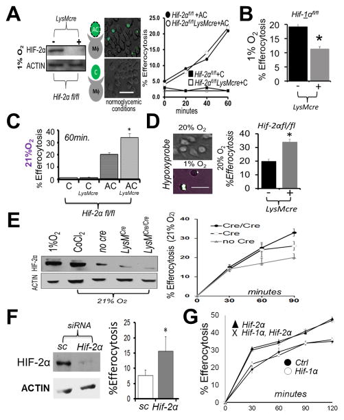
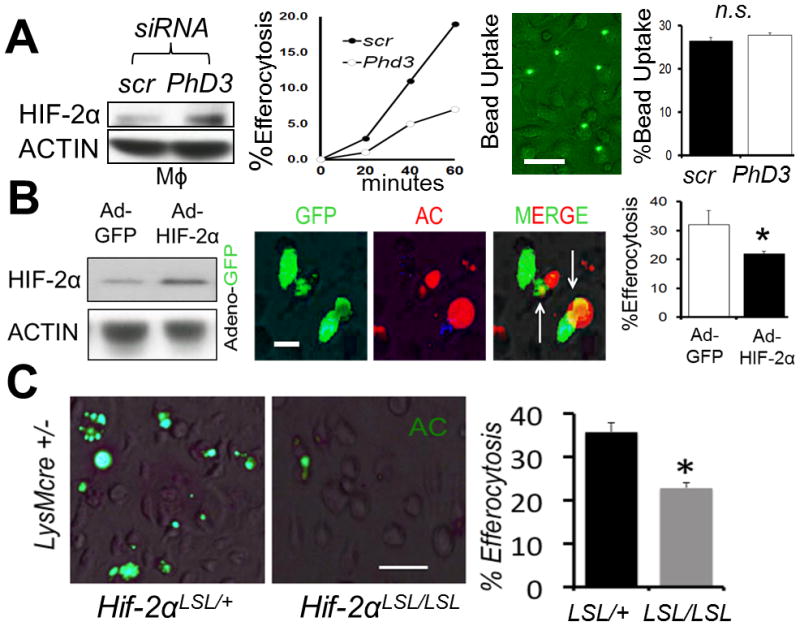
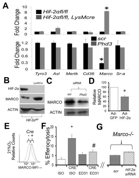
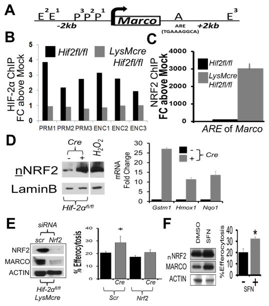
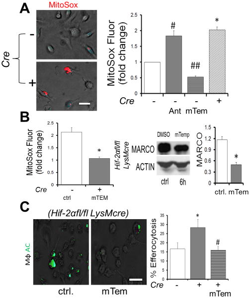
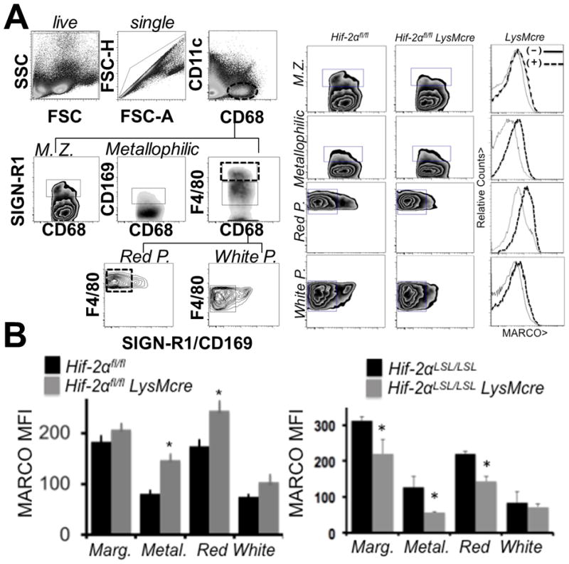
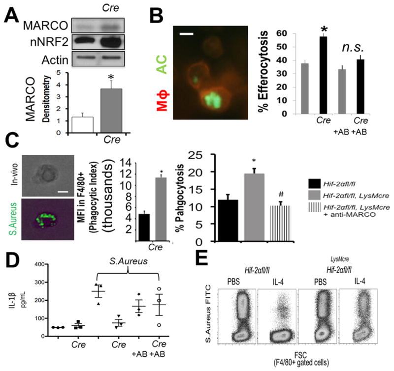
Hypoxia-inducible factor (HIF)-α isoforms regulate key macrophage (MΦ) functions during ischemic inflammation. HIF-2α drives proinflammatory cytokine production; however, the requirements for HIF-2α during other key MΦ functions, including phagocytosis, are unknown. In contrast to HIF-1α, HIF-2α was not required for hypoxic phagocytic uptake. Surprisingly, basal HIF-2α levels under nonhypoxic conditions were necessary and sufficient to suppress phagocytosis. Screening approaches revealed selective induction of the scavenger receptor MARCO, which was required for enhanced engulfment. Chromatin immunoprecipitation identified the antioxidant NRF2 as being directly responsible for inducing Marco Concordantly, Hif-2α-/- MΦs exhibited reduced antioxidant gene expression, and inhibition of mitochondrial reactive oxygen species suppressed Marco expression and phagocytic uptake. Ex vivo findings were recapitulated in vivo; the enhanced engulfment phenotype resulted in increased bacterial clearance and cytokine suppression. Importantly, natural induction of Hif-2α by IL-4 also suppressed MARCO-dependent phagocytosis. Thus, unlike most characterized prophagocytic regulators, HIF-2α can act as a phagocytic repressor. Interestingly, this occurs in resting MΦs through tempering of steady-state mitochondrial reactive oxygen species. In turn, HIF-2α promotes MΦ quiescence by blocking a MARCO bacterial-response pathway. IL-4 also drives HIF-2α suppression of MARCO, leading to compromised bacterial immunosurveillance in vivo.


Figures

Figure 1
Figure 1. Hif-2α is not required for hypoxic efferocytosis, in contrast to under non-hypoxic conditions, where Hif-2α specifically acts as an efferocytic suppressor
(A) Immunoblot of Hif-2αfl/fl LysMcre bone marrow-derived macrophages (Mϕs) after culture at 1% oxygen. Mϕs were overlaid with fluorescent (calcein-AM) apoptotic Jurkat T-cells (“AC”) or fluorescent viable Jurkat T-cells (“C”) and efferocytosis quantified. Scale bar = 60 micrometers. (B) Efferocytosis carried out similar to as in (A) except during with Hif-1αfl/fl vs. Hif-1αfl/fl LysMcre Mϕs. (C) Efferocytosis similar to as in (A) except under normoxia. (D) Efferocytosis by Hif-2αfl/fl vs Hif-2αfl/fl LysMcre phagocytes cultured on gas permeable plates. Wild type cultures were grown on Lumox plates to optimize gas exchange at indicated oxygen tensions and stained with hypoxyprobe (green) as an indicator of hypoxia. Efferocytosis of fluorescent apoptotic Jurkat cells by Hif-2αfl/fl vs. Hif-2αfl/fl LysMcre phagocytes after quantification by microscopy. (E) Immunoblots of HIF-2α levels as a function of efferocytosis efficiency. (F) Immunoblot of normoxic Mϕ HIF-2α after Hif-2α siRNA in mature Mϕs and % Efferocytosis at 30min. Sc = scrambled siRNA. (G) Normoxic efferocytosis kinetics of Hif-2αfl/fl LysMcre vs. Hif-1αfl/fl Hif-2αfl/fl LysMcre vs. Hif-1αfl/fl LysMcre vs. control (Hif- 1αfl/fl Hif-2αfl/fl). * indicates < 0.05 relative to control.
Figure 2
Figure 2. Strategies that induce HIF-2α are sufficient to suppress efferocytosis
(A) Bone marrow-derived Mϕs were treated with scrambled (scr) vs. Phd3 siRNA and HIF-2α levels revealed by immunoblot, as a function of efferocytosis and engulfment of control latex beads. (B) Adeno-HIF-2α-GFP (green) was transduced into Mϕs and immunoblots and efferocytosis of red apoptotic cells (ACs) quantified. (C) Non-stained primary Hif-2α LSL/+ LysMcre vs. Hif-2α LSL/LSL LysMcre Mϕs were challenged with ACs and efferocytosis imaged and quantified. Scale bar = 60 micrometers. * indicates < 0.05 relative to control.
Figure 3
Figure 3. Hif-2α deficiency enhances MARCO expression in primary mouse Mϕs, which is required for enhanced efferocytosis
(A) qPCR data of indicated receptors from Hif-2αfl/fl LysMcre (TOP) vs after Phd3 siRNA from Mϕs under normoxic conditions. (B) Immunoblot of MARCO in Hif-2αfl/fl vs. Hif-2αfl/fl LysMcre Mϕs under normoxia. (C) Immunoblot of MARCO after PhD3 siRNA in Mϕs under normoxia. (D) Densitometry of MARCO protein after adenoviral Hif-2α expression under normoxia. (E) Cell surface flow cytometry of MARCO in Hif-2αfl/fl vs. Hif-2αfl/fl LysMcre Mϕs. (F) Hif-2αfl/fl versus Hif2αfl/fl LysMcre (CRE) primary Mϕs were treated with 10μg/mL MARCO blocking antibody (clone ED31, azide-free) or IgG1 isotype (ISO) control prior to co-culture with fluorescently-labelled apoptotic cells and efferocytosis enumerated. * indicates p < 0.05 vs. ISO control and # indicates p < 0.04 vs. CRE/ISO control. (G) Marco−/− Mϕs were treated with Hif-2α siRNA and challenged with apoptotic cells (Bar graph axis same as in (F).
Figure 4
Figure 4. Minimal HIF-2α association with Marco, in contrast to NRF2, which is required for enhanced efferocytosis
(A) Schematic of Marco gene regulator regions, including Enhancer (E), Promoter (P), and ARE element. (B) Chromatin precipitation for HIF-2α on 3 P (PRM1-3) and 3 E (ENC1-3) regions each. (C) Chromatin precipitation for NRF2 on Marco. (D) Nuclear translocation of NRF2 in control vs Hif-2αfl/fl LysMcre Mϕs and induction of indicated NRF2 target genes. (E) Nrf2 siRNA tests (vs scrambled/scr) reduces MARCO in Hif-2αfl/fl LysMcre Mϕs. Efferocytosis efficiency in control Hif-2αfl/fl vs. Hif-2αfl/fl LysMcre Mϕs treated with indicated siRNA (Scr vs Nrf2). * indicates < 0.05 relative to control. (F) NRF2 agonist induces MARCO and enhances efferocytosis. WT bone marrow derived macrophages were treated with 20uM Sulforaphane, NRF2 agonist, for 24hrs, prior to blotting (nNRF2 = nuclear NRF2) and efferocytosis.
Figure 5
Figure 5. Elevated mROS increases MARCO & efferocytosis in the absence of Hif-2α
(A) Elevated mROS in Hif-2α-deficient Mϕs was measured vs. control with MitoSox. Antimycin A, an electron transport chain-inhibitor and positive control for mROS formation. mTempo was used to inhibit mROS. # and ## and * indicate p < 0.05 versus control. (B) mTEMPO reduces MitoSox mROS in Hif-2α deficient Mϕs and also suppresses MARCO. (C) Mito-Tempo reduces enhanced efferocytosis. Scale bar indicates 30 micrometers. * indicates p < 0.05 versus WT control. # indicates p < 0.05 versus knockout control.
Figure 6
Figure 6. Hif-2α-MARCO axis is revealed in vivo
(A) Evidence for elevated MARCO protein on splenic macrophages from Hif-2αfl/fl vs Hif-2αfl/fl LysMcre. To the left is the gating strategy for splenic macrophage subsets. To the right are the analyses of the individual populations. (B) Quantification of elevations in MARCO expression (mean fluorescent intensity) in Hif-2αfl/fl vs Hif-2αfl/fl LysMcre mice and Hif-2α LSL/LSL mice vs. Hif-2α LSL/LSL LysMcre mice.
Figure 7
Figure 7. Hif-2α-Marco axis is functional in vivo
(A) Evidence for elevated MARCO protein and nuclear NRF2 in resident peritoneal Mϕs from Hif-2αfl/fl vs. Hif-2αfl/fl LysMcre mice after immunoblotting. (B) Peritoneal resident Mϕ efferocytosis after intraperitoneal injection of fluorescent apoptotic thymocytes (AC) into Hif-2αfl/fl vs. Hif-2αfl/fl LysMcre mice +/− AB (MARCO blocking antibody). (C) Acute phagocytosis of S. Aureus is enhanced in the absence of Hif-2α through MARCO. Heat-killed FITC-labeled S. Aureus were injected into the peritoneum of Hif-2α-deficient mice and % phagocytosis by resident F4/80+ phagocytes enumerated. Anti-MARCO antibody was added prior to bacterial injection. (D) Plasma cytokine levels were examined in Hif-2αfl/fl vs Hif-2αfl/fl LysMcre mice after injection of S. Aureus vs. PBS vehicle and +/− MARCO blocking antibody vs. isotype control. (E) IL-4 suppression of F4/80-gated macrophage phagocytosis under saturating MOI requires Hif-2α.

Similar articles

Cited by

References

    1. Medzhitov R. Origin and physiological roles of inflammation. Nature. 2008;454:428–435. - PubMed
    1. Gordon S. The macrophage: past, present and future. Eur J Immunol. 2007;37:S9–17. - PubMed
    1. Nathan C. Immunology: Oxygen and the inflammatory cell. Nature. 2003 Apr 17;422(6933):675–6. - PubMed
    1. Eltzschig HK, Carmeliet P. Hypoxia and inflammation. N Engl J Med. 2011;364:656–665. - PMC - PubMed
    1. Nizet V, Johnson RS. Interdependence of hypoxic and innate immune responses. Nature reviews Immunology. 2009;9:609–617. - PMC - PubMed

MeSH terms

Substances