

Intraluminal delivery of thrombospondin-2 small interfering RNA inhibits the vascular response to injury in a rat carotid balloon angioplasty model
- PMID: 27671229
- PMCID: PMC5161524
- DOI: 10.1096/fj.201600501R
Intraluminal delivery of thrombospondin-2 small interfering RNA inhibits the vascular response to injury in a rat carotid balloon angioplasty model
Abstract
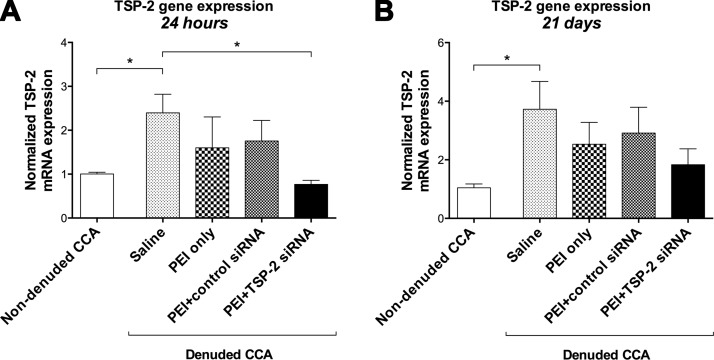
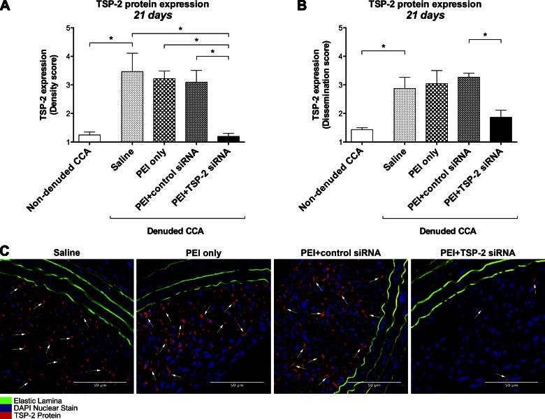
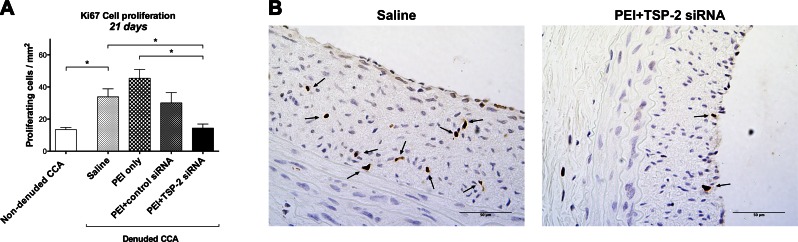
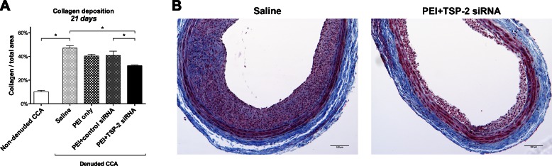
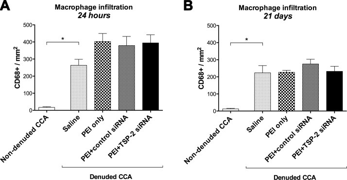
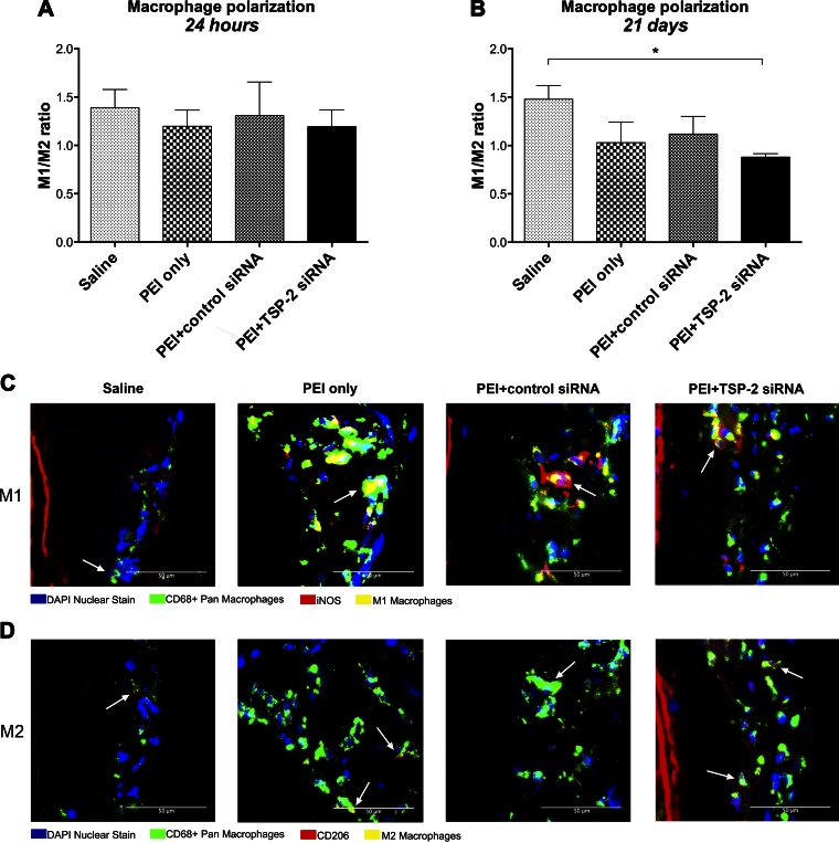
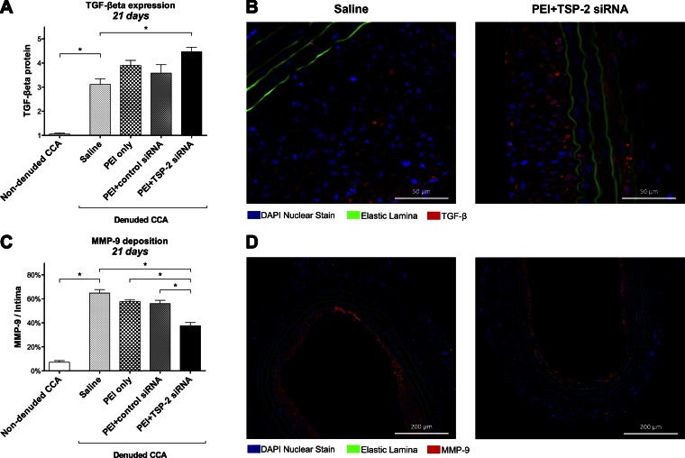
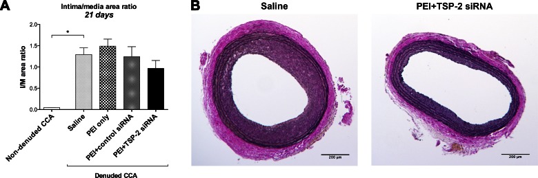
In an effort to inhibit the response to vascular injury that leads to intimal hyperplasia, this study investigated the in vivo efficacy of intraluminal delivery of thrombospondin-2 (TSP-2) small interfering RNA (siRNA). Common carotid artery (CCA) balloon angioplasty injury was performed in rats. Immediately after denudation, CCA was transfected intraluminally (15 min) with one of the following: polyethylenimine (PEI)+TSP-2 siRNA, saline, PEI only, or PEI+control siRNA. CCA was analyzed at 24 h or 21 d by using quantitative real-time PCR and immunohistochemistry. TSP-2 gene and protein expression were significantly up-regulated after endothelial denudation at 24 h and 21 d compared with contralateral untreated, nondenuded CCA. Treatment with PEI+TSP-2 siRNA significantly suppressed TSP-2 gene expression (3.1-fold) at 24 h and TSP-2 protein expression, cell proliferation, and collagen deposition up to 21 d. These changes could be attributed to changes in TGF-β and matrix metalloproteinase-9, the downstream effectors of TSP-2. TSP-2 knockdown induced anti-inflammatory M2 macrophage polarization at 21 d; however, it did not significantly affect intima/media ratios. In summary, these data demonstrate effective siRNA transfection of the injured arterial wall and provide a clinically effective and translationally applicable therapeutic strategy that involves nonviral siRNA delivery to ameliorate the response to vascular injury.-Bodewes, T. C. F., Johnson, J. M., Auster, M., Huynh, C., Muralidharan, S., Contreras, M., LoGerfo, F. W., Pradhan-Nabzdyk, L. Intraluminal delivery of thrombospondin-2 small interfering RNA inhibits the vascular response to injury in a rat carotid balloon angioplasty model.
Keywords: arterial injury; extracellular matrix protein; intimal hyperplasia; siRNA; vascular remodeling.
© FASEB.
Figures
References
-
- Mozaffarian D., Benjamin E. J., Go A. S., Arnett D. K., Blaha M. J., Cushman M., de Ferranti S., Després J. P., Fullerton H. J., Howard V. J., Huffman M. D., Judd S. E., Kissela B. M., Lackland D. T., Lichtman J. H., Lisabeth L. D., Liu S., Mackey R. H., Matchar D. B., McGuire D. K., Mohler E. R. III, Moy C. S., Muntner P., Mussolino M. E., Nasir K., Neumar R. W., Nichol G., Palaniappan L., Pandey D. K., Reeves M. J., Rodriguez C. J., Sorlie P. D., Stein J., Towfighi A., Turan T. N., Virani S. S., Willey J. Z., Woo D., Yeh R. W., Turner M. B.; American Heart Association Statistics Committee and Stroke Statistics Subcommittee (2015) Heart disease and stroke statistics--2015 update: a report from the American Heart Association. Circulation 131, e29–e322 - PubMed
-
- Bradbury A. W., Adam D. J., Bell J., Forbes J. F., Fowkes F. G., Gillespie I., Ruckley C. V., Raab G. M.; BASIL trial Participants (2010) Bypass versus Angioplasty in Severe Ischaemia of the Leg (BASIL) trial: an intention-to-treat analysis of amputation-free and overall survival in patients randomized to a bypass surgery-first or a balloon angioplasty-first revascularization strategy. J. Vasc. Surg. 51, 5S–17S - PubMed
-
- Suckow B. D., Kraiss L. W., Stone D. H., Schanzer A., Bertges D. J., Baril D. T., Cronenwett J. L., Goodney P. P.; Vascular Study Group of New England (2013) Comparison of graft patency, limb salvage, and antithrombotic therapy between prosthetic and autogenous below-knee bypass for critical limb ischemia. Ann. Vasc. Surg. 27, 1134–1145 - PMC - PubMed
-
- Davies M. G., Hagen P. O. (2011) Reprinted article “Pathophysiology of vein graft failure: a review”. Eur. J. Vasc. Endovasc. Surg. 42, S19–S29 - PubMed
-
- Tseng C. N., Karlöf E., Chang Y. T., Lengquist M., Rotzius P., Berggren P. O., Hedin U., Eriksson E. E. (2014) Contribution of endothelial injury and inflammation in early phase to vein graft failure: the causal factors impact on the development of intimal hyperplasia in murine models. PLoS One 9, e98904 - PMC - PubMed
Publication types
MeSH terms
Substances
Grants and funding
LinkOut - more resources
Full Text Sources
Other Literature Sources
Miscellaneous
