. 2017 Jan;15(1):45-58.
doi: 10.1158/1541-7786.MCR-16-0157. Epub 2016 Sep 26.

AXL Inhibition Suppresses the DNA Damage Response and Sensitizes Cells to PARP Inhibition in Multiple Cancers

Affiliations

AXL Inhibition Suppresses the DNA Damage Response and Sensitizes Cells to PARP Inhibition in Multiple Cancers

Kavitha Balaji et al. Mol Cancer Res. 2017 Jan.

Abstract

Epithelial to mesenchymal transition (EMT) is associated with a wide range of changes in cancer cells, including stemness, chemo- and radio-resistance, and metastasis. The mechanistic role of upstream mediators of EMT has not yet been well characterized. Recently, we showed that non-small cell lung cancers (NSCLC) that have undergone EMT overexpress AXL, a receptor tyrosine kinase. AXL is also overexpressed in a subset of triple-negative breast cancers (TNBC) and head and neck squamous cell carcinomas (HNSCC), and its overexpression has been associated with more aggressive tumor behavior and linked to resistance to chemotherapy, radiotherapy, and targeted therapy. Because the DNA repair pathway is also altered in patient tumor specimens overexpressing AXL, it is hypothesized that modulation of AXL in cells that have undergone EMT will sensitize them to agents targeting the DNA repair pathway. Downregulation or inhibition of AXL directly reversed the EMT phenotype, led to decreased expression of DNA repair genes, and diminished efficiency of homologous recombination (HR) and RAD51 foci formation. As a result, AXL inhibition caused a state of HR deficiency in the cells, making them sensitive to inhibition of the DNA repair protein, PARP1. AXL inhibition synergized with PARP inhibition, leading to apoptotic cell death. AXL expression also associated positively with markers of DNA repair across TNBC, HNSCC, and NSCLC patient cohorts.

Implications: The novel role for AXL in DNA repair, linking it to EMT, suggests that AXL can be an effective therapeutic target in combination with targeted therapy such as PARP inhibitors in several different malignancies. Mol Cancer Res; 15(1); 45-58. ©2016 AACR.


Figures

Figure 1
Figure 1. AXL is a direct mediator of EMT
A) TNBC and NSCLC cell lines infected with scramble shRNA (Scr) or AXL-specific shRNA (sh#1 and sh#2) and subjected to immunoblot analysis of AXL and apoptosis markers, cleaved PARP and cleaved caspase-7. Actin was used as a loading control. B) HCC1806 (TNBC) and SKLU1 (NSCLC) cell lines infected with scramble shRNA or AXL shRNA and counted (with trypan blue for viablility) over the indicated time points. Data represent average of cell numbers from three independent experiments. Doubling times of the cells were calculated and presented in Supp. Table 1. C) TNBC (MDA-MB-157 and HCC1806) and NSCLC cell lines (Calu1 and SKLU1) infected with scramble shRNA (Scr) or AXL-specific shRNA (AXL KD) and subjected to qRT-PCR analysis for EMT transcription factors, Slug, Twist, ZEB1 and mesenchymal markers, vimentin and N-cadherin. GAPDH was used as loading control for normalization. Data is representative of three independent experiments, each performed in triplicate. D) MDA-MB-157 (TNBC) cells infected with scramble shRNA (Scr) or AXL-specific shRNA (sh#1 and sh#2) and immunoblotted for AXL and EMT markers, E-cadherin and ZEB-1. Actin was used as a loading control. E) HCC1806 (TNBC) and SKLU1 (NSCLC) cell lines infected with scramble or AXL shRNA and plated in Matrigel coated Boyden chambers. Invasion was measured by crystal violet staining over a 24 hour time point as described in the Methods section and quantified.
Figure 2
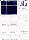
Figure 2. AXL inhibition causes DNA damage and reduces levels of DNA repair markers
A) (Left): HCC1806 (TNBC), SKLU1 (NSCLC) and 584 (HNSCC) cell lines were treated with 25 nM of the AXL inhibitor (TP0903) for 96 hours, following which they were fixed and stained for γH2AX. Data represents percentage cells exhibiting >10 γH2AX foci measured using Slidebook. (Right): Images corresponding to γH2AX staining in DMSO or 25 nM TP0903 treated cells. Blue: DAPI, Green: γH2AX. B) Reverse phase protein array (RPPA) analysis corresponding to the indicated DNA repair markers in TNBC, NSCLC and HNSCC cell lines, which were treated with vehicle (DMSO) or 25 nM TP0903 for 24 or 48 hours. n=3, FDR<0.05. C) Reverse phase protein array (RPPA) analysis of RAD51 expression in scramble-shRNA or AXL-shRNA infected cell lines. Data represent average from three independent biological replicates; p<0.05, FDR<0.05, n=3.
Figure 3
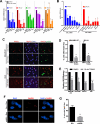
Figure 3. AXL inhibition diminishes homologous recombination (HR)
A) TNBC (MDA-MB-157 and HCC1806), NSCLC (SKLU1) and HNSCC (584) cell lines were treated with vehicle or 25 nM TP0903 for 48 hours and subjected to qRT-PCR analysis of the indicated HR genes. Data is representative of three independent experiments, each performed in triplicate. B) TNBC (MDA-MB-157 and HCC1806), NSCLC (SKLU1 and Calu1) and HNSCC (584 and 1386-LN) cell lines infected with scramble or AXL shRNA and subjected to qRT-PCR analysis to measure RNA levels of the HR genes, RAD51 and BRCA2. Data is representative of three independent experiments each performed in triplicate C, D) Images (C) corresponding to formation of RAD51 and γH2AX foci in TNBC (MDA-MB-157) and NSCLC (SKLU1) following treatment with doxorubicin and DMSO or 25 nM TP0903 for 96 hours (red: RAD51, blue: DAPI, green: γH2AX) and its quantification (percentage of cells containing >20 RAD51 foci) (D). Data is representative of three independent experiments. E) TNBC (MDA-MB-157 and HCC1806), NSCLC (SKLU1) and HNSCC (584) cell lines, transfected with the two-plasmid system (DR-GFP and pSceI) were treated with vehicle or 25 nM TP0903 for 48 hours. HR efficiency assay was performed on the cell lines as described in the Methods section. Data represents fold-decrease in GFP+ cells in 25 nM TP0903 treated cells compared to DMSO treated cells from three independent experiments. F, G) Images (F) corresponding to formation of RAD51 foci in TNBC (MDA-MB-157) and NSCLC (SKLU1) following infection with Scrambled or AXL shRNA and treatment with doxorubicin for 48 hours (red: RAD51, blue: DAPI); G) Quantification of RAD51 foci (percentage of cells containing >5 foci calculated from 3 fields with min of 75 cells per field).
Figure 4
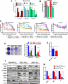
Figure 4. AXL inhibition is synergistic with PARP inhibition
A) Combination index calculated using Calcusyn for the AXL and PARP inhibitors (TP0903 and olaparib) in the indicated mesenchymal cell lines. Data is representative of three independent experiments. B) Combination index calculated in epithelial cell lines using Calcusyn for the AXL and PARP inhibitors (TP0903 and olaparib). Data is representative of three independent experiments. C) High throughput survival assay (HTSA - Methods) showing concentration dependent proliferation curves in the TNBC (MDA-MB-157), NSCLC (SKLU1) and HNSCC (584) cell lines. MCF10A cells were used as a normal mammary epithelial cell control. Cells were treated with single agent (TP0903 or olaparib) or indicated concentration of the drug combinations. Data represents average of three independent experiments. D). Clonogenic assay in MDA-MB-157 (157) and SKLU1 cells treated with TP0903, olaparib or combination of TP0903 and olaparib for 7 days and recovery in drug free media for 5 days. (Right) Quantification of the clonogenic assay in the indicated cell lines. E) Combination index calculated from HTSA performed in TNBC (MDA-MB-157) and NSCLC (SKLU1) cell lines, following treatment with the AXL inhibitor, R428 alone, 1 uM olaparib or the combination of R428 and olaparib. F) TNBC (MDA-MB-157 and HCC1806), NSCLC (SKLU1) and HNSCC (584) cell lines were treated with vehicle, 25 nM TP0903, 1 μM olaparib or the combination of both TP0903 and olaparib for 7 days. Cells were lysed and immunoblot analysis was performed for markers of DNA repair (RAD51, MRE11, E2F1, c-Myc) and apoptosis (cleaved PARP). Actin was used as a loading control. G. Scramble or AXL shRNA infected TNBC (MDA-MB-157), NSCLC (SKLU1) and HNSCC (584) cell lines were treated with DMSO or 1 μM olaparib and cell number was counted on day 7. Data is representative of normalized mean cell number from three independent experiments.
Figure 5
Figure 5. AXL inhibition in combination with PARP inhibition causes apoptosis
A) AnnexinV +ve PI+ve population measured in the indicated cell lines treated with DMSO, 25 nM TP0903, 0.5 μM olaparib or 25 nM TP0903+0.5 μM olaparib. Data is representative of three independent experiments. B) Sub-G0 population measured by propidium iodide staining in the indicated cell lines treated with DMSO, 25 nM TP0903, 0.5 μM olaparib or 25 nM TP0903+0.5 μM olaparib. Data is representative of three independent experiments. C) Cell cycle analysis from three independent experiments measured by flow cytometry in the indicated cell lines treated with DMSO, 25 nM TP0903, 0.5 μM olaparib or 25 nM TP0903+0.5 μM olaparib. D) Control (Scr) and AXL-shRNA (AXL KD) infected MDA-MB-157 and SKLU1 cells were treated with DMSO or 0.5 μM olaparib and Annexin-V+ve / PI+ve population was measured. Data is representative of three independent experiments. E) Cell cycle analysis from three independent experiments measured by flow cytometry in MDA-MB-157 and SKLU1 cells infected with control (Scr) or AXL-shRNA (AXL KD) and treated with DMSO or 0.5 μM olaparib. Data is representative of three independent experiments.
Figure 6
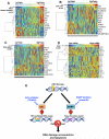
Figure 6. AXL expression corresponds to expression of DNA repair proteins and EMT markers in patient cohorts
A-C) Correlation analysis of AXL protein with other protein markers in NSCLC PROSPECT cohort (A), the TCGA LUSC cohort (B) and the TCGA HNSCC cohorts (C). Heat maps show the top significant markers that correlate with AXL expression (FDR<5%, Spearman Rho value > +/− 0.35). D) Correlation analysis of AXL gene expression with other genes in the breast cancer patient cohort, with heat map showing the top significant markers that correlate with AXL expression. E) Model depicting how AXL inhibition causes HR deficiency and synthetic lethality with PARP inhibitor. In mesenchymal cell lines and tissues, AXL expression leads to higher expression of HR DNA repair proteins, facilitating higher HR-DNA repair efficiency, which is inhibited in the presence of an AXL inhibitor (TP0903). This makes the cells sensitive to inhibition of an alternate DNA repair pathway that utilizes PARP (base excision repair). Thus, a synergy occurs when AXL and PARP are inhibited simultaneously, leading to accumulation of DNA damage and apoptotic cell death.

References

    1. Lemke G. Biology of the TAM receptors. Cold Spring Harb Perspect Biol. 2013;5:a009076. - PMC - PubMed
    1. Zhang YX, Knyazev PG, Cheburkin YV, Sharma K, Knyazev YP, Orfi L, et al. AXL is a potential target for therapeutic intervention in breast cancer progression. Cancer research. 2008;68:1905–15. - PubMed
    1. Li Y, Ye X, Tan C, Hongo JA, Zha J, Liu J, et al. Axl as a potential therapeutic target in cancer: role of Axl in tumor growth, metastasis and angiogenesis. Oncogene. 2009;28:3442–55. - PubMed
    1. Koorstra JB, Karikari CA, Feldmann G, Bisht S, Rojas PL, Offerhaus GJ, et al. The Axl receptor tyrosine kinase confers an adverse prognostic influence in pancreatic cancer and represents a new therapeutic target. Cancer biology & therapy. 2009;8:618–26. - PMC - PubMed
    1. Reichl P, Dengler M, van Zijl F, Huber H, Fuhrlinger G, Reichel C, et al. Axl activates autocrine transforming growth factor-beta signaling in hepatocellular carcinoma. Hepatology. 2015;61:930–41. - PMC - PubMed

Publication types

MeSH terms

Substances