. 2016 Sep 26;16(1):185.
doi: 10.1186/s12883-016-0707-z.

Decreased IL-8 levels in CSF and serum of AD patients and negative correlation of MMSE and IL-1β

Affiliations

Decreased IL-8 levels in CSF and serum of AD patients and negative correlation of MMSE and IL-1β

Raphael Hesse et al. BMC Neurol. .

Abstract

Background: It is widely accepted that neuroinflammatory processes play an important role in the pathogenesis of Alzheimer's disease (AD) and high levels of cytokines and chemokines are detected around Aβ plaques.

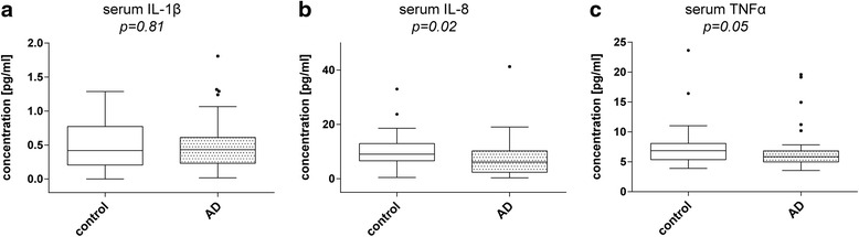
Methods: As neuroinflammation is involved in the development and progression of AD, we measured the pro-inflammatory cytokines interleukin 1β (IL-1β), IL-8 and tumor necrosis factor α (TNF-α) in serum and cerebrospinal fluid (CSF) samples from 45 AD patients and 53 age-matched control subjects using a highly sensitive multiplex electrochemiluminescence assay. To address the association with disease progression we correlated cognitive status with cytokine levels.

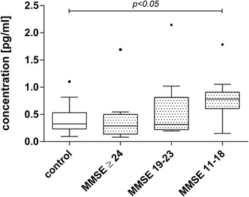
Results: CSF as well as serum IL-8 levels were found to be significantly lower in AD patients than in controls (p = 0.02). A statistically significant inverse correlation was observed between the CSF level of IL-1β and the MMSE score (rs = -0.03, p = 0.02). We therefore stratified the AD patients by their MMSE scores into three equal groups and found that in the AD group with the most severe cognitive impairment CSF-IL-1β was significantly increased compared to age-matched controls (p < 0.05), whereas in the other investigated groups the increase was not statistically significant.

Conclusion: Our results confirm data suggesting that cytokine alterations are involved in AD pathogenesis and may be helpful as a biomarker for monitoring disease progression.

Keywords: Alzheimer’s disease; Cytokines; Neuroinflammation.


Figures

Fig. 1
Fig. 1
Cytokine levels in CSF samples of AD patients compared to controls. Box plots comparing CSF IL-1β, IL-8 and TNF-α levels between AD patients and age-matched controls. a IL-1β levels were not altered in AD patients compared to controls (p = 0.12). b IL-8 levels, by comparison, were significantly reduced in AD patients (p = 0.02). c TNF-α showed no significant difference (p = 0.83). Dark horizontal lines represent the mean, with the box representing the 25th and 75th percentiles of the observed data, the whiskers representing the 5th and 95th percentiles, and outliers represented by dots. P values were calculated using the Mann-Whitney Rank sum test
Fig. 2
Fig. 2
Cytokine levels in serum of AD patients and age-matched controls. Box plots comparing Serum IL-1β, IL-8 and TNF-α levels between AD patients and age-matched controls. a IL-1β levels were unchanged (p = 0.81). b IL-8 levels were significantly reduced in AD patients (p = 0.02). c TNF-α showed a slight reduction in AD patients but the observed effect was not significant (p = 0.05). Dark horizontal lines represent the mean, with the box representing the 25th and 75th percentiles of the observed data, the whiskers representing the 5th and 95th percentiles, and outliers represented by dots. P values were calculated using the Mann-Whitney Rank sum test
Fig. 3
Fig. 3
Analysis of IL-1β levels in CSF of stratified patients by MMSE. CSF IL-1β levels inversely correlated with MMSE score. To further analyze this correlation, AD patients were grouped according to their MMSE score and IL-1β levels were compared to age-matched controls. Box plots represent median levels of CSF IL-1β with the box representing the 25th and 75th percentiles of the observed data, the whiskers showing the 5th and 95th percentiles and dots representing outliers. The MMSE (11-18) group IL-1β was significantly increased compared to the other investigated groups (p < 0.05, ANOVA on Ranks, Dunn’s method)

References

    1. Blennow K, de Leon MJ, Zetterberg H. Alzheimer's disease. Lancet. 2006;368(9533):387–403. doi: 10.1016/S0140-6736(06)69113-7. - DOI - PubMed
    1. Huang Y, Mucke L. Alzheimer mechanisms and therapeutic strategies. Cell. 2012;148(6):1204–1222. doi: 10.1016/j.cell.2012.02.040. - DOI - PMC - PubMed
    1. Dickson DW. The pathogenesis of senile plaques. J Neuropathol Exp Neurol. 1997;56(4):321–339. doi: 10.1097/00005072-199704000-00001. - DOI - PubMed
    1. Rozemuller JM, Eikelenboom P, Stam FC. Role of microglia in plaque formation in senile dementia of the Alzheimer type. An immunohistochemical study. Virchows Arch B Cell Pathol Incl Mol Pathol. 1986;51(3):247–254. doi: 10.1007/BF02899034. - DOI - PubMed
    1. Selkoe DJ. Alzheimer's disease: genes, proteins, and therapy. Physiol Rev. 2001;81(2):741–766. - PubMed

LinkOut - more resources