. 2016 Oct 11;113(41):E6271-E6280.
doi: 10.1073/pnas.1601506113. Epub 2016 Sep 26.

SARM1-specific motifs in the TIR domain enable NAD+ loss and regulate injury-induced SARM1 activation

Affiliations

SARM1-specific motifs in the TIR domain enable NAD+ loss and regulate injury-induced SARM1 activation

Daniel W Summers et al. Proc Natl Acad Sci U S A. .

Abstract

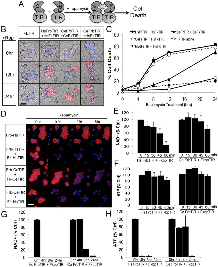
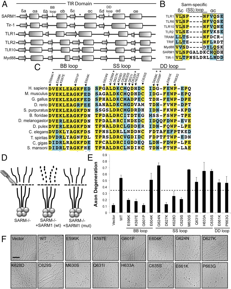
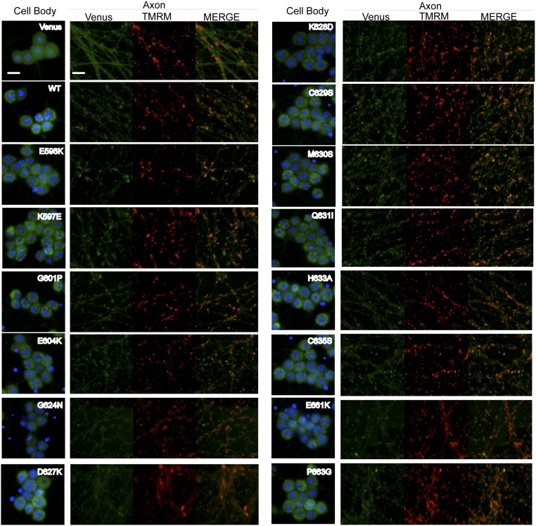
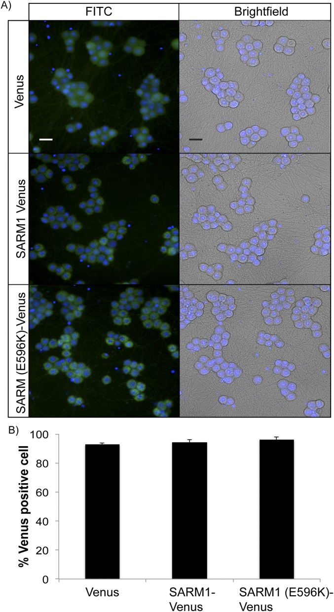
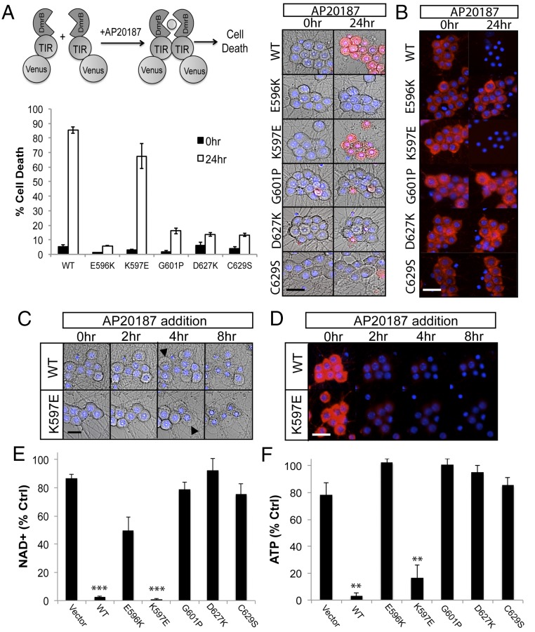
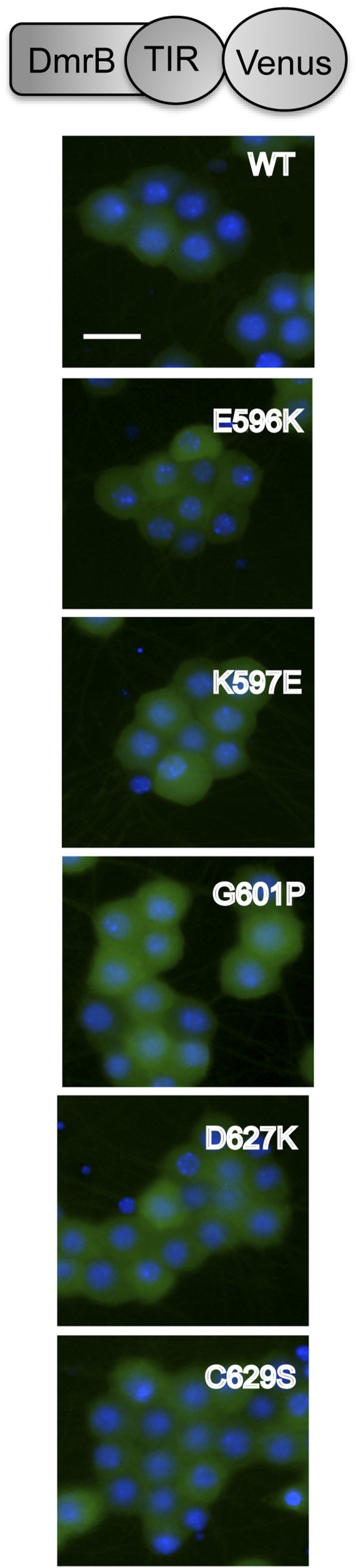
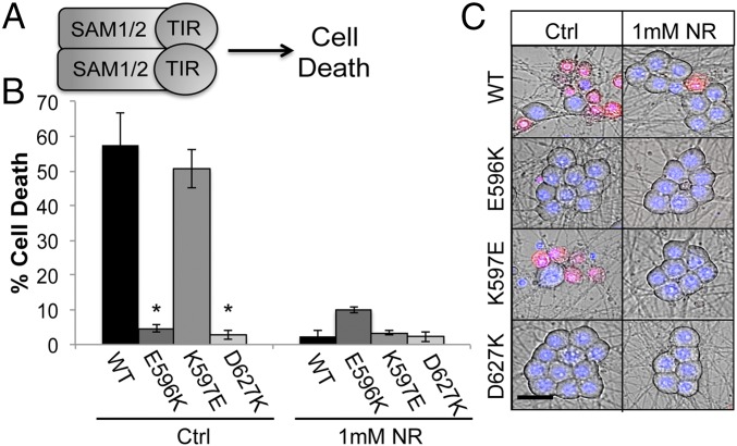
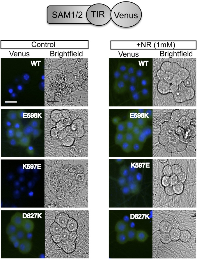
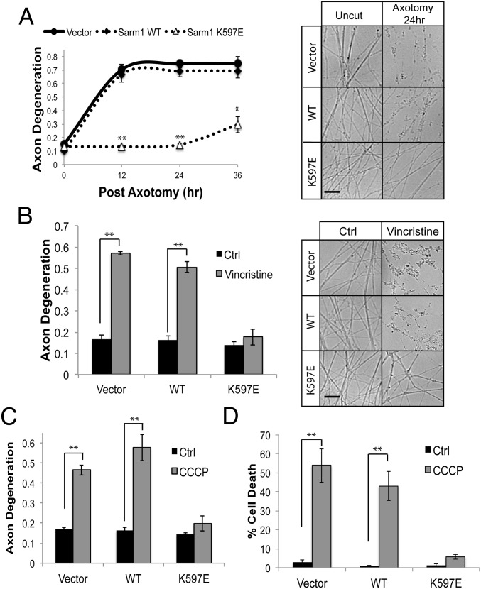
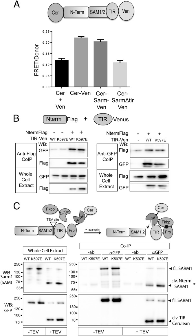
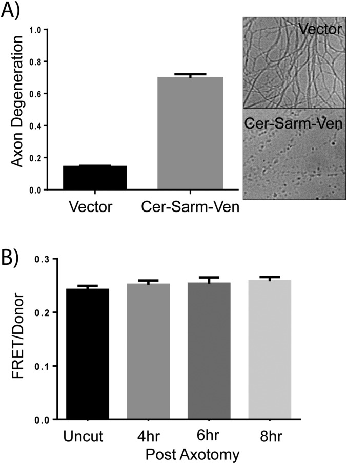
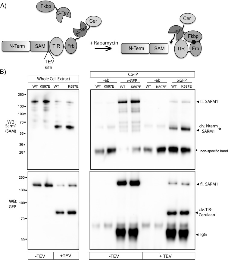
Axon injury in response to trauma or disease stimulates a self-destruction program that promotes the localized clearance of damaged axon segments. Sterile alpha and Toll/interleukin receptor (TIR) motif-containing protein 1 (SARM1) is an evolutionarily conserved executioner of this degeneration cascade, also known as Wallerian degeneration; however, the mechanism of SARM1-dependent neuronal destruction is still obscure. SARM1 possesses a TIR domain that is necessary for SARM1 activity. In other proteins, dimerized TIR domains serve as scaffolds for innate immune signaling. In contrast, dimerization of the SARM1 TIR domain promotes consumption of the essential metabolite NAD+ and induces neuronal destruction. This activity is unique to the SARM1 TIR domain, yet the structural elements that enable this activity are unknown. In this study, we identify fundamental properties of the SARM1 TIR domain that promote NAD+ loss and axon degeneration. Dimerization of the TIR domain from the Caenorhabditis elegans SARM1 ortholog TIR-1 leads to NAD+ loss and neuronal death, indicating these activities are an evolutionarily conserved feature of SARM1 function. Detailed analysis of sequence homology identifies canonical TIR motifs as well as a SARM1-specific (SS) loop that are required for NAD+ loss and axon degeneration. Furthermore, we identify a residue in the SARM1 BB loop that is dispensable for TIR activity yet required for injury-induced activation of full-length SARM1, suggesting that SARM1 function requires multidomain interactions. Indeed, we identify a physical interaction between the autoinhibitory N terminus and the TIR domain of SARM1, revealing a previously unrecognized direct connection between these domains that we propose mediates autoinhibition and activation upon injury.

Keywords: NAD; SARM; axon degeneration; cell death; sarmoptosis.


Conflict of interest statement

The authors declare no conflict of interest.

Figures

Fig. 1.
Fig. 1.
Dimerization of CeTIR stimulates NAD+ loss and neuronal cell death. (A) Scheme for TIR dimerization via fusing Frb and Fkbp moieties to the TIR domain. Rapamycin stimulates TIR dimerization and cell death. (B) Images of embryonic DRGs expressing the indicated Frb/Fkbp TIR constructs after rapamycin addition. Dying cells are labeled with ethidium homodimer and costained with Hoescht to label nuclei. (C) Quantification of cellular death after rapamycin addition. (D) ΔΨ was monitored with tetramethylrhodamine methyl ester in DRG neurons expressing the indicated Frb/Fkbp TIR construct after rapamycin addition. (E and F) NAD+ and ATP levels were measured from DRGs expressing the indicated Frb/Fkbp TIR construct after rapamycin addition. (G and H) NAD+ and ATP were measured at more extended time points after rapamycin addition to assess metabolite levels after long-term TIR dimerization. Error bars reflect ± SEM (n = 3). (Scale bars, 25 μM.)
Fig. 2.
Fig. 2.
Residues within the TIR domain are required for SARM1-dependent axon degeneration. (A) Secondary structure of TIR domains from human SARM1 and C. elegans TIR-1 was predicted with three algorithms. The secondary structures of other human TIR domains are shown below. Secondary structures are derived from solved crystal structures. The β-sheets and α-helices are labeled using conventional nomenclature applied to these elements in TIR domains. (B) Extended loop region between the βc and αc elements in the TIR domain from SARM1 and TIR-1 is largely absent in other human TIR domains. We are calling this motif the SS loop. (C) Sequence alignments of TIR motifs in SARM1 orthologs. Residues highlighted in yellow are conserved. Residues highlighted in blue are amino acid changes to similar residues. Residues that not highlighted are poorly conserved. Residues chosen for mutagenesis are indicated in the alignment. *SARM1-Venus expression was very low. (D) Diagram of rescue strategy in SARM1−/− DRGs. SARM1−/− DRGs expressing full-length SARM1 restore axon degeneration, whereas SARM1 containing loss-of-function mutations (mut) does not rescue axon degeneration. (E and F) Axons from SARM1−/− DRGs expressing the indicated SARM1-Venus construct were transected with a razor blade. Axon degeneration was measured 24 h later. Degenerated axons manifest in an axon degeneration score of 0.35 or greater (indicated by dashed line). Error bars reflect ± SEM (n = 3). (Scale bar, 25 μM.)
Fig. S1.
Fig. S1.
Expression of SARM1-Venus constructs in DRG cell bodies and axons. SARM1−/− DRGs were transduced with lentivirus expressing Venus or the indicated SARM1-Venus construct. DRGs were also incubated with Hoechst 33342 to label nuclei as well as tetramethylrhodamine methyl ester (TMRM) to label mitochondria. Representative images of cell bodies and distal axons are shown. (Scale bars, 25 μM.)
Fig. S2.
Fig. S2.
Transduction efficiency in embryonic DRGs. (A) Images of SARM1−/− DRGs expressing Venus, WT SARM1-Venus, or SARM1 (E596K)-Venus. Cells were preincubated with Hoechst 33342 to stain nuclei. (Left) Cells were imaged with an FITC filter to detect Venus. (Right) Same field of cells was bright-field imaged. (B) Quantification of percentage of cells expressing Venus from three independent cultures and lentiviral preparations. (Scale bars, 25 μM.)
Fig. 3.
Fig. 3.
Mutagenesis of TIR motifs abrogates NAD+ loss upon TIR dimerization. (A) Point mutations were made in homodimerizable (DmrB) TIR, wherein AP20187 induces dimerization and neuronal death. Nonfunctional point mutations in the BB loop and SS loop prevent DmrB-TIR–dependent death 24 h after AP20187 addition. Images of ethidium homodimer-labeled DRG neurons (Right) and quantification (Lower) are shown. (B) Tetramethylrhodamine methyl ester is preserved in DRGs expressing loss-of-function TIR mutations 24 h after AP20187 addition. DmrB-TIR (K597E) induces cell death as well as WT, even though this mutation abolishes activity of full-length SARM1. (C and D) Kinetics of membrane blebbing and TMRM loss upon dimerization of WT TIR or K597E TIR. (E and F) NAD+ and ATP levels are substantially reduced 10 h after AP20187 treatment in DRG cells expressing WT and K597E DmrB-TIR; however, levels are preserved in the presence of loss-of-function TIR mutants. Error bars reflect ± SEM (n = 3). (Scale bars, 25 μM.) **P < 0.01.
Fig. S3.
Fig. S3.
Expression of DmrB-TIR-Venus constructs. Embryonic DRG neurons expressing the indicated DmrB-TIR-Venus constructs were imaged with Hoechst 33342 to identify nuclei. Representative images from multiple experiments are shown. (Scale bar, 25 μM.)
Fig. 4.
Fig. 4.
K597E does not impair SAM-TIR–dependent neuronal death. (A) Expression of fragment containing the SAM domains and TIR domain from SARM1 induces neuronal cell death. (B) Quantification of neuronal death in cells expressing the indicated SAM-TIR protein. DRGs were also preincubated with 1 mM NR, which suppresses SARM1-dependent cell death. (C) Images of DRG neurons labeled with ethidium homodimer. *P < 0.05. Error bars reflect ± SEM (n = 3). Ctrl, control. (Scale bar, 25 μM.)
Fig. S4.
Fig. S4.
Expression of SAM-TIR-Venus constructs. Embryonic DRG neurons were preincubated with 1 mM NR and then infected with the indicated lentivirus expressing SAM-TIR-Venus (WT or mutant). Shown are representative images of Venus fluorescence to illustrate SAM-TIR-Venus expression as well as bright-field images illustrating cell death upon expression of WT SAM-TIR-Venus and K597E SAM-TIR-Venus. Venus expression of WT and K597E SAM-TIR-Venus is largely undetectable unless DRGs are preincubated with NR. However, SAM-TIR-Venus with E596K or D627K point mutations does not induce toxicity and does express in the presence or absence of NR. (Scale bar, 25 μM.)
Fig. 5.
Fig. 5.
Sarm1 (K597E) is a potent dominant negative, blocking axon degeneration after axotomy. (A) Axons from WT DRG neurons expressing the indicated Sarm1 construct were transected, and the degeneration of distal axons monitored over time. Expression of Sarm1 (K597E) delays axon degeneration for ∼36 h postaxotomy. (B) Expression of SARM1 (K597E) also preserves axon integrity in response to the chemotherapeutic agent vincristine. Axons were treated with 40 nM vincristine, and axon degeneration was measured 36 h postexposure. Expression of SARM1 (K597E) suppresses axon degeneration (C) and cell death (D) in response to the mitochondrial uncoupling agent CCCP (50 μM, 24 h). *P < 0.05; **P < 0.01. Error bars reflect ± SEM (n = 3). (Scale bars, 25 μM.)
Fig. S5.
Fig. S5.
SARM1 (K597E) preserves ΔΨ in severed distal axons. WT DRGs expressing the indicated construct were loaded with TMRM to label mitochondria with intact membrane potential. Axons were severed, and images were collected of distal axons at the indicated time points.
Fig. S6.
Fig. S6.
SARM1 K597E does not prevent cell death in response to NGF withdrawal. DRGs expressing the indicated construct were deprived of NGF for 24 h and then loaded with ethidium homodimer to label dead cells and Hoechst to label nuclei. (Scale bar, 25 μM.)
Fig. 6.
Fig. 6.
SARM1 (K597E) expression prevents axonal NAD+ loss after axotomy. (A and B) Axonal NAD+ and ATP levels were measured from transected SARM1−/− DRGs expressing an empty vector, WT SARM1, or SARM1 (K597E). There is a significant drop in axonal NAD+ in the presence of SARM1; however, there is no significant decrease in the presence of SARM1 (K597E). (C and D) Axonal NAD+ and ATP levels were measured from transected WT DRGs expressing the same constructs as described in A and B. Expression of SARM1 (K597E) delays axonal loss of NAD+ and ATP after axotomy. *P < 0.05. Error bars reflect ± SEM (n = 3). N.D, not detectable.
Fig. 7.
Fig. 7.
SARM1 N terminus and TIR domain form a physical complex. (A) FRET pairs Cerulean and Venus were attached to the N terminus and C terminus of SARM1, respectively (CerSARMVen). The FRET/donor ratio in DRGs from CerSARMVen was higher than Cerulean and Venus expressed in trans (Cer + Ven) and comparable to a direct Cerulean-Venus fusion. Deletion of the SARM1 TIR domain reduces FRET signal to background levels. (B) N terminus (Nterm) of SARM1 tagged with a Flag epitope was expressed in HEK293T cells with the TIR domain (WT or K597E) tagged with Venus. Error bars reflect ± SEM (n = 3). TIR-Venus was immunoprecipitated from HEK293T cell extracts, and the coimmunoprecipitated Nterm was assessed by Western immunoblotting (WB) for Flag. Nterm Flag was immunoprecipitated from cell extracts, and coimmunoprecipitated TIR-Venus was assessed by Western immunoblotting for GFP. Western blots are reflective of four independent experiments. (C) SARM1ps was transfected in HEK293T cells with or without Fkbp-C-TEV to cleave an engineered TEV site upstream of the TIR domain. (Left) WB of whole-cell extracts shows efficient proteolysis to generate cleaved (clv.) Nterm SARM1 containing Nterm and SAM domains, detected with antibody that recognizes peptide in the SAM2 domain of SARM1. Proteolysis also generates a fragment containing the SARM1 TIR domain tagged with Cerulean (detected with GFP antibody). TIR-Cerulean was immunoprecipitated with GFP antisera or antibody (−ab) as a control. In the presence of Fkbp-C-TEV (+TEV), a bound SARM1 fragment containing Nterm/SAM domains is detected with anti-SARM1 antibody by Western blot (*clv. Nterm SARM1). (Lower) GFP WB detecting immunoprecipitated TIR-Cerulean. Western blots are representative of five independent experiments. Co-IP, coimmunoprecipitation; f.l., full length.
Fig. S7.
Fig. S7.
Characterization of Cer-SARM-Ven FRET sensor in SARM1−/− DRG axons. (A) Cer-SARM-Ven FRET construct rescues axon degeneration in SARM1−/− DRGs. SARM1−/− DRGs were transduced with an empty vector or lentivirus expressing Cer-SARM-Ven. Axons were severed, and axon degeneration was measured 24 h later. Representative images of distal axons are shown with quantification of axon degeneration from three independent experiments. (B) FRET ratio was measured in distal axons from SARM1−/− at the indicated time after axotomy. Error bars reflect ± SEM (n = 3).
Fig. S8.
Fig. S8.
SARM1 TIR domain physically associates with the N terminus/SAM domains of SARM1. (A) Diagram of biochemical strategy to identify the complex between the TIR domain and N terminus/SAM domains from SARM1. SARMps is expressed in HEK293T cells in the presence or absence of FKBP-C-TEV. When cells are treated with rapamycin, the reconstituted TEV protease cleaves SARM1 at an engineered TEV protease site upstream of the TIR domain. In cell extracts, the liberated TIR domain is immunoprecipitated via a Cerulean tag at the C terminus. The N-terminal portion of SARM1 is detected in TIR immunoprecipitates by Western immunoblotting (WB) with SARM1 antisera generated against a peptide from the SARM1/SAM2 domain. (B) Full blots from Fig. 7C. Nonspecific bands in blots from GFP immunoprecipitates are noted with an asterisk (*). clv., cleaved; Co-IP, coimmunoprecipitation; f.l., full length.

Similar articles

Cited by

References

    1. Gerdts J, Summers DW, Milbrandt J, DiAntonio A. Axon self-destruction: New links among SARM1, MAPKs, and NAD+ metabolism. Neuron. 2016;89(3):449–460. - PMC - PubMed
    1. Osterloh JM, et al. dSarm/Sarm1 is required for activation of an injury-induced axon death pathway. Science. 2012;337(6093):481–484. - PMC - PubMed
    1. Gerdts J, Summers DW, Sasaki Y, DiAntonio A, Milbrandt J. Sarm1-mediated axon degeneration requires both SAM and TIR interactions. J Neurosci. 2013;33(33):13569–13580. - PMC - PubMed
    1. Gerdts J, Brace EJ, Sasaki Y, DiAntonio A, Milbrandt J. SARM1 activation triggers axon degeneration locally via NAD⁺ destruction. Science. 2015;348(6233):453–457. - PMC - PubMed
    1. Sasaki Y, Araki T, Milbrandt J. Stimulation of nicotinamide adenine dinucleotide biosynthetic pathways delays axonal degeneration after axotomy. J Neurosci. 2006;26(33):8484–8491. - PMC - PubMed

Publication types

MeSH terms