. 2016 Oct 11;113(41):E6045-E6054.
doi: 10.1073/pnas.1604807113. Epub 2016 Sep 26.

Tyrosine phosphorylation stimulates activity of human RAD51 recombinase through altered nucleoprotein filament dynamics

Affiliations

Tyrosine phosphorylation stimulates activity of human RAD51 recombinase through altered nucleoprotein filament dynamics

Shyamal Subramanyam et al. Proc Natl Acad Sci U S A. .

Abstract

The DNA strand exchange protein RAD51 facilitates the central step in homologous recombination, a process fundamentally important for accurate repair of damaged chromosomes, restart of collapsed replication forks, and telomere maintenance. The active form of RAD51 is a nucleoprotein filament that assembles on single-stranded DNA (ssDNA) at the sites of DNA damage. The c-Abl tyrosine kinase and its oncogenic counterpart BCR-ABL fusion kinase phosphorylate human RAD51 on tyrosine residues 54 and 315. We combined biochemical reconstitutions of the DNA strand exchange reactions with total internal reflection fluorescence microscopy to determine how the two phosphorylation events affect the biochemical activities of human RAD51 and properties of the RAD51 nucleoprotein filament. By mimicking RAD51 tyrosine phosphorylation with a nonnatural amino acid, p-carboxymethyl-l-phenylalanine (pCMF), we demonstrated that Y54 phosphorylation enhances the RAD51 recombinase activity by at least two different mechanisms, modifies the RAD51 nucleoprotein filament formation, and allows RAD51 to compete efficiently with ssDNA binding protein RPA. In contrast, Y315 phosphorylation has little effect on the RAD51 activities. Based on our work and previous cellular studies, we propose a mechanism underlying RAD51 activation by c-Abl/BCR-ABL kinases.

Keywords: RAD51 recombinase; c-Abl tyrosine kinase; homologous recombination; phosphorylation; single-molecule total internal reflection fluorescence microscopy.


Conflict of interest statement

The authors declare no conflict of interest.

Figures
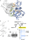
Fig. 1.
Fig. 1.
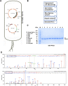
Incorporation of pCMF into human RAD51. (A) Homology model of two adjacent human RAD51 monomers. RAD51 DNA binding loops L1 and L2 are shown in orange and green, respectively. Residues Y54, Y315, and F195 are shown in a ball-and-stick rendering. (B) Structures of tyrosine, phosphorylated tyrosine, and pCMF. (C) Incorporation of pCMF incorporation using the amber suppressor system: RAD51 expression in the absence of pCMF leads to translation of a truncated product, whereas full-length protein is produced in the presence of pCMF. (D) Western blot (WB) showing expression of full-length RAD51Y54pCMF and RAD51Y315pCMF proteins using mouse anti-RAD51 (3C10) antibody. Truncated products are observed in the absence of pCMF (Y54 truncations are too small to be verified by standard gel electrophoresis). (E) SDS/PAGE gel showing purified RAD51, RAD51Y54pCMF, and RAD51Y315pCMF. The RAD51Y315pCMF has a very small amount of truncated product (<18%) that could not be separated using our purification scheme (Fig. S1B).
Fig. S1.
Fig. S1.
Incorporation of a nonnatural amino acid into RAD51 protein. (A) Expression system established for incorporation of the pCMF amino acid consists of E. coli Acella cells transformed with two plasmids. The first plasmid, pCH1-RAD51opt, contains a RAD51 ORF optimized for E. coli expression as well as the GroE operon from E. coli. The second plasmid contains components for the amber suppressor system. (B) Purification scheme for obtaining homogeneous RAD51 protein. (C) SDS-PAGE gel (4–15% gradient) showing purified RAD51 and phosphomimetic mutants (112.5 pmol in 15 µL of buffer containing 20 mM Hepes (pH 7.5), 10% glycerol, 150 mM NaCl, 1 mM DTT, 1 mM EDTA). All proteins were purified to near homogeneity (except RAD51Y315pCMF, which contained a small amount of truncated protein). The electrophoretic mobility of the wild-type RAD51 and all mutants was as expected for the 37-kDa protein, except for the RAD51Y315F, which consistently runs slightly faster. (D) Data from MALDI-TOF/MS experiments confirming incorporation of pCMF amino acid at the Y54 (Top) and Y315 (Bottom) residues, respectively.
Fig. 2.
Fig. 2.
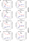
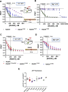
Y54 phosphorylation stimulates DNA strand exchange activity of RAD51. (A) RAD51-first DNA strand exchange reaction: Circular ϕX174 ssDNA is incubated with RAD51 in reaction conditions permitting ATP hydrolysis (Materials and Methods), followed by addition of RPA, which helps remove secondary structures in the ssDNA, allowing stable nucleoprotein filament formation. The RAD51 nucleoprotein then invades ϕX174 linear dsDNA to form nicked circular (NC) dsDNA products through several joint-molecule (JM) intermediates showing various stages of branch migration. Strand exchange reactions carried out by RAD51Y54pCMF (B) and RAD51Y315pCMF (C), both compared with the RAD51 wild-type protein. For each protein, the respective panel shows reactions stopped at 0, 30, 60, 120, and 180 min. All substrates, joint molecules, and nicked circular products are observed on a 0.8% Tris-Acetate-EDTA (TAE) agarose gel stained with SYBR Gold. (D and E) Quantitative analysis of formed nicked circular products in RAD51Y54pCMF and RAD51Y315pCMF strand exchange reactions. The RAD51Y54pCMF mutant (red) is able to convert ∼75% of linear dsDNA substrate into nicked circular products compared with ∼40% of products formed for RAD51 wild-type (gray) and RAD51Y315pCMF mutant (orange) converts ∼27% of linear dsDNA substrate into nicked circular products compared with 33% for the wild-type protein. All reactions were performed in triplicate, with data represented as mean ± SEM. The results for both mutants were compared with the wild-type protein, using a two-way ANOVA to verify the significance of results.
Fig. S2.
Fig. S2.
Quantitative analysis of strand exchange reactions with RAD51 Y54 and Y315 pCMF mutants The appearance of the nicked circular (NC) products (black), disappearance of dsDNA substrates (red), and formation of JM (blue) were quantitated using ImageJ software normalized to the amount of initial linear dsDNA substrate. The intensities obtained for the mutants were then statistically compared with the wild-type strand exchange reaction. Comparison of strand exchange reactions between RAD51Y54pCMF and wild-type RAD51 proteins when RPA was added after (A) and before (B) the addition of RAD51 protein. Comparison of strand exchange reactions between RAD51Y315pCMF and wild-type RAD51 proteins when RPA was added after (C) and before (D) the addition of RAD51 protein.
Fig. S3.
Fig. S3.
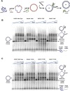
Strand exchange reactions with RAD51 Y54 and Y315 mutants. (A) Schematic showing the in vitro reconstituted RAD51-first DNA strand exchange reaction (Fig. 2). JM, joint molecules; NC, nicked circular. (B) Strand exchange reactions comparing RAD51 wild-type with RAD51Y54D, RAD51Y54E, and RAD51Y54F mutants, respectively. (C) Strand exchange reactions comparing RAD51 wild-type with RAD51Y315D, RAD51Y315E, and RAD51Y315F mutants, respectively.
Fig. 3.
Fig. 3.
RAD51Y54pCMF performs efficient RPA-first DNA strand exchange. (A) RPA-first reaction: Circular ϕX174 ssDNA is incubated with RPA protein, followed by the addition of RAD51 protein, which displaces the bound RPA to form nucleoprotein filaments that can then invade ϕX174 linear dsDNA to form nicked circular (NC) products through a series of JM intermediates. (BD) RPA-first DNA strand exchange reactions carried out by RAD51 (Top Left), RAD51Y54pCMF (Top Right), and RAD51Y315pCMF (Bottom Left). For each protein, the respective panel shows reactions stopped at 0, 30, 60, 120, and 180 min. (E and F) Quantitation of product formed in the DNA strand exchange reactions. The RAD51Y54pCMF mutant (blue) converts ∼75% of linear dsDNA substrate into nicked circular products in the presence of RPA compared with ∼25% of products formed for RAD51 wild type (black), whereas RAD51Y315pCMF mutant (cyan) is similar to wild-type protein in both RAD51-first and RPA-first reactions, where it is able to convert 20% of the linear substrate into nicked circular products. All reactions were performed in triplicate, with data represented as mean ± SEM. The results for both mutants were compared with the wild-type protein, using a two-way ANOVA to verify significance of results.
Fig. S4.
Fig. S4.
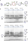
RPA-first strand exchange reactions with RAD51 Y54 and Y315 mutants. (A) Schematic showing the in vitro reconstituted RPA-first DNA strand exchange reaction (Fig. 3). (B) Strand exchange reactions comparing RAD51 wild-type with RAD51Y54D, RAD51Y54E, and RAD51Y54F mutants, respectively. (C) Strand exchange reactions comparing RAD51 wild-type with RAD51Y315D, RAD51Y315E, and RAD51Y315F mutants, respectively.
Fig. 4.
Fig. 4.
RAD51 phosphorylation limits large nucleoprotein complexes on both ssDNA and dsDNA. (A) EMSA RAD51 Y54 mutants binding linear ϕX174 dsDNA. Fifteen micromolar (base pair) dsDNA was incubated with increasing concentrations of 2.5 μM, 5.0 μM, or 7.5 μM RAD51 and loaded on a 0.9% TAE agarose gel and stained with SYBR Gold. RAD51Y54pCMF showed a decrease in dsDNA binding compared with wild-type (WT) protein and RAD51Y315pCMF. Protein/base pair ratios are indicated at the bottom of each lane. (B) In a similar assay containing 30 μM (nucleotide) ϕX174 circular ssDNA, RAD51Y54pCMF is unable to form the higher order nucleoprotein filament complexes formed by the WT protein and RAD51Y315pCMFmutant. Protein/nucleotide ratios are indicated at the bottom of each lane.
Fig. S5.
Fig. S5.
DNA binding properties of traditional RAD51 phosphomimetic mutants differ from both RAD51 wild-type and pCMF mutants when using longer substrates. (A) EMSA for RAD51 Y54D/E/F and Y315D/E/F mutants binding linear ϕX174 dsDNA. Fifteen micromolar (base pair) dsDNA was incubated with increasing concentrations of 2.5 μM, 5.0 μM, or 7.5 μM RAD51 and loaded on a 0.9% TAE agarose gel and stained with SYBR Gold. Aspartate (D) and glutamate (E) mutants showed a difference in dsDNA binding compared with wild-type protein as well as the pCMF mutants. Protein/base pair ratios are indicated at the bottom of each lane. (B) In a similar assay containing 30 μM (nucleotide) ϕX174 circular ssDNA, D and E mutants also showed different ssDNA binding characteristics compared with the wild-type pCMF mutants. Protein/nucleotide ratios are indicated at the bottom of each lane.
Fig. 5.
Fig. 5.
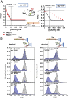
Effects of Y54 phosphorylation on ssDNA binding activity. (A) RAD51 binding to ssDNA was observed by following the extension of the 60-mer oligonucleotide poly(dT)-60 containing Cy3 (FRET donor) and Cy5 (FRET acceptor) fluorophores separated by 25 nucleotides (nt). Binding of RAD51 to 600 nM (nt) ssDNA moves the two dyes apart, which can be seen as a change from high FRET (0.55) to low FRET (0.19). Under conditions preventing ATP hydrolysis (Ca2+), the RAD51Y54pCMF binds and extends the ssDNA substrate similar to wild-type RAD51, with an ∼1:3 stoichiometric RAD51/ssDNA ratio, whereas under reaction conditions permitting ATP hydrolysis (Mg2+), RAD51Y54pCMF binding is nonstoichiometric. Data are represented as mean ± SEM with n = 3. (B) Analysis of the equilibrium ssDNA binding using smTIRFM. Distributions of the FRET states of the DNA substrates in the presence of the indicated concentrations of RAD51 (Left) and RAD51Y54pCMF (Right) overlaid with the distributions of the FRET states in the absence of protein (Materials and Methods and Fig. S7). Unbound ssDNA (gray) yields a histogram centered on a FRET value of ∼0.5, whereas fully extended RAD51 nucleoprotein filament (blue) yields a histogram centered on a FRET value of ∼0.1. Concentrations of RAD51 or RAD51Y54pCMF, as well the number of molecules used to build each histogram, are indicated in each panel.
Fig. S6.
Fig. S6.
Altered ssDNA binding of RAD51 Y54 and Y315 mutants. (A) FRET-based assay to measure binding for RAD51 Y54 mutants to ssDNA. Under conditions inhibiting ATP hydrolysis (Ca2+), RAD51Y54D and RAD51Y54E mutants deviate from ideal RAD51 binding behavior. (B) This effect is significantly exaggerated under conditions permitting ATP hydrolysis (Mg2+). (C and D) Similar FRET-based experiments were conducted for Y315 mutants that were observed to have similar ssDNA binding characteristics under both prohibitive conditions for ATP hydrolysis (Ca2+) and permissive conditions (Mg2+). Data are represented as mean ± SEM with n = 3. (E) Rates of ssDNA-dependent ATP hydrolysis of all RAD51 mutants measured using the coupled NADH assay. Data are represented as mean ± SEM with n ≥ 3.
Fig. S7.
Fig. S7.
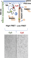
smTIRF ssDNA binding assay. Poly(dT)-60 ssDNA substrate is immobilized to a biotinylated quartz slide using the biotin–Neutravidin interaction (Materials and Methods). Evanescent wave produced by a prism-based total internal reflection illumination and a 532-nm laser is used to excite the Cy3 dye on the surface-tethered DNA substrate. In the absence of protein, the acceptor Cy5 dye on the partial duplex DNA is excited via FRET. Upon binding, RAD51 extends the ssDNA substrate, leading to a decrease in energy transfer and reduction in Cy5 emission, with a corresponding increase in Cy3 intensity. Both Cy3 and Cy5 emission can be tracked simultaneously using a dual-view system. The change in Cy3 and Cy5 intensities on RAD51 binding to ssDNA substrate is tracked over time by recording movies over the course of the experiment.
Fig. 6.
Fig. 6.
HMM analysis shows that RAD51 and RAD51Y54pCMF nucleate on ssDNA as dimers, with RAD51 forming dynamic nuclei distinct from stable RAD51Y54pCMF nuclei. Representative FRET trajectories from preequilibrium experiments visualizing RAD51 (A) and RAD51Y54pCMF (B) nucleation onto ssDNA in real time. RAD51 protein was introduced into the reaction chamber at t ≈ 10 s. Regions containing transitions were trimmed and used for HMM (ebFRET). The idealized trajectories are overlaid on the raw FRET trajectories showing the fit. (C) Schematic explaining the 3D TDPs. Four distinct FRET states 1, 2, 3, and 4 correspond to respective states in the RAD51 nucleus formation on ssDNA, with three steps between these states required to extend the ssDNA substrate fully. Transitions 1→2→3→4 correspond to the filament formation, whereas filament disassembly is reflected in transitions in the reverse direction. (D) TDPs showing the transition densities corresponding to each state for the wild-type RAD51 protein at increasing concentrations. The transitions were calculated from 85, 50, and 146 molecules for 250 nM, 750 nM, and 2.5 μM concentrations, respectively. Brighter colors represent more frequent transitions. The frequency scale is shown to the right of the graphs. (E) TDP plots showing transition densities for the RAD51Y54pCMF mutant. No transitions were observed at 250 nM concentrations. Transitions were calculated from 63 and 116 molecules for 750 nM and 2.5 μM concentrations, respectively.
Fig. S8.
Fig. S8.
HMM analysis shows that RAD51 and RAD51Y54pCMF nucleate on ssDNA as dimers. Representative Cy3 (donor) and Cy5 (acceptor) trajectories from preequilibrium experiments visualizing RAD51 (A) and RAD51Y54pCMF (B) nucleation onto ssDNA in real time are shown. RAD51 protein was introduced into the reaction chamber at t ≈ 10 s. Trajectories that showed anticorrelated behavior of the Cy3 and Cy5 signals were selected and corrected for background intensities as well as donor leakage. The processed Cy3 and Cy5 trajectories were then converted to FRET trajectories (blue). From each FRET trajectory, we selected the regions that include the first observed transition and all subsequent transitions until one of the dyes photobleaches, the transitions stop occurring (i.e., equilibrium is achieved), or the recording is terminated (gray box); the remainder of the trajectory was excluded from the analysis. The selected regions were collectively fit to the models, with the number of FRET states ranging from two to eight using ebFRET analysis. The black lines overlaying the FRET data (blue) represent the best fit to the trajectory, which uses four FRET states. AU, arbitrary units. Histograms show global distributions of the idealized FRET states from 281 and 179 molecules for RAD51 (C) and RAD51Y54pCMF (D), respectively. Four distinct FRET states were observed with FRET values between 0.5 and 0.1. The peak for each state can be approximated by a Gaussian distribution with the center of each peak well separated from the other peaks/states. Because the Cy3-Cy5 FRET pair is 21 nucleotides apart, it is apparent that the RAD51 molecules bound ssDNA in three steps, where each step is a dimer.

Similar articles

Cited by

References

    1. Ciccia A, Elledge SJ. The DNA damage response: Making it safe to play with knives. Mol Cell. 2010;40(2):179–204. - PMC - PubMed
    1. Couëdel C, et al. Collaboration of homologous recombination and nonhomologous end-joining factors for the survival and integrity of mice and cells. Genes Dev. 2004;18(11):1293–1304. - PMC - PubMed
    1. Heyer W-D. Regulation of recombination and genomic maintenance. Cold Spring Harb Perspect Biol. 2015;7(8):a016501. - PMC - PubMed
    1. Li X, Heyer WD. Homologous recombination in DNA repair and DNA damage tolerance. Cell Res. 2008;18(1):99–113. - PMC - PubMed
    1. Moynahan ME, Jasin M. Mitotic homologous recombination maintains genomic stability and suppresses tumorigenesis. Nat Rev Mol Cell Biol. 2010;11(3):196–207. - PMC - PubMed

Publication types

MeSH terms