. 2016 Oct 11;113(41):11525-11530.
doi: 10.1073/pnas.1613121113. Epub 2016 Sep 26.

Flow-dependent YAP/TAZ activities regulate endothelial phenotypes and atherosclerosis

Affiliations

Flow-dependent YAP/TAZ activities regulate endothelial phenotypes and atherosclerosis

Kuei-Chun Wang et al. Proc Natl Acad Sci U S A. .

Abstract

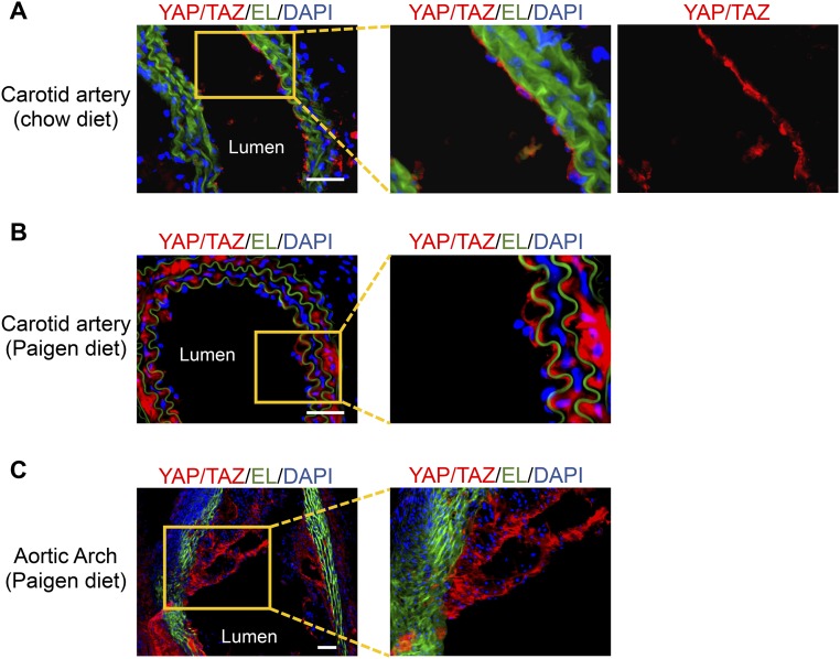
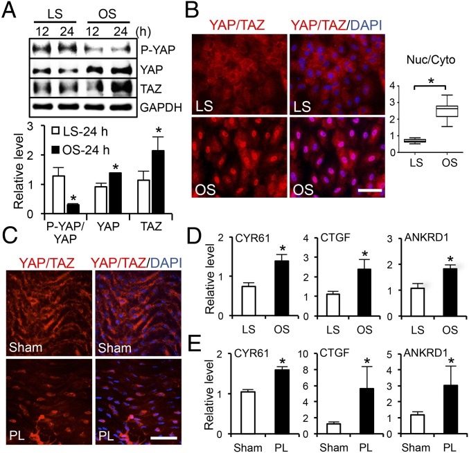
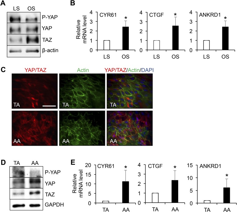
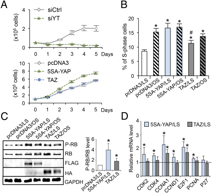
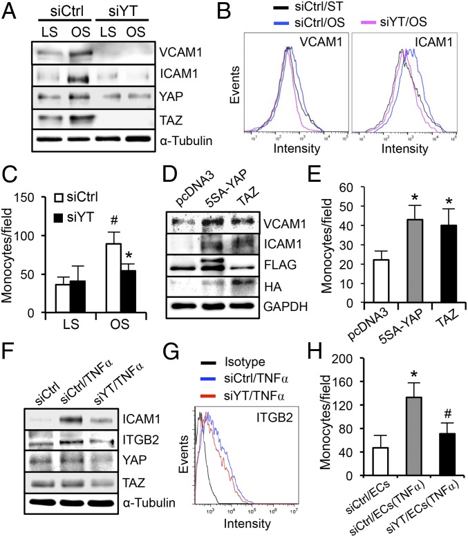
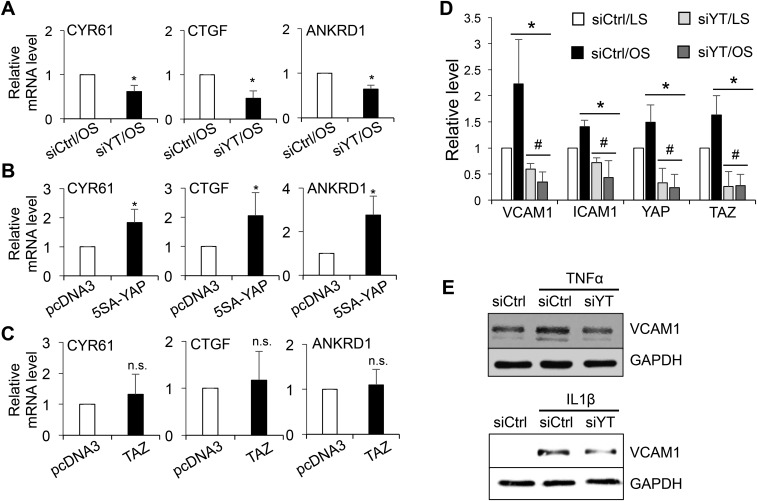
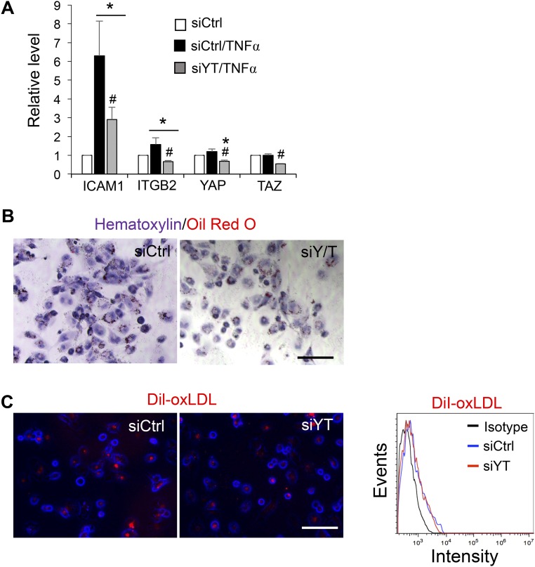
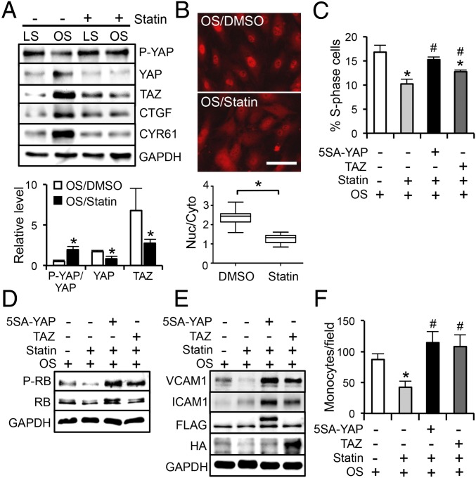
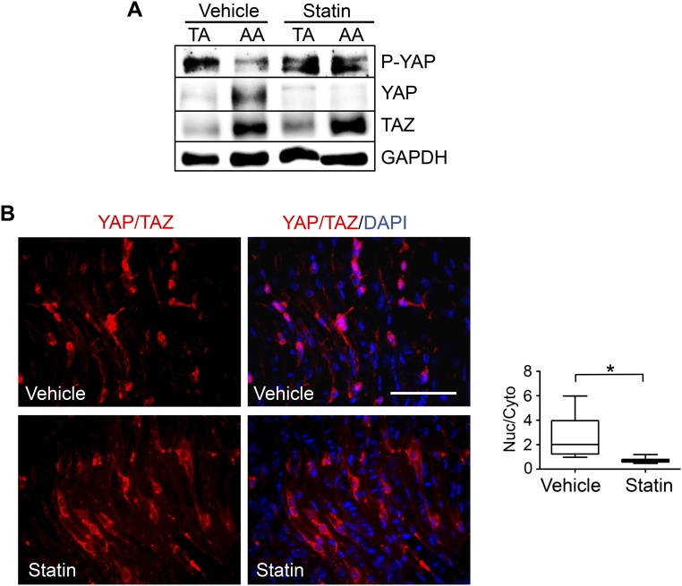
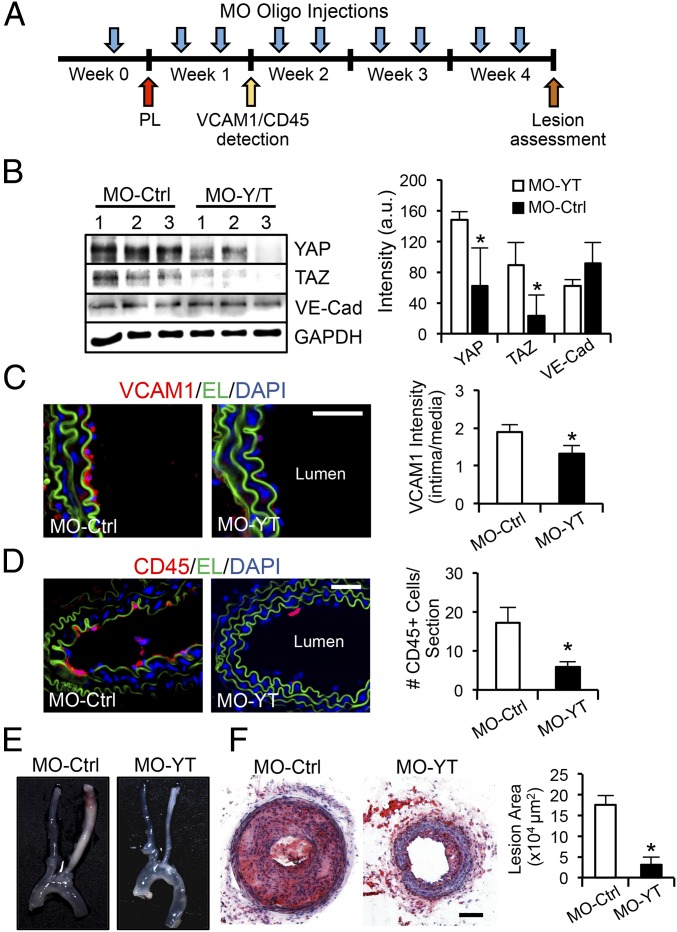
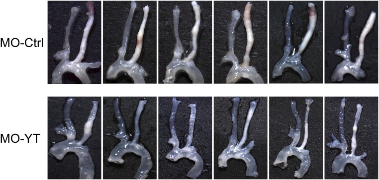
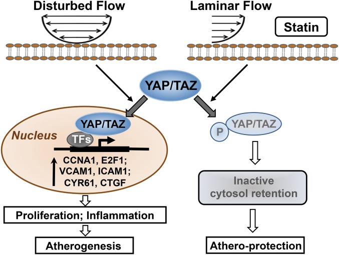
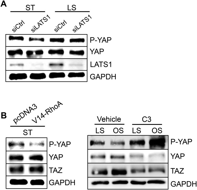
The focal nature of atherosclerotic lesions suggests an important role of local hemodynamic environment. Recent studies have demonstrated significant roles of Yes-associated protein (YAP) and transcriptional coactivator with PDZ-binding motif (TAZ) in mediating mechanotransduction and vascular homeostasis. The objective of this study is to investigate the functional role of YAP/TAZ in the flow regulation of atheroprone endothelial phenotypes and the consequential development of atherosclerotic lesions. We found that exposure of cultured endothelial cells (ECs) to the atheroprone disturbed flow resulted in YAP/TAZ activation and translocation into EC nucleus to up-regulate the target genes, including cysteine-rich angiogenic inducer 61 (CYR61), connective tissue growth factor (CTGF), and ankyrin repeat domain 1 (ANKRD1). In contrast, the athero-protective laminar flow suppressed YAP/TAZ activities. En face analysis of mouse arteries demonstrated an increased nuclear localization of YAP/TAZ and elevated levels of the target genes in the endothelium in atheroprone areas compared with athero-protective areas. YAP/TAZ knockdown significantly attenuated the disturbed flow induction of EC proliferative and proinflammatory phenotypes, whereas overexpression of constitutively active YAP was sufficient to promote EC proliferation and inflammation. In addition, treatment with statin, an antiatherosclerotic drug, inhibited YAP/TAZ activities to diminish the disturbed flow-induced proliferation and inflammation. In vivo blockade of YAP/TAZ translation by morpholino oligos significantly reduced endothelial inflammation and the size of atherosclerotic lesions. Our results demonstrate a critical role of the activation of YAP/TAZ by disturbed flow in promoting atheroprone phenotypes and atherosclerotic lesion development. Therefore, inhibition of YAP/TAZ activation is a promising athero-protective therapeutic strategy.

Keywords: atherogenesis; disturbed flow; endothelial cells; mechanotransduction.


Conflict of interest statement

The authors declare no conflict of interest.

Figures

Fig. S1.
Fig. S1.
The LCA from the control ApoE−/− mice on a chow diet (A) and the LCA and aortic arch from atherosclerotic ApoE−/− mice fed the Paigen diet for 8 wk (B and C) were embedded in O.C.T. compound and subjected to frozen section. The expression of YAP/TAZ in the sections of LCAs (A and B) and aortic arch (C) was demonstrated by immunofluorescence staining of YAP/TAZ (red). Elastic lamina (EL, green) and nuclei (DAPI, blue) were shown to indicate vessel structure. Boxed regions are enlarged in the right images. Results are representative images from four animals with similar staining. (Scale bars: 100 µm.)
Fig. 1.
Fig. 1.
Disturbed flow induces activation and nuclear localization of YAP/TAZ. (A) Western blot analysis of P-YAP, YAP, TAZ, and GAPDH in ECs subjected to LS or OS for the indicated time. The graphic data are mean ± SD of band intensity normalized to its internal control and LS 12-h group, n = 3. *P < 0.05 vs. LS-24 h. (B and C) Immunofluorescence staining of YAP/TAZ in ECs subjected to LS or OS (B) and in the endothelium of PL- or sham-operated carotid arteries (C). (Scale bars: 100 µm.) n = 4. The graphic data in B are the YAP/TAZ intensity ratio (nuclear/cytoplasmic) from cells randomly selected from three independent experiments. *P < 0.05 vs. LS. (D and E) qRT-PCR analysis of YAP/TAZ target genes expression in ECs subjected to OS or LS (D) and in PL or sham groups (E) (mean ± SD, n = 3, *P < 0.05 vs. LS or sham).
Fig. S2.
Fig. S2.
Representative Western blot analysis of P-YAP, YAP, TAZ, and β-actin (A), and qPCR analysis of the expression levels of YAP/TAZ target genes (CYR61, CTGF, and ANKRD1) (B) in HAECs subjected to 24-h LS or OS (mean ± SD, n = 3, *P < 0.05 vs. LS). (CE) The thoracic aorta (TA) and aortic arch (AA) from the control ApoE−/− mice fed a chow diet were dissected for en face staining of YAP/TAZ (red), actin fibers (green), and nuclei (blue) (C); or lysed for Western blot analysis of P-YAP, YAP, TAZ, and GAPDH (D), and qRT-PCR analysis of CYR61, CTGF, and ANKRD1 (E). Results in C and D are representative images from four animals with similar results. The graphic data in E are mean ± SD of the relative expression levels from four animals. *P < 0.05 vs. TA. (Scale bar: 100 µm.)
Fig. 2.
Fig. 2.
YAP/TAZ regulate the EC growth and cell cycle progression. (A) EC growth curves with YAP/TAZ knockdown (Upper) and overexpression (Lower). ECs were transfected with silencing RNAs against YAP/TAZ (siYT) or scramble control (siCtrl) and plasmids expressing FLAG-5SA-YAP (5SA-YAP), HA-TAZ (TAZ), or pcDNA3, and then seeded onto six-well plates. The number of ECs were counted daily for 5 d. n = 3. (B–D) ECs were transfected with 5SA-YAP, TAZ, or pcDNA3 plasmids, followed by OS and LS experiments for the analysis of cell cycle (B), retinoblastoma (RB) phosphorylation (C), and cell cycle regulatory genes expression (D). (BD) Mean ± SD, n = 3, *P < 0.05 vs. pcDNA3/LS and #P < 0.05 vs. 5SA-YAP/LS.
Fig. 3.
Fig. 3.
YAP/TAZ activation promotes the disturbed flow-induced proinflammatory responses. (AC) ECs were transfected with siYT or siCtrl, subjected to 24-h LS or OS, and followed by Western blot analysis of VCAM1, ICAM1, and YAP/TAZ (A), flow-cytometric analysis of VCAM1 and ICAM1 surface expression (B), and THP1 monocyte adhesion assay (C), *P < 0.05 vs. siCtrl/OS and #P < 0.05 vs. siCtrl/LS. (D and E) ECs were transfected with 5SA-YAP, TAZ, or pcDNA3 plasmids and subjected to the Western blot analysis of VCAM1 and ICAM1 (D) and monocyte adhesion assay (E). *P < 0.05 vs. pcDNA3. (FH) THP1 monocytes were transfected with siYT or siCtrl, treated with tumor necrosis factor-α (TNFα, 10 ng/mL) for 6 h, and then subjected to Western blot analysis of ICAM1 and ITGB2 (F), flow cytometric analysis of ITGB2 surface expression (G), and adhesion assay to the TNFα-activated ECs (H). *P < 0.05 vs. siCtrl/EC and #P < 0.05 vs. siCtrl/EC(TNFα).
Fig. S3.
Fig. S3.
The expression levels of CYR61, CTGF, and ANKRD1 in ECs subjected to YAP/TAZ knockdown (A) and overexpression (B and C) were determined by qRT-PCR. The graphic data are mean ± SD of the relative expression levels. n = 3. *P < 0.05 vs. siCtrl/OS (A) or vs. pcDNA3 (B and C). n.s., not significant. (D) Quantification analysis of the Western blot result shown in Fig. 3A (mean ± SD, n = 3, *P < 0.05 vs. siCtrl/LS and #P < 0.05 vs. siCtrl/OS). (E) ECs transfected with siYT or siCtrl were treated with TNFα and IL1β for 6 h and then collected for Western blot analysis of VCAM1 and GAPDH. Results are representative images from three independent experiments with a similar trend.
Fig. S4.
Fig. S4.
(A) THP1 monocytes were transfected with siYT or siCtrl, treated with TNFα (10 ng/mL) for 6 h, and then subjected to Western blot analysis of ICAM1 and ITGB2 (Fig. 3F). The graphic data are the quantification analysis of the Western blot results shown in Fig. 3F and presented as mean ± SD of band intensity normalized to its internal control and the respective control group. (n = 3, *P < 0.05 vs. siCtrl and #P < 0.05 vs. siCtrl/TNFα). (B and C) THP1 monocytes were transfected with siYT or siCtrl, differentiated to macrophages with phorbol myristate acetate (PMA, 25 ng/mL) for 48 h, followed by 0.5% serum starvation for 6 h, and then incubated with oxidized LDL (B) or DiI-oxLDL (C) for 12 h. The LDL uptake levels were determined by Oil Red O staining (B) and fluorescence microscopy and flow cytometric analyses of Dil-positive cells (C). (Scale bars: 100 µm.)
Fig. 4.
Fig. 4.
Statin inhibits YAP/TAZ to exert antiproliferative and antiinflammatory effects on ECs. (A) Western blot analysis of P-YAP, YAP, TAZ, CTGF, and CYR61 in ECs subjected to LS or OS in the presence of 1 µM simvastatin (statin) or DMSO control. *P < 0.05 vs. OS/DMSO. (B) Staining of YAP/TAZ localization in ECs subjected to 24-h OS in the presence of statin or DMSO. (Scale bar: 100 µm.) The graphic data are the nuclear/cytoplasmic ratio of YAP/TAZ intensity from cells randomly selected from three independent experiments. *P < 0.05 vs. OS/DMSO. (C and D) ECs transfected with 5SA-YAP, TAZ, or pcDNA3 were subjected to OS in the presence of statin or DMSO, followed by the analyses of cell cycle (C) and RB phosphorylation (D). (E and F) The transfected ECs were subjected to OS in the presence of statin or DMSO, followed by the analysis of VCAM1, ICAM1, and GAPDH (E) and monocyte adhesion assay (F). (C and F) Mean ± SD, n = 3, *P < 0.05 vs. OS/DMSO/pcDNA3 and #P < 0.05 vs. OS/statin/pcDNA3.
Fig. S5.
Fig. S5.
ApoE−/− mice fed a chow diet were subjected to the PL of LCAs and followed by the i.p. injection of simvastatin (statin) with a daily dose of 15 mg/kg. Three days after PL, the TA and the inner curvature of AA were harvested for the Western blot analysis of P-YAP, YAP, TAZ, and GAPDH (A); and the LCAs were fixed and cut longitudinally for the en face staining of YAP/TAZ (red) and nuclei (blue) (B). Results in (A) and (B) are representative images from three animals with similar results. The graphic data in B are the nuclear/cytoplasmic ratio of the YAP/TAZ intensity in randomly selected cells from three animals, *P < 0.05 vs. vehicle control. (Scale bar: 100 µm.)
Fig. 5.
Fig. 5.
YAP/TAZ play important roles in atherogenesis. (A) Experimental design of morpholino oligo treatments (MO-Ctrl vs. MO-YT, 10 nmol) in ApoE−/− mice fed the Paigen diet. The oligos were i.v. injected into animals 72 h before the PL, immediately after the PL, and twice a week afterward. (B) One-week post-PL, thoracic aortas were harvested for the analysis of YAP, TAZ, VE-Cadherin, and GAPDH expression. n = 3. (C and D) The sections of LCAs were examined by the immunofluorescence staining of VCAM1 (red) (C) and CD45 (red) (D). Elastic lamina (EL, green) and nuclear staining (DAPI, blue) are shown to indicate vessel structure. The VCAM1 intensity ratio (intima/media) and the number of CD45-positive cells attached to intima layer were quantified. n = 4. (E and F) Four-week post-PL, the arterial tissues were isolated to examine the atherosclerotic lesions (E), and the LCAs were sectioned for immunohistochemistry analysis (Oil Red O and Hematoxylin) and quantification of lesion area. n = 6 (F). (BD and F): mean ± SD, *P < 0.05 vs. MO-Ctrl. (Scale bars: 100 µm.)
Fig. S6.
Fig. S6.
Reduction of the PL-induced carotid atherosclerosis with in vivo inhibition of YAP/TAZ. ApoE−/− mice injected with MO oligos (MO-Ctrl and MO-YT, six mice each group) were subjected to PL and followed by the Paigen diet for 4 wk, as described in Fig. 5A. The arterial tissues (from carotid bifurcation to descending aorta) were isolated and examined for atherosclerotic lesions.
Fig. 6.
Fig. 6.
Schematic diagram of YAP/TAZ signaling and their modulation of EC phenotypes in response to disturbed flow, laminar flow, and statin.
Fig. S7.
Fig. S7.
(A) ECs transfected with silencing RNA against LATS1 (siLATS1) or siCtrl were subjected to 24-h LS or kept under static condition, followed by the Western blot analysis of P-YAP, YAP, LATS1, and GAPDH. (B) ECs transfected with the constitutively active form of RhoA (V14-RhoA) or a Rho inhibitor (C3 exoenzyme) followed by 24-h LS or OS, were subjected to the Western blot analysis of P-YAP, YAP, TAZ, and GAPDH. Results are representative images from three independent experiments with similar trend.

References

    1. Libby P, Ridker PM, Maseri A. Inflammation and atherosclerosis. Circulation. 2002;105(9):1135–1143. - PubMed
    1. Davignon J, Ganz P. Role of endothelial dysfunction in atherosclerosis. Circulation. 2004;109(23) Suppl 1:III27–III32. - PubMed
    1. Chiu JJ, Chien S. Effects of disturbed flow on vascular endothelium: Pathophysiological basis and clinical perspectives. Physiol Rev. 2011;91(1):327–387. - PMC - PubMed
    1. Hahn C, Schwartz MA. Mechanotransduction in vascular physiology and atherogenesis. Nat Rev Mol Cell Biol. 2009;10(1):53–62. - PMC - PubMed
    1. Zhao B, Lei QY, Guan KL. The Hippo-YAP pathway: New connections between regulation of organ size and cancer. Curr Opin Cell Biol. 2008;20(6):638–646. - PMC - PubMed

Publication types

MeSH terms