Review
. 2016 Oct 15;129(20):3685-3693.
doi: 10.1242/jcs.188490. Epub 2016 Sep 26.

Autophagy in adhesion and migration

Affiliations
Review

Autophagy in adhesion and migration

Candia M Kenific et al. J Cell Sci. .

Abstract

Autophagy, a pathway for lysosomal-mediated cellular degradation, has recently been described as a regulator of cell migration. Although the molecular mechanisms underlying autophagy-dependent motility are only beginning to emerge, new work demonstrates that selective autophagy mediated by the autophagy cargo receptor, NBR1, specifically promotes the dynamic turnover of integrin-based focal adhesion sites during motility. Here, we discuss the detailed mechanisms through which NBR1-dependent selective autophagy supports focal adhesion remodeling, and we describe the interconnections between this pathway and other established regulators of focal adhesion turnover, such as microtubules. We also highlight studies that examine the contribution of autophagy to selective degradation of proteins that mediate cellular tension and to integrin trafficking; these findings hint at further roles for autophagy in supporting adhesion and migration. Given the recently appreciated importance of selective autophagy in diverse cellular processes, we propose that further investigation into autophagy-mediated focal adhesion turnover will not only shed light onto how focal adhesions are regulated but will also unveil new mechanisms regulating selective autophagy.

Keywords: Autophagy; Focal adhesion; Migration; NBR1.


Conflict of interest statement

The authors declare no competing or financial interests.

Figures
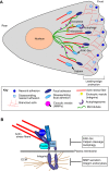
Fig. 1.
Fig. 1.
Cell migration and focal adhesion disassembly. (A) Migrating cells establish front–rear polarity in response to migration-inducing cues. At the front of the cell, branched actin networks support leading-edge lamellipodia protrusions, which are stabilized by the formation of nascent adhesions that mediate integrin-dependent cell–ECM contact. Nascent adhesions rapidly disassemble through unclear mechanisms, or alternatively, linkage of nascent adhesions to actin stress fibers results in maturation to stable focal adhesions, which transmit the forces generated by the actin cytoskeleton to promote forward motility (see B for detailed structural information). Focal adhesions disassemble through multiple mechanisms to allow for efficient cell displacement. Microtubules serve as tracks for both the exocytic and endocytic pathways that contribute to focal adhesion disassembly. Exocytic vesicles transport and enable secretion of matrix metalloproteinases (MMPs) near to focal adhesions to promote localized cleavage of ECM. This pathway of proteolysis disrupts the linkage between integrins and the ECM, leading to focal adhesion turnover. Endocytosis of integrins also compromises the stable architecture of focal adhesions by directly weakening their interaction with the ECM through removal of integrins. Autophagic sequestration of focal adhesion components also facilitates focal adhesion disassembly by sequestering focal adhesion proteins away from focal adhesions. Focal adhesion disassembly is also induced by calpain-mediated cleavage of the focal adhesion protein talin (of which there are two isoforms), which connects integrins to the actin cytoskeleton. Finally, signaling mediated by the focal-adhesion-associated kinase FAK and cytoplasmic tyrosine kinases, such as Src, also promotes focal adhesion disassembly, although the molecular details of this pathway have not been elucidated. (B) Focal adhesions are multiprotein complexes comprising integrins and scaffolding and signaling proteins. Integrins interact directly with the ECM. Intracellularly, focal adhesion proteins interact with each other, and they directly bind to the cytoplasmic tails of integrins (e.g. paxillin, FAK and talin) and/or actin fibers (e.g. talin, vinculin, zyxin). Together, these interactions form a stable connection spanning the plasma membrane from the intracellular actin cytoskeleton to the ECM that anchors the cell to its substratum. Multiple mechanisms (boxed text) disrupt this stable structure of focal adhesions to promote their disassembly (see A for details) by interfering with the connection between focal adhesion proteins and actin or the connection between integrins and the ECM.
Fig. 2.
Fig. 2.
The mammalian autophagy pathway. (A) During extreme stress, such as nutrient starvation, non-selective, bulk autophagy is activated. The autophagosome is a double-membraned vesicle that captures cellular constituents as it elongates. Fully formed autophagosomes eventually fuse with lysosomes, where lysosomal proteases degrade autophagy substrates. The resulting degradation products are fed back to the cell to serve as metabolic building blocks to sustain the cell during stress. (B) Selective autophagy is important for cellular sequestration of foreign pathogens and serves crucial housekeeping functions to maintain cellular homeostasis. Selective autophagy cargos, such as pathogens, protein aggregates and damaged mitochondria, are encapsulated by the growing autophagosome through interaction with autophagy cargo receptors (ACRs). These receptors interact with LC3 proteins (LC3) on the autophagosome membrane through an LC3-interacting region (LIR) motif. Cargo recognition is often mediated by the ubiquitin-binding domain (UBD), which interacts with ubiquitylated cargos. Note that cargo recognition might also occur in a ubiquitin-independent manner (not shown).
Fig. 3.
Fig. 3.
Potential mechanisms of focal adhesion disassembly mediated by NBR1-dependent selective autophagy. NBR1-dependent selective autophagy promotes focal adhesion disassembly. Interaction between the LC3-interacting region (LIR) of NBR1 and LC3 proteins (LC3) promotes the targeting of autophagosomes to focal adhesions for sequestration of focal adhesion proteins. Ubiquitylation (Ub) of focal adhesion proteins could enable their binding to the UBD of NBR1, and thus their targeting to autophagosomes. Phosphorylation of ubiquitin (pUb) could serve as a crucial mechanism that further fine-tunes the recognition of focal adhesions by NBR1, but the E3 ligases and kinases activating these potential pathways remain unknown. Microtubules (MTs) might also have an important role in promoting recruitment of autophagy proteins, including LC3 and NBR1, to focal adhesions for their disassembly.
Fig. 4.
Fig. 4.
Regulation of cellular tension and adhesion by autophagy. (A) Contraction of actin stress fibers, which is mediated by myosin II, imposes tension on focal adhesions and contributes to their maturation. Binding of the head domains of myosin II to actin cross-links actin stress fibers. Phosphorylation of the myosin light chain leads to conformational changes in myosin II, which result in contraction as actin filaments slide past each other in opposite directions, thereby exerting tensional forces on focal adhesions. Phosphorylation of myosin light chain is regulated by RhoA GTPase. GTP-bound RhoA activates Rho-associated protein kinase (ROCK, of which there are two isoforms), which directly phosphorylates myosin light chain. Ubiquitylation (Ub) of active RhoA targets it for p62-mediated autophagic degradation; degradation of RhoA by autophagy might, therefore, lead to loss of tension and disassembly of focal adhesions. (B) Filamin A supports focal adhesion formation by mediating the linkage of focal adhesions to actin. In response to tension-induced conformational changes of filamin A, BAG3, a co-chaperone of the chaperone-assisted selective autophagy (CASA) pathway, recruits an E3-ligase-containing complex to filamin A; this results in ubiquitylation of filamin A and binding of p62 for targeting of filamin A for autophagic degradation. Autophagy-dependent filamin A degradation might destabilize focal adhesions. (C) During cell migration, integrins are endocytosed and recycled back to the plasma membrane through either early endosomes, late endosomes or lysosomes, or are degraded at the lysosome. Autophagy has also been proposed to specifically direct trafficking of integrins to the lysosome for their degradation in order to influence cell motility. AP, autophagosome.

Similar articles

Cited by

References

    1. Abercrombie M., Heaysman J. E. M. and Pegrum S. M. (1971). The locomotion of fibroblasts in culture. IV. Electron microscopy of the leading lamella. Exp. Cell Res. 67, 359-367. 10.1016/0014-4827(71)90420-4 - DOI - PubMed
    1. Ao X., Zou L. and Wu Y. (2014). Regulation of autophagy by the Rab GTPase network. Cell Death Differ. 21, 348-358. 10.1038/cdd.2013.187 - DOI - PMC - PubMed
    1. Arndt V., Dick N., Tawo R., Dreiseidler M., Wenzel D., Hesse M., Fürst D. O., Saftig P., Saint R., Fleischmann B. K. et al. (2010). Chaperone-assisted selective autophagy is essential for muscle maintenance. Curr. Biol. 20, 143-148. 10.1016/j.cub.2009.11.022 - DOI - PubMed
    1. Barth A. I. M., Caro-Gonzalez H. Y. and Nelson W. J. (2008). Role of adenomatous polyposis coli (APC) and microtubules in directional cell migration and neuronal polarization. Semin. Cell Dev. Biol. 19, 245-251. 10.1016/j.semcdb.2008.02.003 - DOI - PMC - PubMed
    1. Belaid A., Cerezo M., Chargui A., Corcelle-Termeau E., Pedeutour F., Giuliano S., Ilie M., Rubera I., Tauc M., Barale S. et al. (2013). Autophagy plays a critical role in the degradation of active RHOA, the control of cell cytokinesis, and genomic stability. Cancer Res. 73, 4311-4322. 10.1158/0008-5472.CAN-12-4142 - DOI - PMC - PubMed

Publication types