. 2016 Dec;29(12):583-594.
doi: 10.1093/protein/gzw049. Epub 2016 Sep 26.

Engineering a minimal G protein to facilitate crystallisation of G protein-coupled receptors in their active conformation

Affiliations

Engineering a minimal G protein to facilitate crystallisation of G protein-coupled receptors in their active conformation

Byron Carpenter et al. Protein Eng Des Sel. 2016 Dec.

Abstract

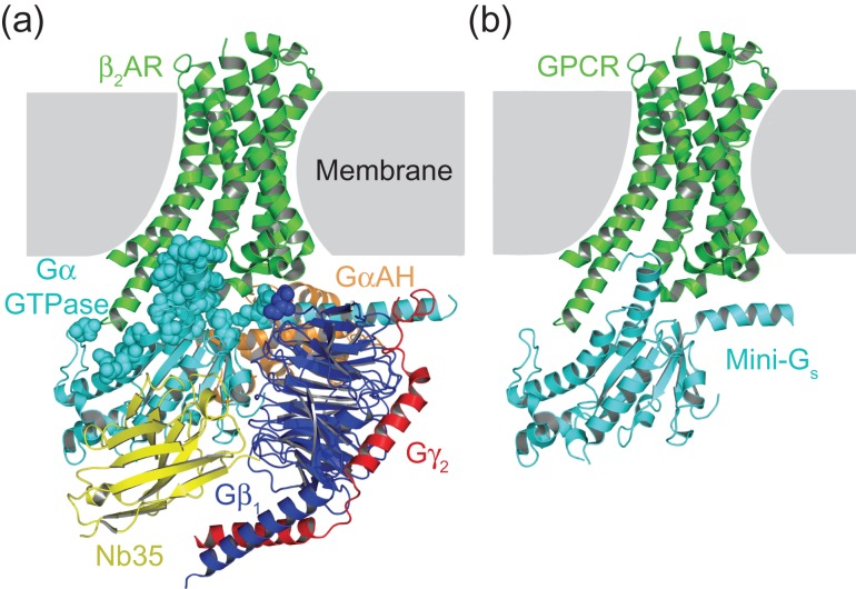
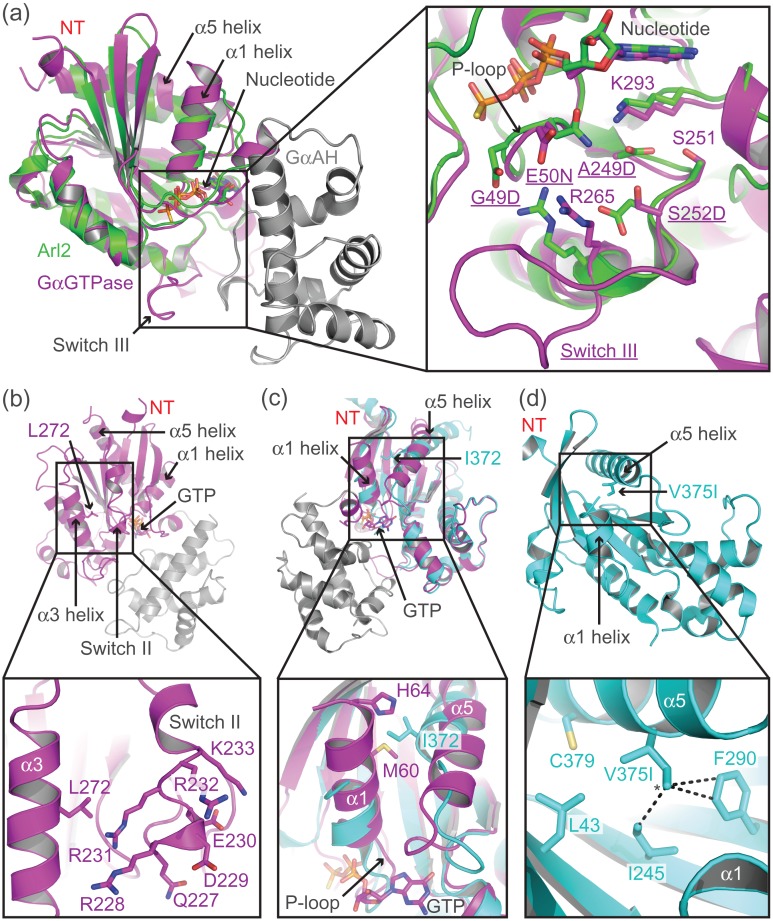
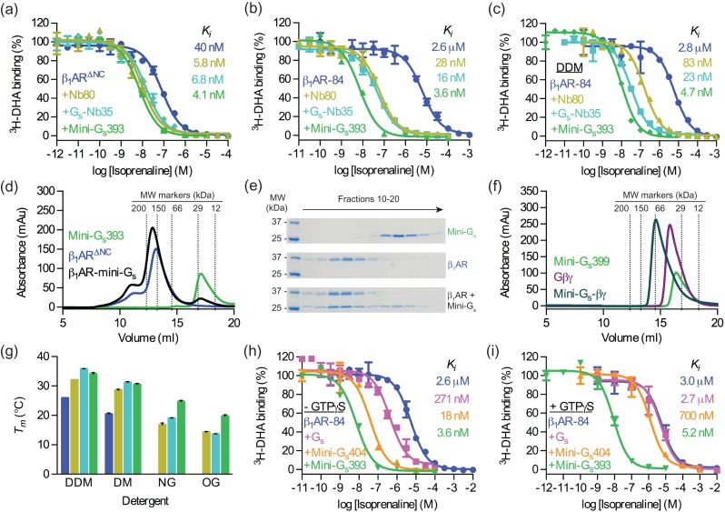
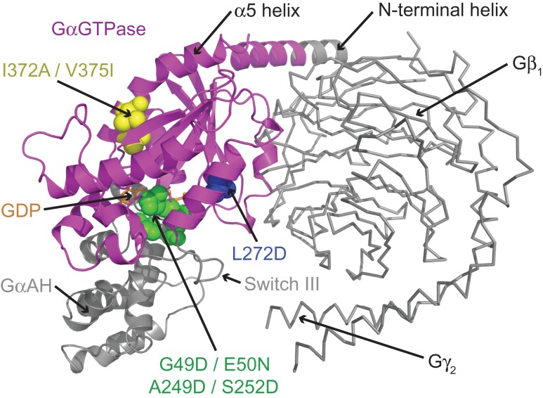
G protein-coupled receptors (GPCRs) modulate cytoplasmic signalling in response to extracellular stimuli, and are important therapeutic targets in a wide range of diseases. Structure determination of GPCRs in all activation states is important to elucidate the precise mechanism of signal transduction and to facilitate optimal drug design. However, due to their inherent instability, crystallisation of GPCRs in complex with cytoplasmic signalling proteins, such as heterotrimeric G proteins and β-arrestins, has proved challenging. Here, we describe the design of a minimal G protein, mini-Gs, which is composed solely of the GTPase domain from the adenylate cyclase stimulating G protein Gs Mini-Gs is a small, soluble protein, which efficiently couples GPCRs in the absence of Gβγ subunits. We engineered mini-Gs, using rational design mutagenesis, to form a stable complex with detergent-solubilised β1-adrenergic receptor (β1AR). Mini G proteins induce similar pharmacological and structural changes in GPCRs as heterotrimeric G proteins, but eliminate many of the problems associated with crystallisation of these complexes, specifically their large size, conformational dynamics and instability in detergent. They are therefore novel tools, which will facilitate the biochemical and structural characterisation of GPCRs in their active conformation.

Keywords: G protein; G protein-coupled receptor; GPCR; Gs; complex; mini G protein; mini-Gs.


Figures

Fig. 1
Fig. 1
Design of a minimal G protein. (a) Crystal structure of the β2AR–Gs complex (PDB code 3SN6; Rasmussen et al., 2011b). The intracellular component of this complex, which is composed of Gαs, Gβ1, Gγ2 and Nb35, totals over 100 kDa in molecular weight. However, over 97% of direct contacts (3.9 Å cut-off) between β2AR and Gs are formed by the GαGTPase domain (cyan). Residues from Gs that form contacts with β2AR are shown as spheres. (b) Model of the proposed complex between a GPCR and mini-Gs (isolated GαGTPase domain). The intracellular component of this complex is a single protein with a molecular weight of approximately 27 kDa. Figures were prepared using PyMOL (The PyMOL Molecular Graphics System, Version 1.7.4 Schrödinger, LLC)
Fig. 2
Fig. 2
Rational design of mutations to stabilise mini-Gs. (a) Structural alignment of Gαs (PDB code 1AZT; Sunahara et al., 1997), coloured magenta and grey, and Arl2 (PDB code 1KSH; Hanzal-Bayer et al., 2002), coloured green. The Gαs GTPase domain aligns to Arl2 with an RMSD of 1.9 Å, despite sharing sequence identity of only 25%, determined using the Dali server (Holm and Rosenstrom, 2010). See Supplementary Fig. S4 for a sequence alignment between Gαs and Arl-2. The inset shows an expanded view of mini-Gs residues (shown as sticks and underlined name) that were mutated (G49D, E50N, A249D, and S252D) to match the corresponding residue in Arl2. Residues with which the mutations potentially interact are shown as sticks. (b) Mutation of Leu272, which is located within the α3 helix of Gαs (PDB code 1AZT; Sunahara et al., 1997), to aspartic acid allows potential interactions with a cluster of charged and polar residues (227–233) in the N-terminal region of switch II. (c) Alignment of Gαs in its GTP-bound conformation (PDB code 1AZT; Sunahara et al., 1997), coloured magenta, and GPCR-bound conformation (PDB code 3SN6; Rasmussen et al., 2011b), coloured cyan. In the GPCR-bound conformation Ile372 (α5 helix) sterically clashes with Met60 and His64 (α1 helix), preventing close packing of the α1 helix against the core of the GαGTPase domain. (d) The V375I mutation (modelled using PyMOL) was designed to increase hydrophobic contacts between the core of the GαGTPase domain and the α5 helix in its GPCR-bound conformation (PDB code 3SN6; Rasmussen et al., 2011b). Residues that interact with Val375 are shown as sticks, additional contacts (less than 4.2 Å), which are predicted to be formed by the δ-carbon (*) of the isoleucine mutation are displayed as dashed lines.
Fig. 3
Fig. 3
Validation of mini-Gs (a–c) A competition binding assay was used to measure the change in affinity (Ki) of isoprenaline induced by Nb80, Gs–Nb35, or mini-Gs393 coupling to: (a) membrane-embedded β1AR∆NC, (b) membrane-embedded β1AR-84 and (c) DDM-solubilised β1AR-84. (d) Analytical gel filtration analysis of β1AR∆NC binding to mini-Gs393. The apparent molecular weight of mini-Gs393 was 23 kDa (17.1 ml), which compares well with the theoretical value of 27 kDa. The apparent molecular weight of β1AR∆NC was 139 kDa (13.2 ml), which is consistent with the 45 kDa receptor being associated with a large detergent micelle; the shoulder at 11 ml (> 300 kDa) probably represents aggregated receptor. A mixture of β1AR∆NC and mini-Gs393 (1.2-fold molar excess) resolved as a predominant peak with an apparent molecular weight of 160 kDa (12.9 ml). The 21 kDa increase in the apparent molecular weight of the β1AR∆NC–mini-Gs393 complex compared to uncoupled β1AR∆NC is consistent with mini-Gs393 binding with 1:1 stoichiometry. (e) SDS-PAGE analysis of the gel filtration eluate confirmed the presence of both β1AR∆NC and mini-Gs393 in the peak fractions. (f) Analytical gel filtration analysis of Gβγ binding to mini-Gs399. The apparent molecular weights of mini-Gs399 (Supplementary Table SIII) and Gβγ were 32 kDa (16.4 ml) and 42 kDa (15.8 ml), respectively, which is in close agreement with the theoretical values of 29 kDa and 46 kDa, respectively. An equimolar mixture of mini-Gs399 and Gβγ resolved as a single peak with an apparent molecular weight of 73 kDa (14.6 ml). The 31 kDa increase in the apparent molecular weight of the mini-Gs399–Gβγ complex compared to Gβγ is consistent with mini-Gs399 binding with 1:1 stoichiometry. (g) Thermostability of detergent-solubilised β1AR∆NC alone or in complex with Nb80, Gs–Nb35, or mini-Gs393, in different detergents (Supplementary Fig. S10). Uncoupled β1AR∆NC did not survive solubilisation in NG or OG. Colours correspond to those used in (a). (h–i) GTP-mediated dissociation of β1AR-84 complexes, measured by competition binding assay. The response in isoprenaline Ki induced by Gs, mini-Gs404 (Supplementary Table SIII), or mini-Gs393 coupling to β1AR-84 was measured in the presence or absence of GTPγS (250 μM). (a–c,h,i) Data are representative of at least two independent experiments, each performed in duplicate, with error bars ± SEM. (g) Data represent mean ± SEM of at least two independent experiments, each performed in duplicate.
Fig. 4
Fig. 4
A model of heterotrimeric Gs highlighting the region that corresponds to mini-Gs (magenta). The model of heterotrimeric Gs was constructed by superposition of the crystal structures of Gαs (PDB code 1AZT; Sunahara et al., 1997) and heterotrimeric Gαt/i1 (PDB code 1GOT; Lambright et al., 1996). Residues that were mutated in mini-Gs (shown as spheres) were clustered in three regions of the protein: the nucleotide-binding pocket (green), switch II (blue), and the α5 helix (yellow). Regions of Gαs that were deleted in mini-Gs (GαAH, switch III and half of the N-terminal helix) are coloured grey. The Gβγ subunits, which are not required for mini-Gs coupling to GPCRs are shown as ribbons and coloured grey. GDP is shown as sticks and coloured orange.

References

    1. Alexander N.S., Preininger A.M., Kaya A.I., Stein R.A., Hamm H.E. and Meiler J. (2014) Nat. Struct. Mol. Biol., 21, 56–63. - PMC - PubMed
    1. Bai X.C., McMullan G. and Scheres S.H. (2015) Trends Biochem. Sci., 40, 49–57. - PubMed
    1. Baker J.G., Proudman R.G. and Tate C.G. (2011) Naunyn Schmiedebergs Arch. Pharmacol., 384, 71–91. - PMC - PubMed
    1. Bornancin F., Pfister C. and Chabre M. (1989) Eur. J. Biochem., 184, 687–698. - PubMed
    1. Carpenter B., Nehme R., Warne T., Leslie A.G. and Tate C.G. (2016) Nature, 536, 104–107. - PMC - PubMed