. 2016 Nov 5;371(1707):20150505.
doi: 10.1098/rstb.2015.0505.

Convergent evolution of pathogenicity islands in helper cos phage interference

Affiliations

Convergent evolution of pathogenicity islands in helper cos phage interference

Nuria Carpena et al. Philos Trans R Soc Lond B Biol Sci. .

Abstract

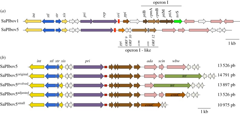
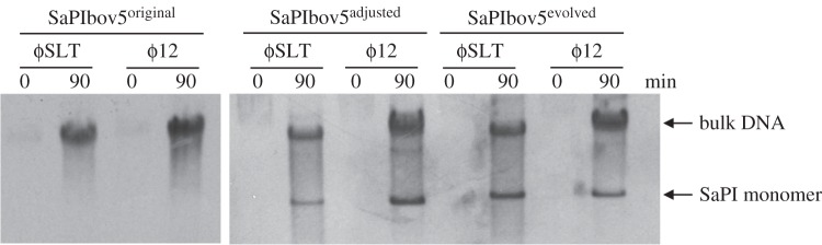
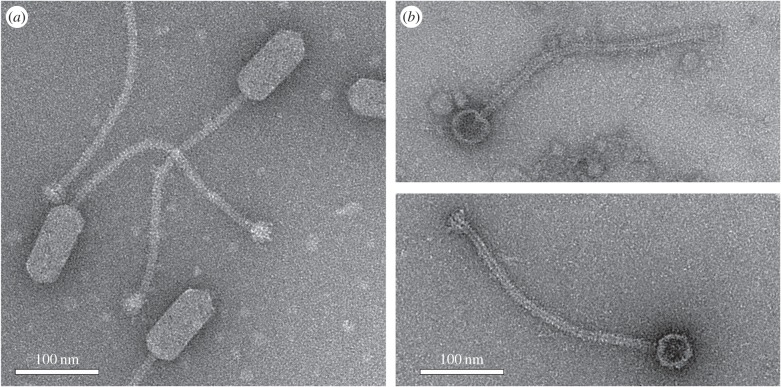
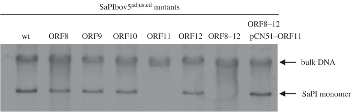
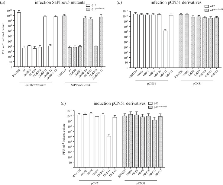
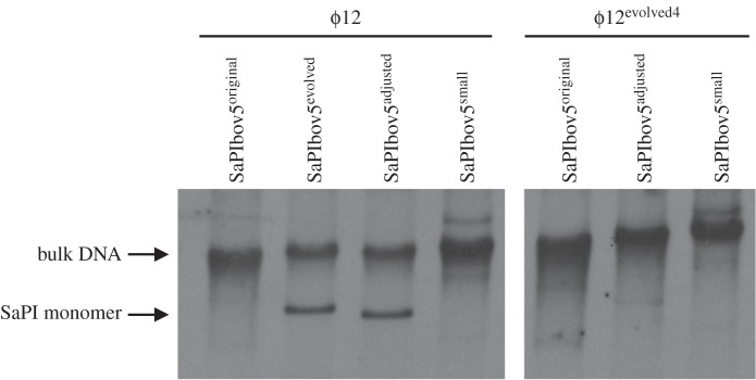
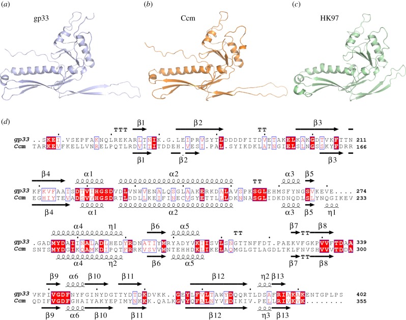
Staphylococcus aureus pathogenicity islands (SaPIs) are phage satellites that exploit the life cycle of their helper phages for their own benefit. Most SaPIs are packaged by their helper phages using a headful (pac) packaging mechanism. These SaPIs interfere with pac phage reproduction through a variety of strategies, including the redirection of phage capsid assembly to form small capsids, a process that depends on the expression of the SaPI-encoded cpmA and cpmB genes. Another SaPI subfamily is induced and packaged by cos-type phages, and although these cos SaPIs also block the life cycle of their inducing phages, the basis for this mechanism of interference remains to be deciphered. Here we have identified and characterized one mechanism by which the SaPIs interfere with cos phage reproduction. This mechanism depends on a SaPI-encoded gene, ccm, which encodes a protein involved in the production of small isometric capsids, compared with the prolate helper phage capsids. As the Ccm and CpmAB proteins are completely unrelated in sequence, this strategy represents a fascinating example of convergent evolution. Moreover, this result also indicates that the production of SaPI-sized particles is a widespread strategy of phage interference conserved during SaPI evolution.This article is part of the themed issue 'The new bacteriology'.

Keywords: PICIs; SaPIs; bacteriophage packaging; bacteriophage resistance; capsid morphogenesis; small capsids.


Figures

Figure 1.
Figure 1.
Genomic structure of the cos SaPIs. (a) Comparison of the pac (SaPIbov1) and cos (SaPIbov5) SaPIs. (b) Alignment of selected SaPIbov5 size adjustment. Genomes are aligned according to the prophage convention with the integrase gene at the left end. Gene colour code: int and xis, yellow; transcription regulators, blue; replication genes, purple; replication origin, red; genes affecting expression (pti) or assembly (cpm) of helper phage virion components are dark brown and medium brown, respectively; the terminase small subunit gene (terS) is green; pip (phage interference) orange, the two variant subsets are distinguished by dark versus light fill; superantigen and other accessory genes, pink. Genes encoding hypothetical proteins, white. In (a), the cos site is shown in grey. In (b), the tetracycline resistance gene is light green, and the erythromycin resistance gene is dark red.
Figure 2.
Figure 2.
Replication analysis of the different SaPIbov5 derivative islands. Southern blot of ϕ12 and ϕSLT lysates, from strains carrying SaPIbov5original, SaPIbov5adjusted and SaPIbov5evolved as indicated (see text for details). Samples were isolated 0 or 90 min after induction with mitomycin C, separated on agarose gels and blotted with a SaPIbov5-specific probe. Upper band is ‘bulk’ DNA, and represents replicating SaPIbov5. SaPI monomer represents SaPI DNA packaged in small capsids.
Figure 3.
Figure 3.
Electron microscopy of ϕ12 and SaPIbov5 particles. Electron micrographs of negatively stained wt ϕ12 virions (a), and particles produced by induction of a ϕ12 lysogen containing SaPIbov5adjusted (b). Scale bars are 100 nm.
Figure 4.
Figure 4.
Replication analysis of SaPIbov5 mutants. Southern blot of ϕ12 lysates, from strains carrying the wt or the different SaPIbov5 mutants (carrying ochre mutations in the SaPIbov5 genes 8–12). Samples were isolated 90 min after induction with mitomycin C, separated on agarose and blotted with a SaPIbov5-specific probe. Upper band is ‘bulk’ DNA, and represents replicating SaPIbov5. SaPI monomer represents SaPI DNA packaged in small capsids. SaPIbov5 ORF11 corresponds to ccm.
Figure 5.
Figure 5.
SaPIbov5 Ccm-mediated interference. (a) Strain RN4220 containing wt or the different SaPIbov5 mutants were infected with ϕ12 or ϕ12evolved4, plated on phage bottom agar, and incubated for 48 h at 32°C. (b) Phage interference mediated by cloned SaPIbov5 genes. The indicated genes were cloned into plasmid pCN51. Strain RN4220 containing the indicated plasmids was infected with phages 12 or ϕ12evolved4, plated on phage bottom agar containing 5 µM CdCl2 (induces the expression of the cloned genes) and incubated for 48 h at 32°C. (c) Effect of the different pCN51 cloned genes in phage reproduction. The lysogenic strains for ϕ12 or ϕ12evolved4, containing the different pCN51 derivative plasmids, were SOS induced and the lysates plated on phage bottom agar for 48 h at 32°C.
Figure 6.
Figure 6.
Replication analysis of the different sized SaPIbov5 islands induced by phages ϕ12 or ϕ12evolved4. Southern blot of ϕ12 and ϕ12evolved4 lysates, from strains carrying SaPIbov5original, SaPIbov5evolved, SaPIbov5adjusted or SaPIbov5small, as indicated. Samples were taken 90 min after induction with mitomycin C, separated on agarose and blotted with a SaPIbov5-specific probe. Upper band is ‘bulk’ DNA, and represents replicating SaPIbov5. SaPI monomer represents SaPI DNA packaged in small capsids.
Figure 7.
Figure 7.
C-terminal portion of gp33 and Ccm proteins are predicted to adopt the characteristic HK97-fold of phage coat proteins. Cartoon representation of the C-terminal portion of (a) ϕ12 gp33 (residues 127–402) and (b) SaPIbov5 Ccm (residues 83–355), generated by RaptorX [31]. Both proteins show similar folding to the prototypical coat protein from phage HK97 (c; PDB 1OHG). (d) Structural alignment of ϕ12 gp33 (a) and SaPIbov5 Ccm (b) models carried out with Mustang [33]. Identical residues are highlighted on a red background and conserved residues are in a blue box with red text. The elements of secondary structure for each model are shown above (gp33) or below (Ccm) the corresponding sequence.

References

    1. Penadés JR, Christie GE. 2015. The phage-inducible chromosomal islands: a family of highly evolved molecular parasites. Annu. Rev. Virol. 2, 181–201. (10.1146/annurev-virology-031413-085446) - DOI - PubMed
    1. Frígols B, Quiles-Puchalt N, Mir-Sanchis I, Donderis J, Elena SF, Buckling A, Novick RP, Marina A, Penadés JR. 2015. Virus satellites drive viral evolution and ecology. PLoS Genet. 11, e1005609 (10.1371/journal.pgen.1005609) - DOI - PMC - PubMed
    1. Ubeda C, Tormo MA, Cucarella C, Trotonda P, Foster TJ, Lasa I, Penadés JR. 2003. Sip, an integrase protein with excision, circularization and integration activities, defines a new family of mobile Staphylococcus aureus pathogenicity islands. Mol. Microbiol. 49, 193–210. (10.1046/j.1365-2958.2003.03577.x) - DOI - PubMed
    1. Mir-Sanchis I, Martínez-Rubio R, Martí M, Chen J, Lasa I, Novick RP, Tormo-Más MÁ, Penadés JR. 2012. Control of Staphylococcus aureus pathogenicity island excision. Mol. Microbiol. 85, 833–845. (10.1111/j.1365-2958.2012.08145.x) - DOI - PubMed
    1. Ubeda C, Barry P, Penadés JR, Novick RP. 2007. A pathogenicity island replicon in Staphylococcus aureus replicates as an unstable plasmid. Proc. Natl Acad. Sci. USA 104, 14 182–14 188. (10.1073/pnas.0705994104) - DOI - PMC - PubMed

Publication types

MeSH terms

Substances