Review
. 2016 Sep 7;22(33):7486-99.
doi: 10.3748/wjg.v22.i33.7486.

Aberrant regulation of Wnt signaling in hepatocellular carcinoma

Affiliations
Review

Aberrant regulation of Wnt signaling in hepatocellular carcinoma

Li-Juan Liu et al. World J Gastroenterol. .

Abstract

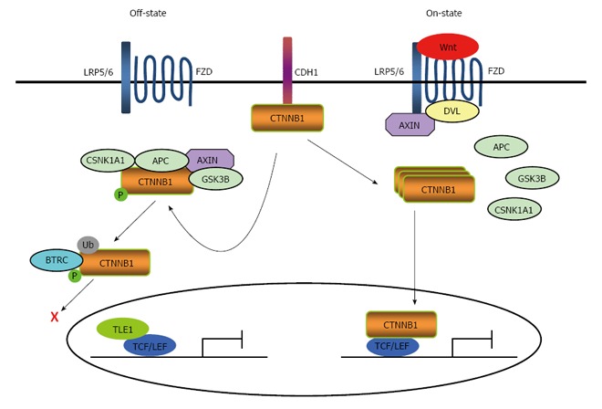
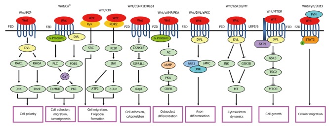
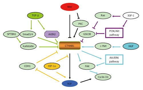
Hepatocellular carcinoma (HCC) is one of the most lethal malignancies in the world. Several signaling pathways, including the wingless/int-1 (Wnt) signaling pathway, have been shown to be commonly activated in HCC. The Wnt signaling pathway can be triggered via both catenin β1 (CTNNB1)-dependent (also known as "canonical") and CTNNB1-independent (often referred to as "non-canonical") pathways. Specifically, the canonical Wnt pathway is one of those most frequently reported in HCC. Aberrant regulation from three complexes (the cell-surface receptor complex, the cytoplasmic destruction complex and the nuclear CTNNB1/T-cell-specific transcription factor/lymphoid enhancer binding factor transcriptional complex) are all involved in HCC. Although the non-canonical Wnt pathway is rarely reported, two main non-canonical pathways, Wnt/planar cell polarity pathway and Wnt/Ca(2+) pathway, participate in the regulation of hepatocarcinogenesis. Interestingly, the canonical Wnt pathway is antagonized by non-canonical Wnt signaling in HCC. Moreover, other signaling cascades have also been demonstrated to regulate the Wnt pathway through crosstalk in HCC pathogenesis. This review provides a perspective on the emerging evidence that the aberrant regulation of Wnt signaling is a critical mechanism for the development of HCC. Furthermore, crosstalk between different signaling pathways might be conducive to the development of novel molecular targets of HCC.

Keywords: Canonical wingless/int-1 signaling; Catenin β1; Crosstalk; Hepatocellular carcinoma; Non-canonical wingless/int-1 signaling; Wingless/int-1.


Conflict of interest statement

Conflict-of-interest statement: The authors declare that they have no conflicts of interest.

Figures

Figure 1
Figure 1
Canonical wingless/int-1signaling pathway. Three complexes are involved in the dynamic activating event: (1) the cell-surface receptor complex; (2) the destruction complex in the cytoplasm; and (3) the CTNNB1/TCF/LEF transcriptional complex in the nucleus. In a normal steady state, there are two pools for CTNNB1 in cells. One is known to interact with CDH1 at the cell-cell junction. The second is present in the destruction complex in cytoplasm, which is assembled by the scaffold proteins AXIN, APC, GSK3β, and CSNK1A1. CSNK1A1 and GSK3β phosphorylate CTNNB1 in the AXIN complex. Phosphorylated CTNNB1 is subsequently recognized and ubiquitinated by BTRC. In the absence of nuclear CTNNB1 translocated from the cytoplasm, TCF/LEF proteins bind to DNA and act as transcriptional repressors by binding to TLE1 proteins. These in turn interact with histone deacetylases whose activities lead to the transcriptional silence of chromatin. The binding of Wnts to FZDs, which form the cell-surface receptor complex, promotes the binding of scaffold proteins such as DVL to the FZD intracellular domains. This subsequently induces the aggregation and phosphorylation of LRP6 and the translocation of AXIN. Phosphorylated LRP6 also recruits AXIN to LRP6 on the plasma membrane. This allows AXIN to be inactivated, which then inhibits CTNNB1 phosphorylation. This in turn allows CTNNB1 to escape degradation, accumulate in the cytoplasm, and translocate to the nucleus. In the nucleus, CTNNB1 interacts primarily with members of the TCF/LEF family of transcription factors and triggers the activation of multiple intracellular signaling cascades. This results in the regulation of various cellular functions. CTNNB1: Catenin beta 1; TCF/LEF: T-cell-specific transcription factor/lymphoid enhancer binding factor; CDH1: Cell adhesion molecule cadherin 1; APC: Adenomatous polyposis coli; GSK3β: GSK3B, glycogen synthase kinase 3 beta; CSNK1A1: Casein kinase 1 alpha 1; FZD: Frizzled class receptor; BTRC: Beta-transducin repeat containing E3 ubiquitin protein ligase.
Figure 2
Figure 2
Non-canonical wingless/int-1 signaling pathway. Non-canonical Wnt pathways are mediated by several possible mechanisms which are independent of the CTNNB1-TCF/LEF transcriptional function, including: (1) Wnt/PCP pathway; (2) Wnt/Ca2+ pathway; (3) Wnt/RTK pathway; (4) Wnt/CSNK1E/Rap1 pathway; (5) Wnt/cAMP/PKA pathway; (6) Wnt/DVL/aPKC pathway; (7) Wnt/GSK3β/MT pathway; (8) Wnt/MTOR pathway; and (9) Wnt/FYN/STAT3 pathway. Lines ending with arrows or bars indicate activating or inhibitory effects respectively. Wnt: Wingless/int-1; CTNNB1: Catenin beta 1; TCF/LEF: T-cell-specific transcription factor/lymphoid enhancer binding factor; PCP: Planar cell polarity; RTK: Receptor tyrosine kinases; CSNK1E: Casein kinase I epsilon; cAMP: Cyclic adenosine monophosphate; PKA: Protein kinase A; DVL: Disheveled; aPKC: Atypical protein kinase C; GSK3β: GSK3B, glycogen synthase kinase 3 beta; MTOR: Mechanistic target of rapamycin; FYN: FYN proto-oncogene, Src family tyrosine kinase; STAT3: Signal transducer and activator of transcription 3.
Figure 3
Figure 3
Regulation of wingless/int-1 signaling by crosstalk in hepatocellular carcinoma. The crosstalk between other signaling cascades and the Wnt signaling pathways involved in hepatocarcinogenesis are shown (see text). Lines ending with arrows or bars indicate activating or inhibitory effects respectively. The distinct line colors indicate the different pathways that crosstalk with Wnt signaling, including: Wnt signaling pathway (black), TGF-β pathway (green), HGF/c-Met pathway (blue), HIF-1α/EMT pathway (yellow), and IGF-1 pathway (purple). Wnt: Wingless/int-1; TGF-β: Transforming growth factor beta; HGF: Hepatocyte growth factor; HIF-1α: Hypoxia- inducible factor-1 alpha; EMT: Epithelial-mesenchymal transition; IGF-1: Insulin/insulin-like growth factor-1.

Similar articles

Cited by

References

    1. Ferlay J, Soerjomataram I, Dikshit R, Eser S, Mathers C, Rebelo M, Parkin DM, Forman D, Bray F. Cancer incidence and mortality worldwide: sources, methods and major patterns in GLOBOCAN 2012. Int J Cancer. 2015;136:E359–E386. - PubMed
    1. Tornesello ML, Buonaguro L, Izzo F, Buonaguro FM. Molecular alterations in hepatocellular carcinoma associated with hepatitis B and hepatitis C infections. Oncotarget. 2016 Epub ahead of print. - PMC - PubMed
    1. Kawai-Kitahata F, Asahina Y, Tanaka S, Kakinuma S, Murakawa M, Nitta S, Watanabe T, Otani S, Taniguchi M, Goto F, et al. Comprehensive analyses of mutations and hepatitis B virus integration in hepatocellular carcinoma with clinicopathological features. J Gastroenterol. 2016;51:473–486. - PubMed
    1. Revill K, Wang T, Lachenmayer A, Kojima K, Harrington A, Li J, Hoshida Y, Llovet JM, Powers S. Genome-wide methylation analysis and epigenetic unmasking identify tumor suppressor genes in hepatocellular carcinoma. Gastroenterology. 2013;145:1424–35.e1-25. - PMC - PubMed
    1. Anastas JN, Moon RT. WNT signalling pathways as therapeutic targets in cancer. Nat Rev Cancer. 2013;13:11–26. - PubMed