Review
. 2016 Sep 7;22(33):7507-17.
doi: 10.3748/wjg.v22.i33.7507.

Hepatobiliary and pancreatic ascariasis

Affiliations
Review

Hepatobiliary and pancreatic ascariasis

Mohammad S Khuroo et al. World J Gastroenterol. .

Abstract

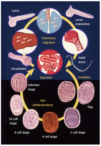
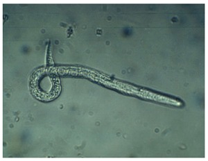
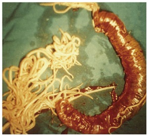
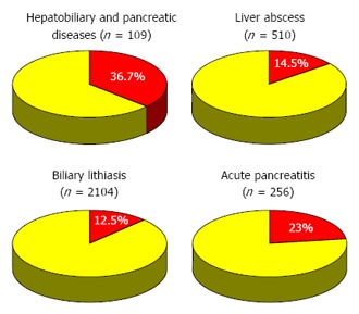
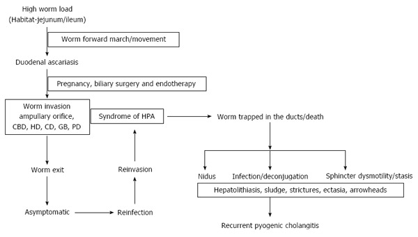
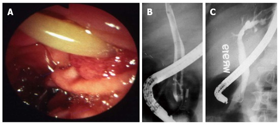
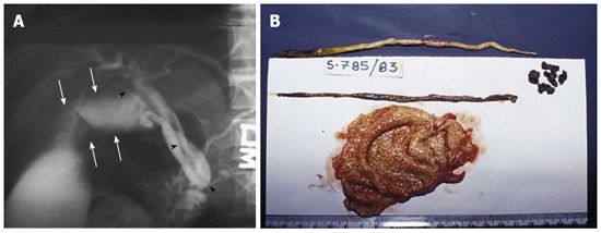
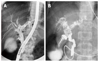
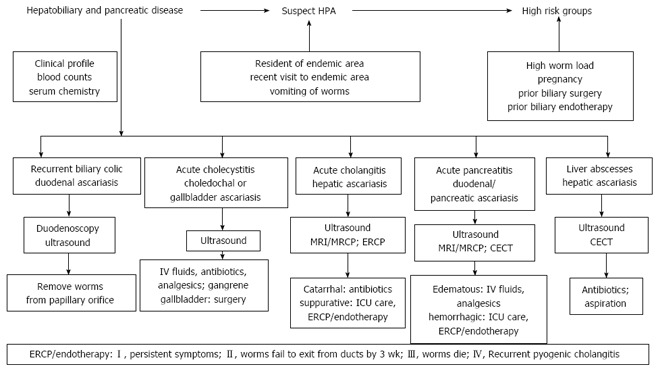
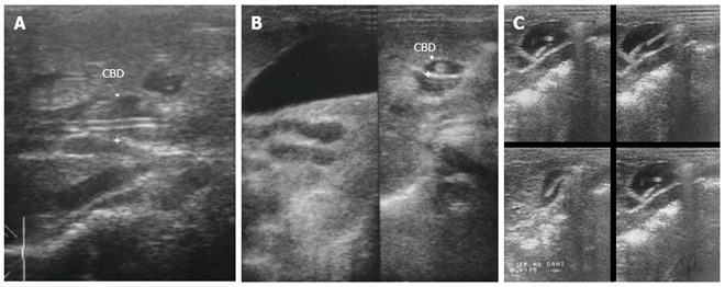
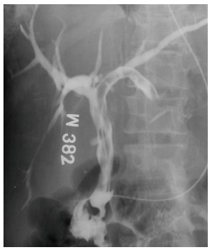
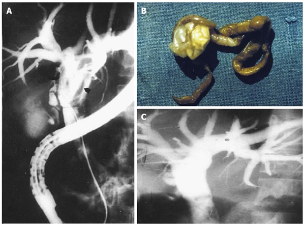
Hepatobiliary and pancreatic ascariasis (HPA) was described as a clinical entity from Kashmir, India in 1985. HPA is caused by invasion and migration of nematode, Ascaris lumbricoides, in to the biliary tract and pancreatic duct. Patients present with biliary colic, cholangitis, cholecystitis, hepatic abscesses and acute pancreatitis. Ascarides traverse the ducts repeatedly, get trapped and die, leading to formation of hepatolithiasis. HPA is ubiquitous in endemic regions and in Kashmir, one such region, HPA is the etiological factor for 36.7%, 23%, 14.5% and 12.5% of all biliary diseases, acute pancreatitis, liver abscesses and biliary lithiasis respectively. Ultrasonography is an excellent diagnostic tool in visualizing worms in gut lumen and ductal system. The rational treatment for HPA is to give appropriate treatment for clinical syndromes along with effective anthelmintic therapy. Endotherapy in HPA is indicated if patients continue to have symptoms on medical therapy or when worms do not move out of ductal lumen by 3 wk or die within the ducts. The worms can be removed from the ductal system in most of the patients and such patients get regression of symptoms of hepatobiliary and pancreatic disease.

Keywords: Ascaris lumbricoides; Biliary calculi; Cholangitis; Cholecystitis; Pancreatitis; Recurrent pyogenic cholangitis.


Conflict of interest statement

Conflict-of-interest statement: The authors report no conflict of interest.

Figures

Figure 1
Figure 1
Life cycle of Ascaris lumbricoides. Adapted from khuroo et al[5].
Figure 2
Figure 2
Sputum examination of a child with ascaris pneumonia showing filariform larva.
Figure 3
Figure 3
Massive aggregate of ascarides within jejunum causing obstruction, infarction and gangrene of bowel necessitating bowel resection. Adapted from Khuroo[5].
Figure 4
Figure 4
Impact of hepatobiliary and pancreatic ascariasis as an etiologic factor in biliary disease, hepatic abscesses, biliary lithiasis and acute pancreatitis. Numbers in parenthesis shows number of patients with disease and percentages are those caused by ascariasis. Adapted from Ref. [1,3,6,37].
Figure 5
Figure 5
Pathogenesis and natural course of hepatobiliary and pancreatic ascariasis in high endemic regions.
Figure 6
Figure 6
Duodenal ascariasis presenting as biliary colic. A: Duodenoscopy showing adult ascaride in the ampullary orifice; B: Endoscopic retrograde cholangiogram showing long linear filling defect in the common bile duct; C: Cholangiogram after extraction of worms from bile duct. Patient had immediate relief of biliary colic. Adapted from Khuroo[5].
Figure 7
Figure 7
Choledochus ascariasis blocking orifice of cystic duct, causing acute obstructive cholecystitis, needing emergency cholecystectomy. A: Endoscopic retrograde cholangiogram showing linear filling defect in common bile duct (black arrows). Cystic duct and gall bladder is grossly dilated (white arrows); B: Gall bladder showing inflamed gall bladder with 2 adult ascarides and few stones around macerated dead worm. Adapted from Khuroo[5].
Figure 8
Figure 8
Hepatic ascariasis presenting as suppurative cholangitis. A: Endoscopic retrograde cholangiogram showing palisading of ascarides in common bile ducts and hepatic ducts; B: Duodenoscopic view showing pus exuding from ampullary orifice.
Figure 9
Figure 9
Pancreatic ascariasis presenting as necrotizing pancreatitis. A linear filling defect is seen all along the pancreatic duct (straight arrows). A stricture is seen in the tail region (curved arrow) reminiscent of pancreatic necrosis. Adapted from Khuroo et al[2].
Figure 10
Figure 10
Recurrent pyogenic cholangitis aftermath of biliary ascariasis. A: Endoscopic retrograde cholangiogram obtained 5 years prior to present admission revealing two long linear smooth filling defect in the common and left hepatic duct (arrows); B: Cholangiogram performed during present admission. Cholangiogram revealed biliary dilatation with multiple filling defects and cholangitic changes in the common hepatic, right and left hepatic duct. Naso-biliary tube is in place to treat pyogenic cholangitis. Adapted from Khuroo et al[4].
Figure 11
Figure 11
Flow chart showing diagnostic and management algorithm.
Figure 12
Figure 12
Ultrasonography images in hepatobiliary and pancreatic ascariasis. A: Biliary ultrasound depicting four-line sign; B: Ultrasound showing tube like structure within common bile duct with distended gall bladder; C: Ascarides in gall bladder showing active movements as seen in serial images. Adapted from Khuroo[5].
Figure 13
Figure 13
Hepatic ascariasis presenting as suppurative cholangitis managed with nasobiliary tube drainage. Note ascaride placed in common bile duct and left hepatic duct. Nasobiliary tube is in place to decompress and drain the duct.
Figure 14
Figure 14
Endotherapy in hepatobiliary ascariasis. A: Cholangiogram showing dilated ducts multiple linear filling defects; B: Dead ascaride extracted from biliary tract; C: Cholangiogram obtained after extraction of dead ascaris. Ducts are free of filling defects. Adapted from Khuroo et al[15].

Similar articles

Cited by

References

    1. Khuroo MS, Zargar SA. Biliary ascariasis. A common cause of biliary and pancreatic disease in an endemic area. Gastroenterology. 1985;88:418–423. - PubMed
    1. Khuroo MS, Zargar SA, Mahajan R. Hepatobiliary and pancreatic ascariasis in India. Lancet. 1990;335:1503–1506. - PubMed
    1. Khuroo MS, Dar MY, Yattoo GN, Khan BA, Boda MI, Zargar SA, Javid G, Allai MS. Serial cholangiographic appearances in recurrent pyogenic cholangitis. Gastrointest Endosc. 1993;39:674–679. - PubMed
    1. Khuroo MS, Khuroo NS, Khuroo MS. Biliary ascariasis in the etiology of recurrent pyogenic cholangitis in an endemic area. International Journal of Hepatobiliary and Pancreatic Diseases. 2015;5:22.
    1. Khuroo MS. Ascariasis. Gastroenterol Clin North Am. 1996;25:553–577. - PubMed