

Differences in coronary artery blood velocities in the setting of normal coronary angiography and normal stress echocardiography
- PMID: 27672435
- PMCID: PMC4946382
- DOI: 10.5301/heartint.5000221
Differences in coronary artery blood velocities in the setting of normal coronary angiography and normal stress echocardiography
Abstract
Background: Normal left anterior descending (LAD) coronary artery as determined by coronary angiography is considered not only to reflect normal angiography but also to correlate with normal anatomy and function. However, subjects who undergo coronary angiography may differ from those who do not need to have invasive evaluation even if their functional noninvasive studies like dobutamine stress echocardiography (DSE) were normal.
Aim: LAD velocities in subjects with normal angiography and those with normal DSE are equal.
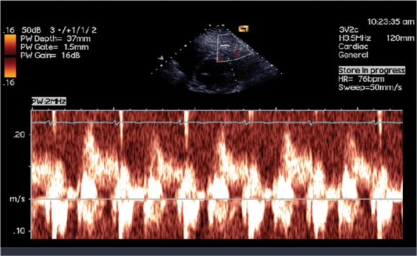
Methods: A total of 244 subjects were evaluated, 78 had normal LAD by angiography and 166 had normal LAD by DSE. All had Doppler sampling of LAD velocities by transthoracic echocardiography.
Results: Velocity was higher in the angiographic subgroup in diastole 41 ± 23 vs 33 ± 14 cm/s, p = 0.0078; systole 18 ± 14 vs 13 ± 7 cm/s, p = 0.012; diastolic integral 12.6 ± 5 vs 9.8 ± 3.8 cm, p = 3.15 × 10(-5); systolic velocity integral 4 ± 2.9 vs 2.8 ± 1.9, p = 0.0014. While heart rate was similar in both groups, the product of diastolic velocity integral and heart rate of the LAD in the angiographic group was higher: 902 ± 450 vs 656 ± 394, p = 0.00599. Diastolic velocity deceleration time was similar in both groups. Coronary flow reserve defined as diastolic velocity ratio before and immediately after DSE correlated negatively with baseline velocity, r = -0.4.
Conclusions: Mode of defining normality of coronary artery affects velocity behavior of the vessel, reflecting functional differences possibly related to microvasculature and vasodilatation.
Keywords: Coronary angiography; Dobutamine stress echocardiography; Doppler; Left anterior descending coronary artery.
Conflict of interest statement
Conflict of interest: None.
Figures
Similar articles
-
Evaluation of left anterior descending coronary artery stenosis of intermediate severity using transthoracic coronary flow reserve and dobutamine stress echocardiography.J Am Soc Echocardiogr. 2005 Dec;18(12):1233-40. doi: 10.1016/j.echo.2005.05.011. J Am Soc Echocardiogr. 2005. PMID: 16376748 Clinical Trial.
-
Coronary Flow Reserve of the Non-Ischemia Related Coronary Artery During Dobutamine Stress Echocardiography.Cardiol Res. 2011 Aug;2(4):174-180. doi: 10.4021/cr57w. Epub 2011 Jul 25. Cardiol Res. 2011. PMID: 28352387 Free PMC article.
-
Noninvasive assessment of significant left anterior descending coronary artery stenosis by coronary flow velocity reserve with transthoracic color Doppler echocardiography.Circulation. 1998 Apr 28;97(16):1557-62. doi: 10.1161/01.cir.97.16.1557. Circulation. 1998. PMID: 9593560
-
Changes in left anterior descending coronary artery flow profiles after coronary artery bypass grafting examined by means of transthoracic Doppler echocardiography.J Thorac Cardiovasc Surg. 2003 Nov;126(5):1531-6. doi: 10.1016/s0022-5223(03)00972-3. J Thorac Cardiovasc Surg. 2003. PMID: 14666029
-
Transthoracic Doppler echocardiographic assessment of left anterior descending coronary artery and intramyocardial small coronary artery flow in patients with hypertrophic cardiomyopathy.J Cardiol. 2001;37 Suppl 1:115-20. J Cardiol. 2001. PMID: 11433814
Cited by
-
A fractal physics explanation for acute thrombotic occlusion in an apparently healthy coronary artery.Anatol J Cardiol. 2017 Aug;18(2):155-157. doi: 10.14744/AnatolJCardiol.2017.7825. Anatol J Cardiol. 2017. PMID: 28766510 Free PMC article. No abstract available.
-
Multifractality through Non-Markovian Stochastic Processes in the Scale Relativity Theory. Acute Arterial Occlusions as Scale Transitions.Entropy (Basel). 2021 Apr 9;23(4):444. doi: 10.3390/e23040444. Entropy (Basel). 2021. PMID: 33918896 Free PMC article.
-
Examination of cardiac functions during acute attack and remission period in children with familial Mediterranean fever.Eur J Pediatr. 2024 Jul;183(7):3137-3145. doi: 10.1007/s00431-024-05570-y. Epub 2024 Apr 26. Eur J Pediatr. 2024. PMID: 38668795 Free PMC article.
-
Forward-viewing estimation of 3D blood flow velocity fields by intravascular ultrasound: Influence of the catheter on velocity estimation in stenoses.Ultrasonics. 2021 Dec;117:106558. doi: 10.1016/j.ultras.2021.106558. Epub 2021 Aug 23. Ultrasonics. 2021. PMID: 34461527 Free PMC article.
References
-
- Hansson GK. Inflammation, atherosclerosis, and coronary artery disease. N Engl J Med. 2005;352(16):1685–1695. - PubMed
-
- Geleijnse ML, Fioretti PM, Roelandt JR. Methodology, feasibility, safety and diagnostic accuracy of dobutamine stress echocardiography. J Am Coll Cardiol. 1997;30(3):595–606. - PubMed
-
- Picano E. Stress echocardiography: a historical perspective. Am J Med. 2003;114(2):126–130. - PubMed
-
- Voci P, Testa G, Plaustro G. Imaging of the distal left anterior descending coronary artery by transthoracic color-Doppler echocardiography. Am J Cardiol. 1998;81(12 12A):74G–78G. - PubMed
LinkOut - more resources
Full Text Sources
Other Literature Sources