. 2016 Sep 20:4:16027.
doi: 10.1038/boneres.2016.27. eCollection 2016.

Cytocompatibility with osteogenic cells and enhanced in vivo anti-infection potential of quaternized chitosan-loaded titania nanotubes

Affiliations

Cytocompatibility with osteogenic cells and enhanced in vivo anti-infection potential of quaternized chitosan-loaded titania nanotubes

Ying Yang et al. Bone Res. .

Abstract

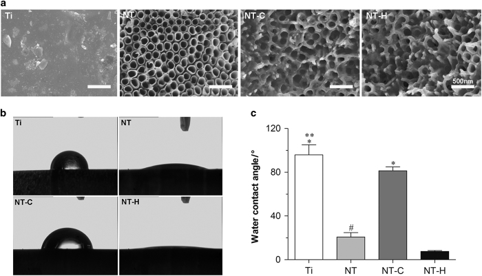
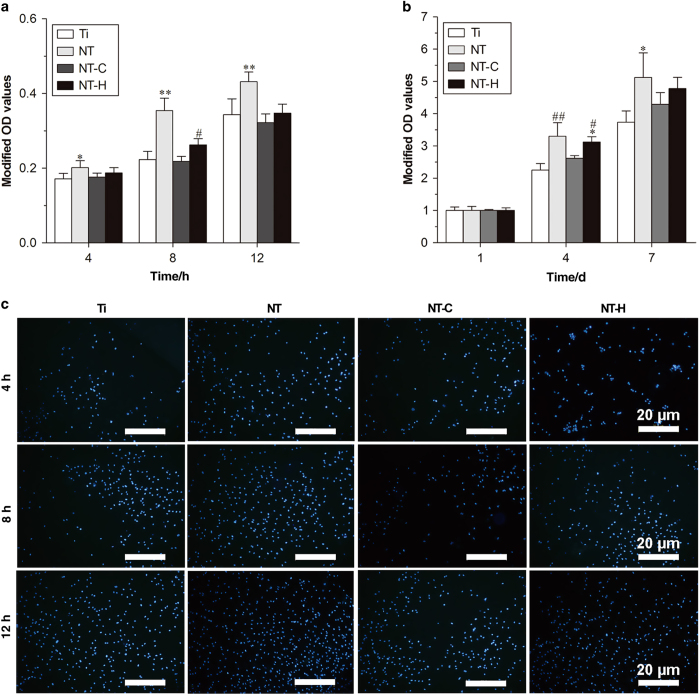
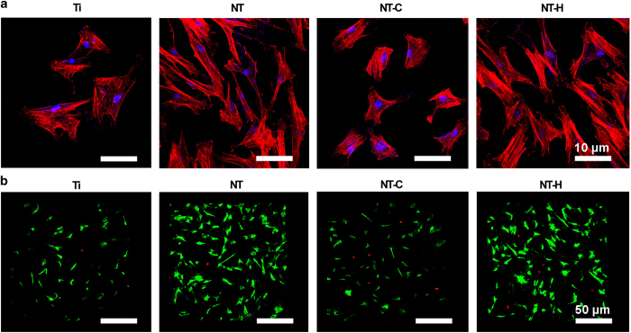
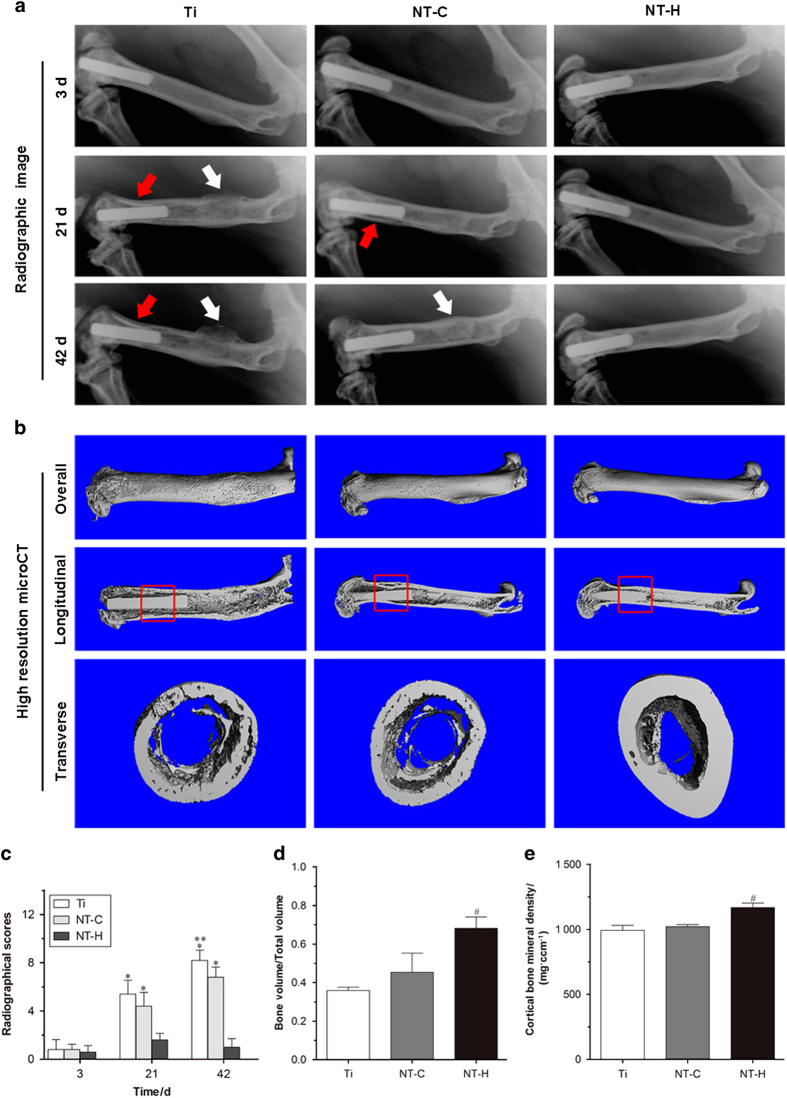
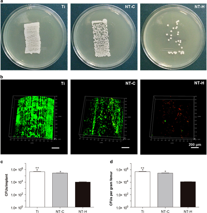
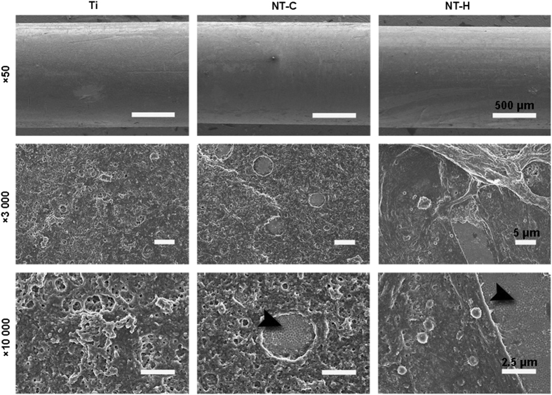
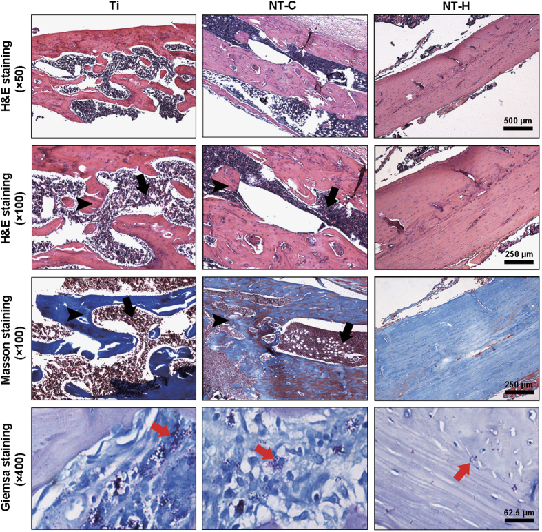
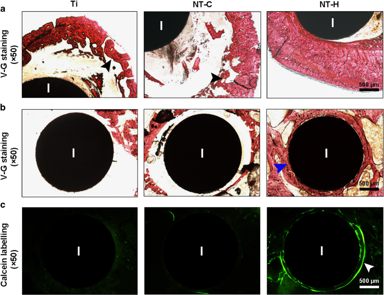
Infection is one of the major causes of failure of orthopedic implants. Our previous study demonstrated that nanotube modification of the implant surface, together with nanotubes loaded with quaternized chitosan (hydroxypropyltrimethyl ammonium chloride chitosan, HACC), could effectively inhibit bacterial adherence and biofilm formation in vitro. Therefore, the aim of this study was to further investigate the in vitro cytocompatibility with osteogenic cells and the in vivo anti-infection activity of titanium implants with HACC-loaded nanotubes (NT-H). The titanium implant (Ti), nanotubes without polymer loading (NT), and nanotubes loaded with chitosan (NT-C) were fabricated and served as controls. Firstly, we evaluated the cytocompatibility of these specimens with human bone marrow-derived mesenchymal stem cells in vitro. The observation of cell attachment, proliferation, spreading, and viability in vitro showed that NT-H has improved osteogenic activity compared with Ti and NT-C. A prophylaxis rat model with implantation in the femoral medullary cavity and inoculation with methicillin-resistant Staphylococcus aureus was established and evaluated by radiographical, microbiological, and histopathological assessments. Our in vivo study demonstrated that NT-H coatings exhibited significant anti-infection capability compared with the Ti and NT-C groups. In conclusion, HACC-loaded nanotubes fabricated on a titanium substrate show good compatibility with osteogenic cells and enhanced anti-infection ability in vivo, providing a good foundation for clinical application to combat orthopedic implant-associated infections.


Figures

Figure 1
Figure 1
Surface characterization of the four different specimens. (a) Surface morphology using scanning electron microscopy (SEM). The surfaces of the drug-loaded nanotubes reserve the nanotubular structure. Magnification, ×50  000. The scale bar for the row is shown in the last image. (b), (c) Water contact angles of various specimens. *P<0.01, compared with titania nanotubes without drug-loading (NT) and HACC-loaded titania nanotubes (NT-H); **P<0.05, compared with chitosan-loaded titania nanotubes (NT-C); #P<0.05, compared with NT-H.
Figure 2
Figure 2
Attachment and proliferation assay of the human marrow-derived mesenchymal stem cells (hMSCs) on the four different surfaces. (a) Cell attachment on the samples assessed by the cell counting kit-8 assay. (b) Cell proliferation on various specimens. (c) Cell attachment on titanium without modification (Ti), titania nanotubes without drug-loading (NT), chitosan-loaded titania nanotubes (NT-C) and HACC-loaded titania nanotubes (NT-H) assessed by 4,6-diamidino-2-phenylindole staining after 4 8, and 12 h of culture. Magnification, ×100. The scale bar for the row is shown in the last image. *P<0.05, compared with Ti; **P<0.01, compared with the other groups; #P<0.05, compared with NT-C; ##P<0.01, compared with Ti and NT-C.
Figure 3
Figure 3
Cell spreading and viability on various specimens. (a) Cell morphology on the samples at 24 h, as observed by confocal laser scanning microscope (CLSM). Cells were stained with rhodamine phalloidin for the actin filaments (red) and 4,6-diamidino-2-phenylindole for the nucleus (blue). Magnification, ×400. (b) Cell viability evaluated by the Live/Dead assay after a 24-h incubation. Live cells with esterase activity appeared green, whereas dead cells with compromised plasma membranes appeared red. Magnification, ×100. The scale bar for the row is shown in the last image.
Figure 4
Figure 4
Radiographical images and evaluation. (a) Lateral X-rays of the left femur obtained at 3, 21, and 42 days during the follow-up period. Red arrows mark osteolysis, and white arrows indicate obvious periosteal reaction and new bone formation. (b) 3D micro-CT images of the left femur obtained from the overall, longitudinal, and transverse viewpoints at the time of sacrifice. The micro-CT evaluation of the middle femurs is confined to the red rectangle region. (c) Radiographic scores of the X-ray images. *P<0.01, compared with the HACC-loaded titania nanotubes (NT-H) (n=5); **P<0.05, compared with the titania nanotubes without drug-loading (NT) (n=5). (d) Bone volume/Total volume and (e) cortical bone mineral density of the selected regions of the left femurs evaluated by micro-CT. #P<0.01, compared with the titanium without modification (Ti) and chitosan-loaded titania nanotubes (NT-C) (n=5).
Figure 5
Figure 5
Microbiological evaluation of the implants and bone. (a) Roll-over cultures obtained from explanted rods. (b) Confocal laser scanning microscopy (CLSM) observation of explanted rods. Live bacteria showing green fluorescence were stained with SYTO 9 and dead bacteria showing red fluorescence were stained with propidium iodide. Magnification, ×100. The scale bar for the row is shown in the last image. (c) Amount of the detached adhered bacteria and biofilm after the rods were rolled over trypticase soy agar and (d) quantity of colony-forming units (CFUs) per gram of pulverized femur. *P<0.01, compared with the HACC-loaded titania nanotubes (NT-H) (n=6); **P<0.01, compared with chitosan-loaded titania nanotubes (NT-C) (n=6).
Figure 6
Figure 6
Scanning electron microscopy (SEM) observation of explanted implants. The black arrowheads indicate the intact nanotubular structure on the titanium rods. The scale bar for the row is shown in the last image.
Figure 7
Figure 7
Representative histological images obtained from decalcified longitudinal sections without implants at the middle of the femur. Hematoxylin and eosin staining, and Masson’s trichrome staining were used to assess the changes in bone morphology, and Giemsa staining was used to determine bacterial contamination. Black arrows—intracortical abscesses or inflammatory cells; black arrowheads—massive enlargement and destruction of bone cortex; red arrows—bacteria. The scale bar for the row is shown in the last image.
Figure 8
Figure 8
Representative histological images obtained from undecalcified transverse sections containing implants. Van Gieson staining was used to evaluate the (a) morphological changes in the cortical bone at the middle of the femur, and (b) the osteointegration around the implants at the condyle of the femur. (c) Fluorescent micrographs demonstrating new bone formation around the implants at 42 days after implantation. Black arrowheads, massive enlargement and destruction of the bone cortex; blue arrowheads, osteointegration around the implants; white arrowheads, obvious fluorescent deposition indicating new bone formation around the implants. I, implant. The scale bar for the row is shown in the last image.

Similar articles

Cited by

References

    1. Court-Brown CM, Keating JF, McQueen MM. Infection after intramedullary nailing of the tibia. Incidence and protocol for management. J Bone Joint Surg Br 1992; 74: 770–774. - PubMed
    1. Chen CE, Ko JY, Wang JW et al. Infection after intramedullary nailing of the femur. J Trauma 2003; 55: 338–344. - PubMed
    1. Court-Brown CM. Reamed intramedullary tibial nailing: an overview and analysis of 1106 Cases. J Orthop Trauma 2004; 18: 96–101. - PubMed
    1. Birdsall PD, Milne DD. Toxic shock syndrome due to percutaneous Kirschner wires. Injury 1999; 30: 509–510. - PubMed
    1. Losic D, Aw MS, Santos A et al. Titania nanotube arrays for local drug delivery: recent advances and perspectives. Expert Opin Drug Deliv 2015; 12: 103–127. - PubMed