Review
. 2016 Sep 20:4:16028.
doi: 10.1038/boneres.2016.28. eCollection 2016.

Bone-cartilage crosstalk: a conversation for understanding osteoarthritis

Affiliations
Review

Bone-cartilage crosstalk: a conversation for understanding osteoarthritis

David M Findlay et al. Bone Res. .

Abstract

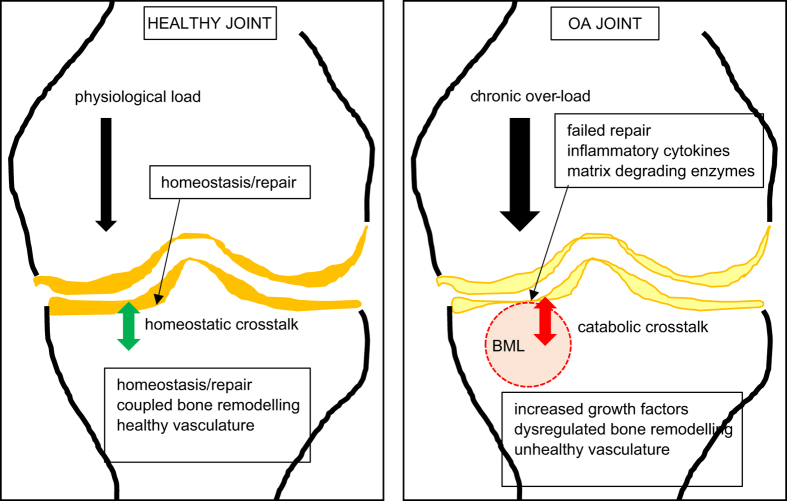
Although cartilage degradation is the characteristic feature of osteoarthritis (OA), it is now recognized that the whole joint is involved in the progression of OA. In particular, the interaction (crosstalk) between cartilage and subchondral bone is thought to be a central feature of this process. The interface between articular cartilage and bone of articulating long bones is a unique zone, which comprises articular cartilage, below which is the calcified cartilage sitting on and intercalated into the subchondral bone plate. Below the subchondral plate is the trabecular bone at the end of the respective long bones. In OA, there are well-described progressive destructive changes in the articular cartilage, which parallel characteristic changes in the underlying bone. This review examines the evidence that biochemical and biomechanical signaling between these tissue compartments is important in OA disease progression and asks whether such signaling might provide possibilities for therapeutic intervention to halt or slow disease development.


Figures

Figure 1
Figure 1
Subchondral bone microarchitecture and osteoid seams characteristic of MRI identified bone marrow lesions (BMLs) in human tibial plateaus obtained at knee arthroplasty surgery for OA. No signal was detected on PDFS and T1-weighted MRI sequences for no-BML. BML signal was detected only by PDFS for BML 1; BML signal was detected by both PDFS and T1 for BML 2. Left panel shows three-dimensional reconstructed micro-computed tomography (CT) images of a 10 mm diameter cylindrical region of interest within the volume of the BML. Left and right middle panels show coronal and axial micro-CT images, respectively. Micro-CT images clearly demonstrate that BML subchondral bone is sclerotic, characterized by thicker subchondral bone plate, and increased trabecular bone volume with more plate-like structure. Right panel shows von Kossa silver/hematoxylin- and eosin-stained subchondral trabecular bone that shows BML tissue has increased osteoid, with increased osteoid seam thickness, and osteoid bridging and in-filling. The sclerotic subchondral bone phenotype characteristic of BMLs was more pronounced for BML 2 versus BML 1.
Figure 2
Figure 2
Cartoon of healthy and OA knee joints. Shown is a healthy joint, in which physiological loading (magnitude of load×frequency of loading) is managed by the joint. Healthy subchondral blood vessels and blood perfusion, and a putative healthy crosstalk between the subchondral bone and articular cartilage lead to coupled bone remodeling to maintain homeostasis and repair microdamage in the bone matrix. In the OA joint, chronic overloading results in failed repair mechanisms. Bone within a bone marrow lesion (BML) zone, imaged by MRI, contains the most severe bony manifestations, in which dysregulated bone remodeling results in both bone attrition and sclerosis. The cartilage overlying the BML is degraded, with increased matrix proteases and decreased production of cartilage matrix. A putative unhealthy crosstalk between the bone and cartilage compartments exacerbates the failure of repair mechanisms, in the face of continued unfavourable biomechanics.

References

    1. Clark JM, Huber JD. The structure of the human subchondral plate. J Bone Joint Surg Br 1990; 72: 866–873. - PubMed
    1. Duncan H, Jundt J, Riddle JM et al. The tibial subchondral plate. A scanning electron microscopic study. J Bone Joint Surg Am 1987; 69: 1212–1220. - PubMed
    1. Lyons TJ, McClure SF, Stoddart RW et al. The normal human chondro-osseous junctional region: evidence for contact of uncalcified cartilage with subchondral bone and marrow spaces. BMC Musculoskelet Disord 2006; 7: 52. - PMC - PubMed
    1. Imhof H, Sulzbacher I, Grampp S et al. Subchondral bone and cartilage disease: a rediscovered functional unit. Invest Radiol 2000; 35: 581–588. - PubMed
    1. Kim HK, Bian H, Aya-ay J et al. Hypoxia and hif-1alpha expression in the epiphyseal cartilage following ischemic injury to the immature femoral head. Bone 2009; 45: 280–288. - PubMed

LinkOut - more resources