

The use of digital periapical radiographs to study the prevalence of alveolar domes
- PMID: 27672614
- PMCID: PMC5035723
- DOI: 10.5624/isd.2016.46.3.179
The use of digital periapical radiographs to study the prevalence of alveolar domes
Abstract
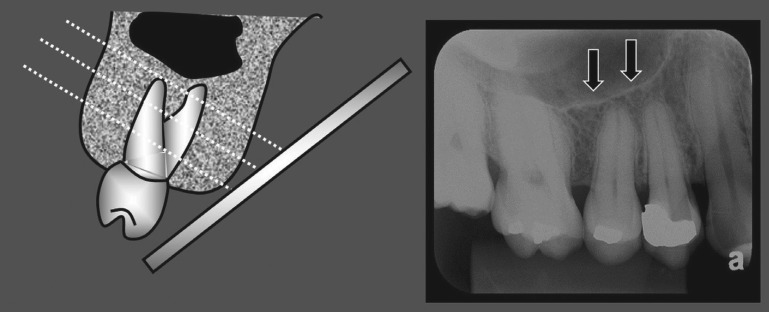
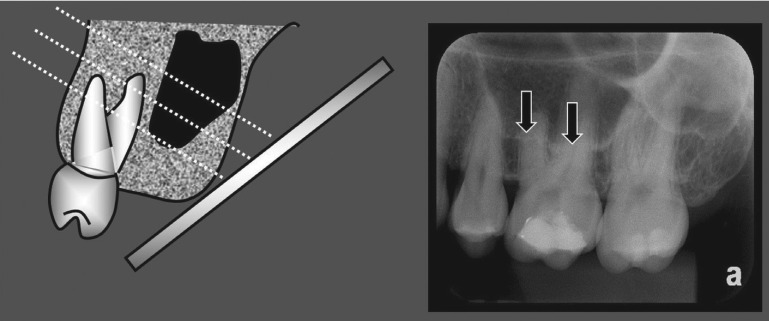
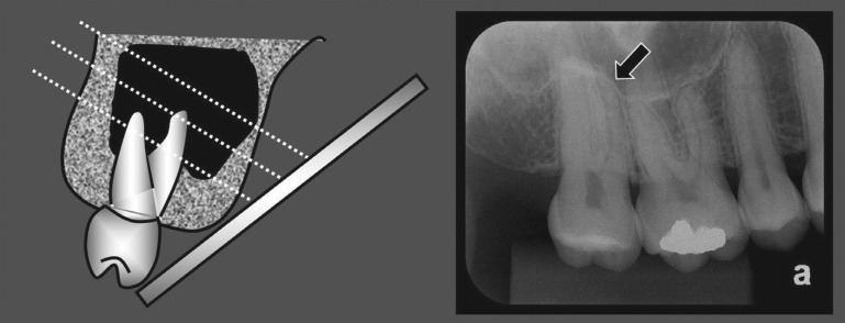
Purpose: In the present study, we coined the term 'alveolar dome' and aimed to demonstrate the prevalence of alveolar domes through digital periapical radiographs.
Materials and methods: This study examined 800 digital periapical radiographs in regard to the presence of alveolar domes. The periapical radiographs were acquired by a digital system using a photostimulable phosphor (PSP) plate. The χ(2) test, with a significance level of 5%, was used to compare the prevalence of alveolar domes in the maxillary posterior teeth and, considering the same teeth, to verify the difference in the prevalence of dome-shaped phenomena between the roots.
Results: The prevalence of alveolar domes present in the first pre-molars was statistically lower as compared to the other maxillary posterior teeth (p<0.05). No statistically significant difference was observed in the prevalence of alveolar domes between the maxillary first and second molars. Considering the maxillary first and second molars, it was observed that the palatal root presented a lower prevalence of alveolar domes when compared to the distobuccal and mesiobuccal roots (p<0.05).
Conclusion: The present study coined the term 'alveolar dome', referring to the anatomical projection of the root into the floor of the maxillary sinus. The maxillary first and second molars presented a greater prevalence of alveolar domes, especially in the buccal roots, followed by the third molars and second pre-molars. Although the periapical radiograph is a two-dimensional method, it can provide dentists with the auxiliary information necessary to identify alveolar domes, thus improving diagnosis, planning, and treatment.
Keywords: Maxillary Sinus; Prevalence; Radiography, Dental, Digital; Tooth Root.
Figures
References
-
- Hauman CH, Chandler NP, Tong DC. Endodontic implications of the maxillary sinus: a review. Int Endod J. 2002;35:127–141. - PubMed
-
- Pagin O, Centurion BS, Rubira-Bullen IR, Alvares Capelozza AL. Maxillary sinus and posterior teeth: accessing close relationship by cone-beam computed tomographic scanning in a Brazilian population. J Endod. 2013;39:748–751. - PubMed
-
- de Oliveira AG, dos Santos Silveira O, Francio LA, de Andrade Marigo Grandinetti H, Manzi FR. Anatomic variations of paranasal sinuses - clinical case report. Surg Radiol Anat. 2013;35:535–538. - PubMed
-
- Lana JP, Carneiro PM, Machado Vde C, de Souza PE, Manzi FR, Horta MC. Anatomic variations and lesions of the maxillary sinus detected in cone beam computed tomography for dental implants. Clin Oral Implants Res. 2012;23:1398–1403. - PubMed
LinkOut - more resources
Full Text Sources
Other Literature Sources
Miscellaneous
