

Huge peripheral primitive neuroectodermal tumor of the small bowel mesentery at nonage: A case report and review of the literature
- PMID: 27672649
- PMCID: PMC5018631
- DOI: 10.12998/wjcc.v4.i9.306
Huge peripheral primitive neuroectodermal tumor of the small bowel mesentery at nonage: A case report and review of the literature
Abstract
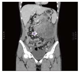
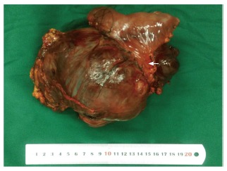
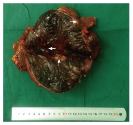
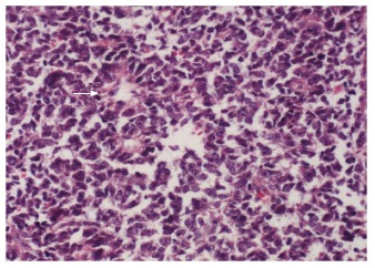
Extraskeletal Ewing's sarcoma/peripheral primitive neuroectodermal tumor (E-EWS/pPNET) is a rare aggressive malignant small round cell tumor. In this report, we present the case of a 15-year-old boy who suffered from acute abdominal pain accompanied by hematemesis and melena, and was eventually diagnosed with E-EWS/pPNET. To date, there have been only five reported cases of E-EWS/pPNET of the small bowel including the patient in this report. To the best of our knowledge, this is the first documentation of a pPNET of the small bowel mesentery at nonage. All these have made this report rare and significant.
Keywords: Extraskeletal Ewing’s sarcoma; Nonage; Peripheral primitive neuroectodermal tumor; Small bowel mesentery; Spontaneous rupture.
Conflict of interest statement
Conflict-of-interest statement: The authors declared that they have no conflicts of interest to this work.
Figures
Similar articles
-
Extraskeletal Ewing's sarcoma/peripheral primitive neuroectodermal tumor of the small bowel presenting with gastrointestinal perforation.Clin Exp Gastroenterol. 2019 Jun 25;12:279-285. doi: 10.2147/CEG.S203697. eCollection 2019. Clin Exp Gastroenterol. 2019. PMID: 31417299 Free PMC article.
-
Ewing's sarcoma/peripheral primitive neuroectodermal tumor with extraskeletal myxoid chondrosarcoma-like areas: a case report.Int J Clin Exp Pathol. 2019 Oct 1;12(10):3940-3943. eCollection 2019. Int J Clin Exp Pathol. 2019. PMID: 31933786 Free PMC article.
-
Congenital Ewing's Sarcoma/Peripheral Primitive Neuroectodermal Tumor: A Case Report and Review of the Literature.Pediatr Neonatol. 2016 Oct;57(5):436-439. doi: 10.1016/j.pedneo.2013.11.002. Epub 2014 Jan 27. Pediatr Neonatol. 2016. PMID: 24480101 Review.
-
Ewing's sarcoma/peripheral primitive neuroectodermal tumor (pPNET) arising in the omentum as a multilocular cyst with intracystic hemorrhage.J Gastroenterol. 2000 Dec;35(12):933-40. doi: 10.1007/s005350070009. J Gastroenterol. 2000. PMID: 11573731
-
Primary intracranial dural-based Ewing sarcoma/peripheral primitive neuroectodermal tumor mimicking a meningioma: A rare tumor with review of literature.Asian J Neurosurg. 2017 Jul-Sep;12(3):351-357. doi: 10.4103/1793-5482.185060. Asian J Neurosurg. 2017. PMID: 28761507 Free PMC article. Review.
Cited by
-
Primary Ewing's sarcoma of the intestine: case report and literature review.Front Oncol. 2024 Jul 30;14:1357945. doi: 10.3389/fonc.2024.1357945. eCollection 2024. Front Oncol. 2024. PMID: 39139288 Free PMC article.
-
Extraskeletal Ewing's sarcoma/peripheral primitive neuroectodermal tumor of the small bowel presenting with gastrointestinal perforation.Clin Exp Gastroenterol. 2019 Jun 25;12:279-285. doi: 10.2147/CEG.S203697. eCollection 2019. Clin Exp Gastroenterol. 2019. PMID: 31417299 Free PMC article.
-
Primitive neuroectodermal tumors: a clinical and radiological analysis of six cases.Quant Imaging Med Surg. 2019 Apr;9(4):722-729. doi: 10.21037/qims.2019.03.16. Quant Imaging Med Surg. 2019. PMID: 31143663 Free PMC article.
-
Primary Ewing's sarcoma in a small intestine - a case report and review of the literature.BMC Surg. 2020 May 25;20(1):113. doi: 10.1186/s12893-020-00774-z. BMC Surg. 2020. PMID: 32450834 Free PMC article. Review.
References
-
- Horie Y, Kato M. Peripheral primitive neuroectodermal tumor of the small bowel mesentery: a case showing perforation at onset. Pathol Int. 2000;50:398–403. - PubMed
-
- Balasubramanian B, Dinakarababu E, Molyneux AJ. Primary primitive neuroectodermal tumour of the small bowel mesentery: case report. Eur J Surg Oncol. 2002;28:197–198. - PubMed
-
- Bala M, Maly A, Remo N, Gimmon Z, Almogy G. Peripheral primitive neuroectodermal tumor of bowel mesentery in adults. Isr Med Assoc J. 2006;8:515–516. - PubMed
-
- Kushner BH, Hajdu SI, Gulati SC, Erlandson RA, Exelby PR, Lieberman PH. Extracranial primitive neuroectodermal tumors. The Memorial Sloan-Kettering Cancer Center experience. Cancer. 1991;67:1825–1829. - PubMed
Publication types
LinkOut - more resources
Full Text Sources
Other Literature Sources
