. 2016 Nov;157(11):4234-4245.
doi: 10.1210/en.2016-1411. Epub 2016 Sep 27.

Excess Testosterone Exposure Alters Hypothalamic-Pituitary-Testicular Axis Dynamics and Gene Expression in Sheep Fetuses

Affiliations

Excess Testosterone Exposure Alters Hypothalamic-Pituitary-Testicular Axis Dynamics and Gene Expression in Sheep Fetuses

Charles E Roselli et al. Endocrinology. 2016 Nov.

Abstract

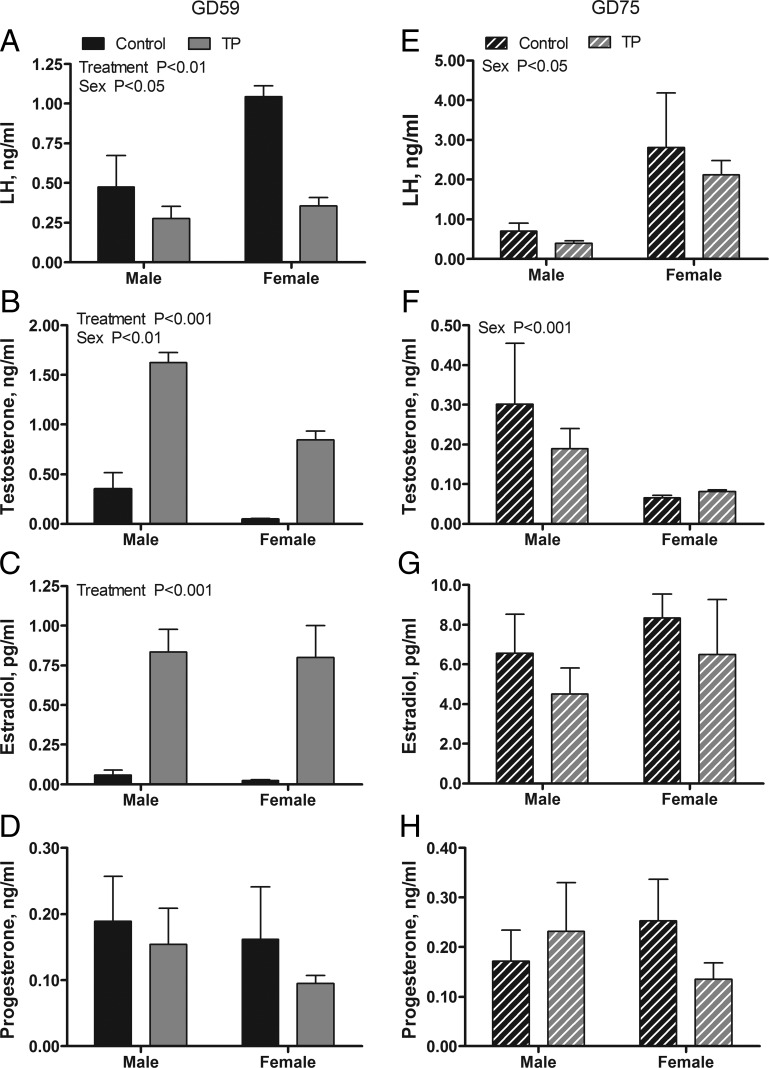
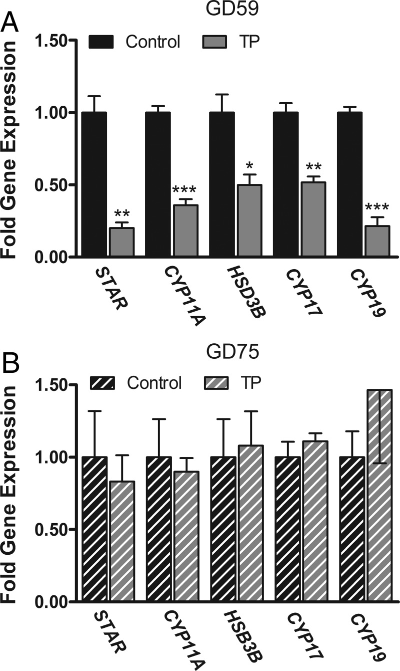
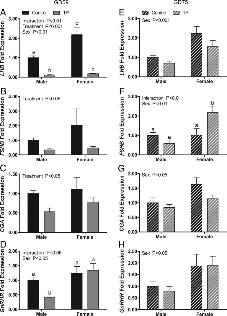
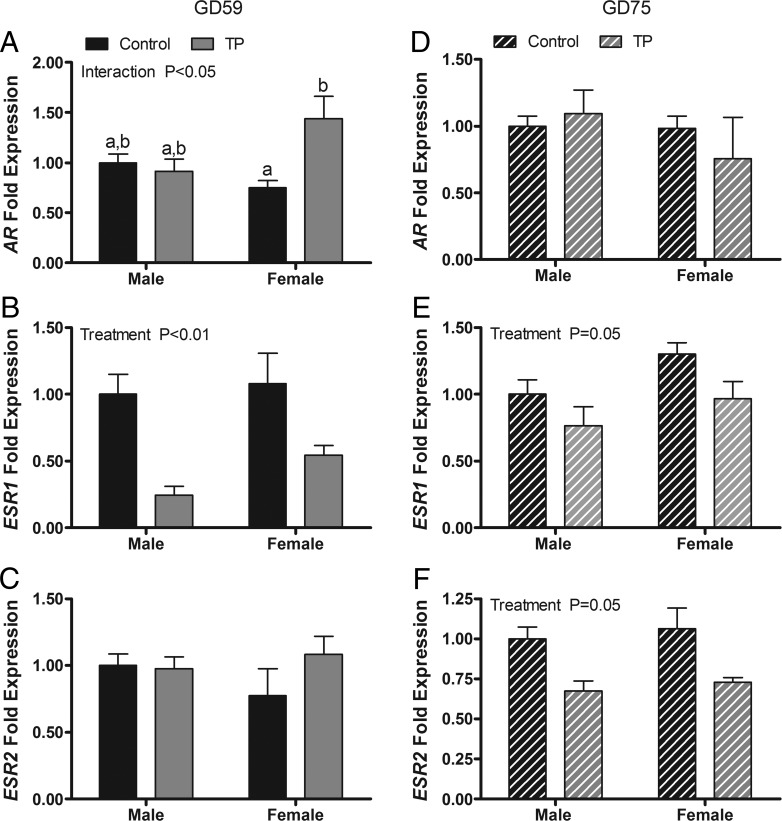
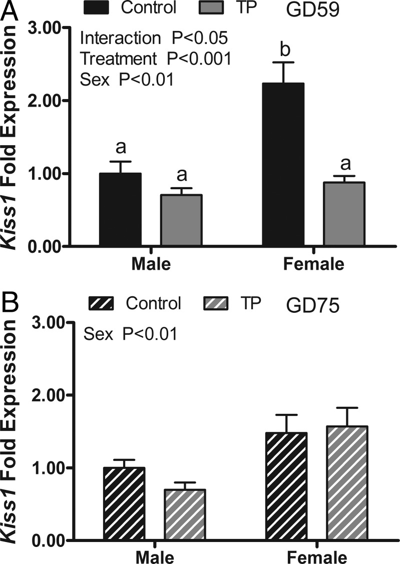
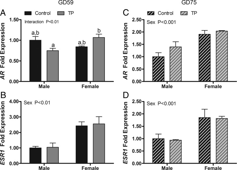
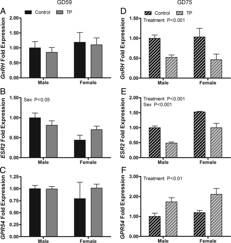
Prenatal exposure to excess androgen may result in impaired adult fertility in a variety of mammalian species. However, little is known about what feedback mechanisms regulate gonadotropin secretion during early gestation and how they respond to excess T exposure. The objective of this study was to determine the effect of exogenous exposure to T on key genes that regulate gonadotropin and GnRH secretion in fetal male lambs as compared with female cohorts. We found that biweekly maternal testosterone propionate (100 mg) treatment administered from day 30 to day 58 of gestation acutely decreased (P < .05) serum LH concentrations and reduced the expression of gonadotropin subunit mRNA in both sexes and the levels of GnRH receptor mRNA in males. These results are consistent with enhanced negative feedback at the level of the pituitary and were accompanied by reduced mRNA levels for testicular steroidogenic enzymes, suggesting that Leydig cell function was also suppressed. The expression of kisspeptin 1 mRNA, a key regulator of GnRH neurons, was significantly greater (P < .01) in control females than in males and reduced (P < .001) in females by T exposure, indicating that hypothalamic regulation of gonadotropin secretion was also affected by androgen exposure. Although endocrine homeostasis was reestablished 2 weeks after maternal testosterone propionate treatment ceased, additional differences in the gene expression of GnRH, estrogen receptor-β, and kisspeptin receptor (G protein coupled receptor 54) emerged between the treatment cohorts. These changes suggest the normal trajectory of hypothalamic-pituitary axis development was disrupted, which may, in turn, contribute to negative effects on fertility later in life.


Figures

Figure 1.
Figure 1.
Effects of twice-weekly maternal TP (100 mg/injection) and control oil vehicle (2 mL) injections from GD 30 to GD 58 on serum LH and steroid concentrations in GD 59 (A–D) and GD 75 (E–H) ovine fetuses. Data for each age (mean ± SEM) were analyzed by a two-way ANOVA.
Figure 2.
Figure 2.
Effects of twice-weekly maternal TP (100 mg/injection) and control oil vehicle (2 mL) injections from GD 30 to GD 58 on steroidogenic gene expression in the testes of GD 59 (A) and GD 75 (B) ovine fetuses. Data (mean ±SEM) were analyzed by a Student's t test. *, P < .05; **, P < .01; ***, P < .001, control vs TP treatments.
Figure 3.
Figure 3.
Effects of twice-weekly maternal TP (100 mg/injection) and control oil vehicle (2 mL) injections from GD 30 to GD 58 on gonadotropin subunit and GnRHR gene expression in the pituitary of GD 59 (A–D) and GD 75 (E–H) ovine fetuses. Data (means ±SEM) were analyzed by a two-way ANOVA. Bars with different superscripts differ significantly (P < .05).
Figure 4.
Figure 4.
Effects of twice-weekly maternal TP (100 mg/injection) and control oil vehicle (2 mL) injections from GD 30 to GD 58 on gene expression in the pituitary of GD 59 (A–C) and GD 75 (D–F) ovine fetuses. Data (mean ±SEM) were analyzed by a two-way ANOVA. Bars with different superscripts are significantly different (P < .05).
Figure 5.
Figure 5.
Effects of twice-weekly maternal TP (100 mg/injection) and control oil vehicle (2 mL) injections from GD 30 to GD 58 on Kiss1 gene expression in the medial basal hypothalamus of GD 59 (A) and GD 75 (B) ovine fetuses. Data (mean ± SEM) were analyzed by a two-way ANOVA. Bars with different superscripts are significantly different (P < .05).
Figure 6.
Figure 6.
Effects of twice-weekly maternal TP (100 mg/injection) and control oil vehicle (2 mL) injections from GD 30 to GD 58 on AR and ESR1 gene expression in the MBH of GD 59 (A and B) and GD 75 (C and D) ovine fetuses. Data (mean ± SEM) were analyzed by a two-way ANOVA. Bars with different superscripts are significantly different (P < .05).
Figure 7.
Figure 7.
Effects of twice-weekly maternal TP (100 mg/injection) and control oil vehicle (2 mL) injections from GD 30 to GD 58 on gene expression in the medial preoptic area of GD 59 (A–C) and GD 75 (D–F) ovine fetuses. Data (mean ± SEM) were analyzed by a two-way ANOVA. Bars with different superscripts are significantly different (P < .05).

Similar articles

Cited by

References

    1. Ford JJ, D'Occhio MJ. Differentiation of sexual behavior in cattle, sheep, and swine. J Anim Sci. 1989;67:1816–1823. - PubMed
    1. Wood RI, Foster DL. Sexual differentiation of reproductive neuroendocrine function in sheep. Rev Reprod. 1998;3:130–140. - PubMed
    1. Roselli CE, Estill C, Stadelman HL, Meaker M, Stormshak F. Separate critical periods exist for testosterone-induced differentiation of the brain and genitals in sheep. Endocrinology. 2011;152:2409–2415. - PMC - PubMed
    1. Roselli CE, Reddy R, Estill C, et al. Prenatal influence of an androgen agonist and antagonist on the differentiation of the ovine sexually dimorphic nucleus in male and female lamb fetuses. Endocrinology. 2014;155:5000–5010. - PMC - PubMed
    1. Clarke IJ, Scaramuzzi RJ, Short RV. Effects of testosterone implants in pregnant ewes on their female offspring. J Embryol Exp Morph. 1976;36:87–99. - PubMed

MeSH terms