. 2016 Dec:109:69-77.
doi: 10.1016/j.biomaterials.2016.09.013. Epub 2016 Sep 19.

Targeted multimodal nano-reporters for pre-procedural MRI and intra-operative image-guidance

Affiliations

Targeted multimodal nano-reporters for pre-procedural MRI and intra-operative image-guidance

Joonseok Lee et al. Biomaterials. 2016 Dec.

Abstract

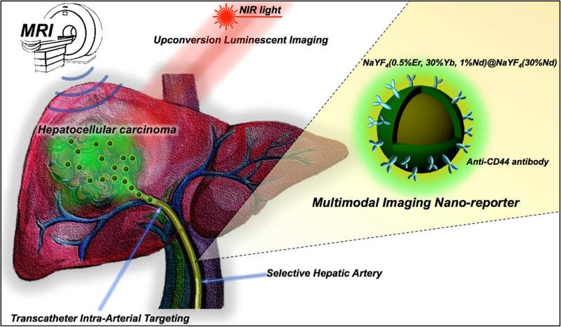
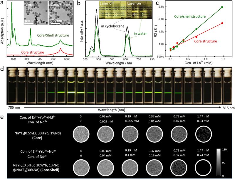
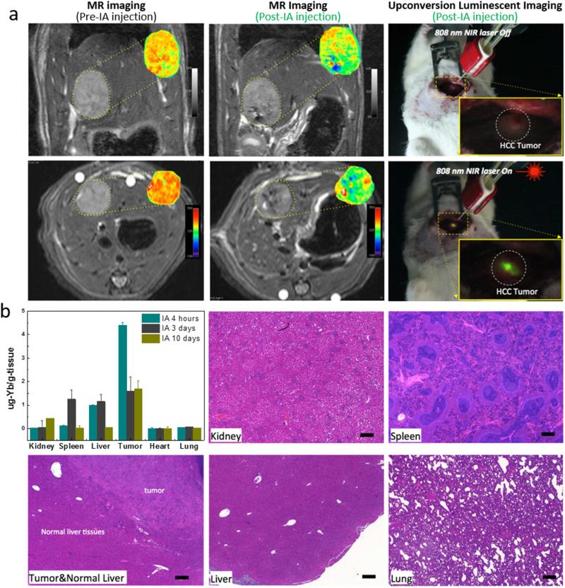
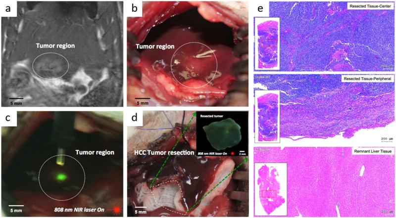
Multimodal-imaging probes offer a novel approach, which can provide detail diagnostic information for the planning of image-guided therapies in clinical practice. Here we report targeted multimodal Nd3+-doped upconversion nanoparticle (UCNP) imaging reporters, integrating both magnetic resonance imaging (MRI) and real-time upconversion luminescence imaging (UCL) capabilities within a single platform. Nd3+-doped UCNPs were synthesized as a core-shell structure showing a bright visible emission upon excitation at the near infrared (minimizing biological overheating and increasing tissue penetration depth) as well as providing strong MRI T2 contrast (high r2/r1 ratio). Transcatheter intra-arterial infusion of Nd3+-doped UCNPs conjugated with anti-CD44-monoclonal antibody allowed for high performance in vivo multimodal UCL and MR imaging of hepatocellular carcinoma (HCC) in an orthotopic rat model. The resulted in vivo multimodal imaging of Nd3+ doped core-shell UCNPs combined with transcatheter intra-arterial targeting approaches successfully discriminated liver tumors from normal hepatic tissues in rats for surgical resection applications. The demonstrated multimodal UCL and MRI imaging capabilities of our multimodal UCNPs reporters suggest strong potential for in vivo visualization of tumors and precise surgical guidance to fill the gap between pre-procedural imaging and intraoperative reality.

Keywords: Cancer; Interventional radiology; Medical imaging; Multimodal probe; Upconversion nanoparticles.


Figures

Figure 1
Figure 1
Schematic of multimodal MR/Upconversion luminescent imaging of HCC tumor using transcatheter hepatic intra-arterial (IA) targeted anti-CD44-Nd-CSUCNPs. Multi-modal imaging reporters, Nd-CSUCNPs conjugated with an anti-CD44 monoclonal antibody, were delivered intra-arterially for the sensitive and rapid detection of HCC in orthotopic HCC rat models. IA delivery of multi-modality Nd-CSUCNPs conjugated with specific tumor targeting molecules facilitates rapid and selective targeting of Nd-CSUCNPs to HCC, providing strong image-contrast between the tumors and normal hepatic tissue. Next, the feasibility of multimodal diagnostic MR and intraoperative UCL image-guided surgery for HCC resection was demonstrated.
Figure 2
Figure 2
(a) Near-IR absorption spectra and TEM images of NaYF4:30%Yb/1%Nd/0.5%Er (core) and NaYF4:30%Yb/1%Nd/0.5%Er@NaYF4:30%Nd (core/shell) nanoparticles showing monodispersed size distribution and strong NIR (around 800 nm) absorption of Nd-CSUCNPs. (b) Photographs and upconversion emission spectra for bare Nd-CSUCNPs in cyclohexane and PAAm-stabilized Nd-CSUCNPs in water. The spectra were recorded under excitation by a 796.4 nm laser at power of 20 mW. (d) Emission photographs of NaYF4:30%Yb/1%Nd/0.5%Er@NaYF4:30%Nd (Nd-CSUNCPs) colloidal solution as a function of excitation wavelength when the excitation laser wavelength changed from 785 nm to 815 nm using uniform step size of ~1.3 nm. The brightest emissions were found for laser excitation wavelengths at roughly 794 nm to 805 nm. (c, e) T2 map images and R2 value plots of agarose phantoms containing different concentration of core (NaYF4:30%Yb/1%Nd/0.5%Er) and core-shell (NaYF4:30%Yb/1%Nd/ 0.5%Er@NaYF4: 30%Nd) structure UCNPs. UCNPs measured from images of phantoms containing 0-1.5 mM of Ln3+ components in UCNPs; the strong linear correlation was observed between R2 and UCNP concentration.
Figure 3
Figure 3
(a) T2-weighted MR images acquired before and after catheter-directed hepatic IA anti-CD44-Nd-CSUCNP infusion and T2 maps (insets) of HCC tumor regions; (right) intraoperative upconversion luminescent imaging of anti-CD44-Nd-CSUCNP imaging reporters was also performed after IA transcatheter infusion. (b) Biodistribution of Nd-CSUCNPs following IA catheter-directed infusion (4 h, 3 days and 10 days follow-up intervals) and histological analysis of primary organs indicating no histologic changes in these rats 30 days after IA infusion of the anti-CD44-Nd-CSUCNP imaging reporters (scale bars=100 um). Retention of Nd-CSUCNPs in HCC tumor by IA infusion was still observed at 10 days post infusion.
Figure 4
Figure 4
Multimodal image-guided surgical resection of HCC using IA targeted anti-CD44-Nd-CSUCNP imaging reporters, (a) MR T2 weighted images of rats after IA delivery confirming HCC detection of anti-CD44-Nd-CSUCNP imaging reporters, (b) digital imaging of HCC lesion, (c) detection of HCC with UCL imaging of IA targeted anti-CD44-Nd-CSUCNPs and NIR light (808 nm), (d) an image of liver after image-guided hepatic electro-cauterization resection, (inset) UCL imaging of resected HCC tissue with NIR light (808 nm), (e) representative H&E histologic images of resected tissues (center and peripheral tissues) with multimodal image guidance using anti-CD44-Nd-CSUCNP imaging reporters and remnant liver tissues.

References

    1. Jung KH, Lee KH. Molecular imaging in the era of personalized medicine. J Pathol Transl Med. 2015;49(1):5–12. - PMC - PubMed
    1. Locke JA, Dal Pra A, Supiot S, Warde P, Bristow RG. Synergistic action of image-guided radiotherapy and androgen deprivation therapy. Nat Rev Urol. 2015;12(4):193–204. - PubMed
    1. Kim DH, Larson AC. Deoxycholate bile acid directed synthesis of branched Au nanostructures for near infrared photothermal ablation. Biomaterials. 2015;56:154–64. - PMC - PubMed
    1. Dupuy DE, Fong Y, McMullen WN. Image-Guided Cancer Therapy. Springer; New York: 2013.
    1. Bouvet M, Hoffman RM. Glowing Tumors Make for Better Detection and Resection. Sci Transl Med. 2011;3(110) - PubMed

Publication types

MeSH terms