. 2016 Dec 15;25(24):1898-1908.
doi: 10.1089/scd.2016.0244. Epub 2016 Oct 27.

Effect of Secreted Molecules of Human Embryonic Stem Cell-Derived Mesenchymal Stem Cells on Acute Hepatic Failure Model

Affiliations

Effect of Secreted Molecules of Human Embryonic Stem Cell-Derived Mesenchymal Stem Cells on Acute Hepatic Failure Model

Majid Lotfinia et al. Stem Cells Dev. .

Abstract

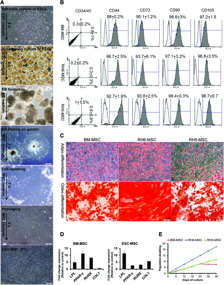
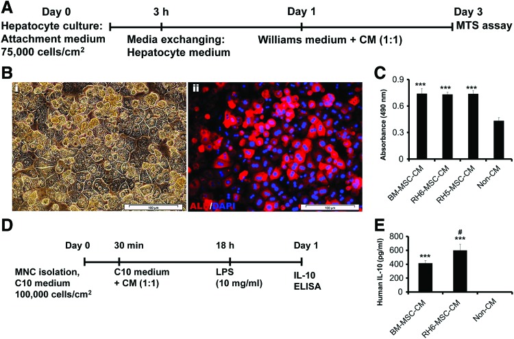
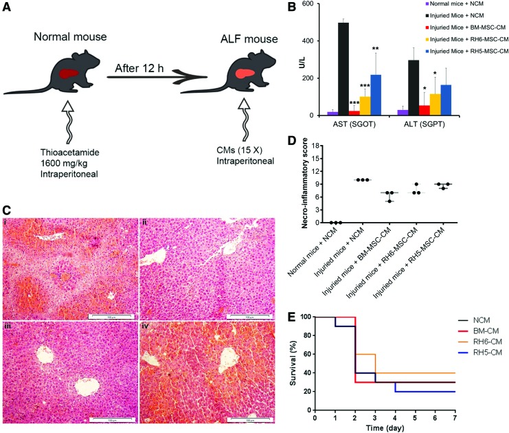
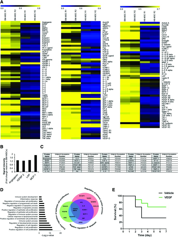
Adult tissue-derived mesenchymal stem cells (MSCs) show tremendous promise for a wide array of therapeutic applications predominantly through paracrine activity. Recent reports showed that human embryonic stem cell (ESC)-derived MSCs are an alternative for regenerative cellular therapy due to manufacturing large quantities of MSCs from a single donor. However, no study has been reported to uncover the secretome of human ESC-MSCs as treatment of an acute liver failure (ALF) mouse model. We demonstrated that human ESC-MSCs showed similar morphology and cell surface markers compared with bone marrow-derived MSCs. ESC-MSCs exhibited a higher growth rate during early in vitro expansion, along with adipogenic and osteogenic differentiation potential. Treatment with ESC-MSC-conditioned medium (CM) led to statistically significant enhancement of primary hepatocyte viability and increased immunomodulatory interleukin-10 secretion from lipopolysaccharide-induced human blood mononuclear cells. Analysis of the MSCs secretome by a protein array screen showed an association between higher frequencies of secretory proteins such as vascular endothelial growth factor (VEGF) and regulation of cell proliferation, cell migration, the development process, immune system process, and apoptosis. In this thioacetamide-induced mouse model of acute liver injury, we observed that systemic infusion of VEGF led to significant survival. These data have provided the first experimental evidence of the therapeutic potential of human ESC-MSC-derived molecules. These molecules show trophic support to hepatocytes, which potentially creates new avenues for the treatment of ALF, as an inflammatory condition.

Keywords: VEGF; acute liver failure; embryonic stem cells; mesenchymal stem cells; secretome; soluble factors.


Conflict of interest statement

Author Disclosure Statement No competing financial interests exist.

Figures

<b>FIG. 1.</b>
FIG. 1.
Generation and characterization of human ESC-MSCs. (A) The protocol for differentiation of human ESC (RH6 and RH5 lines) into MSCs. The detailed differentiation procedure is explained in the Materials and Methods section. Sequential morphological changes of epithelial ESCs into mesenchymal cells during differentiation are shown by inverted phase contrast microscope. (B) Immunophenotyping of RH6-MSCs, RH5-MSCs, and human bone marrow MSCs (BM-MSCs). The three cell types (passages 3–5) are examined by flow cytometry. Analysis of MSC markers, including CD44, CD73, CD90, and CD105, as well as hematopoietic markers CD34 and CD45 (double staining by anti-CD34-PE and anti-CD45-FITC) was performed by Flowing software (version 2.5.1). The white histogram represents the cells stained with isotype control antibodies. Expression of CD markers is presented as the mean ± SD of three different passages. (C) In vitro differentiation of MSCs into mesodermal lineages (adipocytes and osteoblasts). Adipodifferentiation was performed by the serum withdrawal/hypoxia method. Lipid droplets in cells stained with Oil Red O (upper panel) after 2 weeks. For osteodifferentiation of MSCs, we cultured the cells in osteogenic media for 3 weeks. Alizarin Red staining showed deposits of calcium crystals (lower panel). Scale bars: 100 μm. (D) The in vitro differentiation potential of MSCs (BM-MSC and RH6-MSC) was confirmed by studying the expressions of LPL and PPARG for adipodifferentiation and RUNX and COL1 for osteodifferentiation by real-time RT-PCR. BM-MSCs and ESC-MSCs after differentiation were compared with control groups (MSCs cultured in nondifferentiated medium). (E) Growth characteristics of ESC-MSCs. Graph shows PD over time (6 passages) for three different human cells -BM-MSCs, RH6-MSCs, and RH5-MSCs. During cell expansion, PD was calculated based on the cell numbers at the end of every passage in relation to the cell numbers at the first passage. BM, bone marrow; ESC, embryonic stem cell; MSCs, mesenchymal stem cells; PD, population doubling; RT-PCR, reverse transcriptase-polymerase chain reaction; SD, standard deviation. Color images available online at www.liebertpub.com/scd
<b>FIG. 2.</b>
FIG. 2.
In vitro effects of human ESC-MSC-CM and BM-MSC-CM on hepatocyte viability and MNC immunomodulation. (A) Schematic diagram of the experimental procedure to study the trophic potential of MSC-CM on mouse primary hepatocytes. Two-step collagenase perfusion was used for mouse hepatocyte isolation. Isolated cells were initially cultured on plates that contained Williams' medium for 3 h, after which the medium was replaced by hepatocyte medium (HepatoZYME + supplements) for 1 day and then incubated in Williams' medium + CMs for 2 days. (B) Immunostaining for ALB. (i) Cultured hepatocytes represented epithelial morphology and formed a stable monolayer such as polygonal cells with one or two round, prominent nuclei. (ii) Albumin-expressing hepatocytes visualized by immunostaining and fluorescent microscopy. Nuclei stained with DAPI. Scale bar: 100 μm. (C) We used the MTS assay to assess for primary cultured hepatocyte viability in the presence of MSC-CM or NCM. Human BM-MSC-CM and human ESC-derived MSC-CM (RH6-MSC-CM and RH5-MSC-CM) increased hepatocyte survival compared with NCM. (D) Schematic diagram of an in vitro immunomodulation assay. Primary human peripheral blood MNCs were first incubated in the presence of MSC-CM and NCM for 18 h, then stimulated with LPS for 5 h. Finally, we measured the level of IL-10 secreted from MNCs as an indicator for anti-inflammatory activity. (E) ELISA was used to assess the level of IL-10 in MNC culture supernatant. Incubation of MNCs with MSC-CM led to increased secretion of IL-10 from MNCs. Results have been reported as a mean of experiments performed in triplicate. ***P < 0.001 for MSC-CM compared with NCM. #P < 0.05 for ESC-MSC-CM compared with BM-MSC-CM. CM, conditioned medium; DAPI, 4, 6-diamidino-2-phenylindole; IL-10, interleukin-10; LPS, lipopolysaccharide; MNCs, mononuclear cells; NCM, nonconditioned medium. Color images available online at www.liebertpub.com/scd
<b>FIG. 3.</b>
FIG. 3.
In vivo effects of human SCs and their CM on mice with ALF. (A) Schematic representation of the experimental procedure design. (B) Biochemical assessment of AST and ALT in blood serum of ALF mice treated with human MSC-CM (n = 3) after 48 h. Data were reported as mean ± SD. *P < 0.05, **P < 0.01, and ***P < 0.001. (C) Representative micrographs of liver tissue. Liver sections from various groups stained with Hematoxylin and Eosin. Injured mice treated with (i) NCM, (ii) BM-MSC-CM, (iii) RH6-MSC-CM, and (iv) RH5-MSC-CM. Scale bar: 100 μm. (D) Histopathological grading of necrosis and inflammation of the liver (n = 3). Liver injury induced by injection of thioacetamide led to necrosis and inflammation. Treatment with MSC-CMs, unlike NCM, decreased the necroinflammatory score. Each circle represents one mouse. (E) Survival rate of ALF mice treated with CM. After 12 h, 400 μL of 15 × concentrated CM from RH6-MSCs, RH5-MSCs, and BM-MSCs were injected intraperitoneally (i.p.) into thioacetamide-mediated ALF mice (n = 10 per group). NCM was used as the control. ALF, acute liver failure; ALT, alanine aminotransferase; AST, aspartate aminotransferase; Color images available online at www.liebertpub.com/scd
<b>FIG. 4.</b>
FIG. 4.
Secretory profiling of human ESC-MSCs and BM-MSCs. (A) Heat map of cytokine secretion. The cytokine array (C2000; RayBio) was used to interrogate various cytokines and growth factors in human RH6-MSC and BM-MSC-CM. We assayed 174 secreted molecules (60 molecules for array C6, 60 for C7, and 54 for C8) in BM-MSC-CM and ESC-MSC-CM. These assays were performed in duplicate. Protein signals were quantified by NIH ImageJ software and normalized to the signal intensity average of positive controls for each array. Heat maps were generated by MeV software. From left to right: heat map of array C6, C7, and C8. Quantification of the samples revealed that the concentration of most cytokines in BM-MSC-CM was greater than ESC-MSC-CM. (B) Secretory proteins (4 proteins) showed greater expression in ESC-MSCs compared with BM-MSCs (fold change: >1.2). (C) List of 35 secretory proteins present at high levels (normalized signal intensity >0.5) in RH6-MSC-CM. (D) GO analysis of 35 selected cytokines and growth factors from ESC-MSC-CM. These ESC-MSC secretory proteins were analyzed by GO Enrichment Analysis Tools. The graph has indicated a number of biological processes (experimental) in which the 35 selected proteins are involved (P < 0.05). Venn diagram shows overlap between three biological processes—regulation of immune response (red circle), epithelial cell proliferation (green circle), and negative regulation of apoptosis (blue circle). A total of 16 out of 35 proteins were related to these three biological processes, VEGF and BMP4 of which are common in these biological processes. (E) Survival rate of ALF mice treated with rVEGF. Mice in group 1 (n = 8) received i.p. injection of 40 μg/kg rVEGF dissolved in DPBS. Group 2 (n = 6) received DPBS as the vehicle. DPBS, Dulbecco's phosphate-buffered saline; GO, gene ontology; rVEGF, recombinant VEGF; VEGF, vascular endothelial growth factor. Color images available online at www.liebertpub.com/scd

Similar articles

Cited by

References

    1. Dhaliwal HS. and Singh P. (2014). Acute liver failure. N Engl J Med 370:1170 - PubMed
    1. Miwa S, Hashikura Y, Mita A, Kubota T, Chisuwa H, Nakazawa Y, Ikegami T, Terada M, Miyagawa S. and Kawasaki S. (1999). Living-related liver transplantation for patients with fulminant and subfulminant hepatic failure. Hepatology 30:1521–1526 - PubMed
    1. Irfan A. and Ahmed I. (2015). Could stem cell therapy be the cure in liver cirrhosis? J Clin Exp Hepatol 5:142–146 - PMC - PubMed
    1. Owen A. and Newsome PN. (2015). Mesenchymal stromal cell therapy in liver disease: opportunities and lessons to be learnt? Am J Physiol Gastrointest Liver Physiol 309:G791–G800 - PMC - PubMed
    1. Keating A. (2012). Mesenchymal stromal cells: new directions. Cell Stem Cell 10:709–716 - PubMed

MeSH terms

LinkOut - more resources