. 2016 Sep 27:22:3446-3454.
doi: 10.12659/msm.900460.

Protective Effects of Gallic Acid Against NiSO4-Induced Toxicity Through Down-Regulation of the Ras/ERK Signaling Pathway in Beas-2B Cells

Affiliations

Protective Effects of Gallic Acid Against NiSO4-Induced Toxicity Through Down-Regulation of the Ras/ERK Signaling Pathway in Beas-2B Cells

Xuejun An et al. Med Sci Monit. .

Abstract

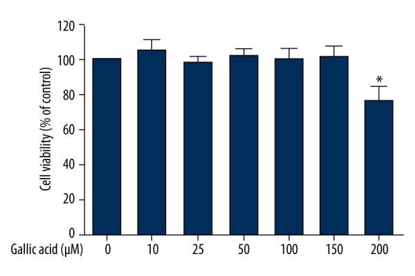
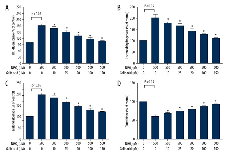
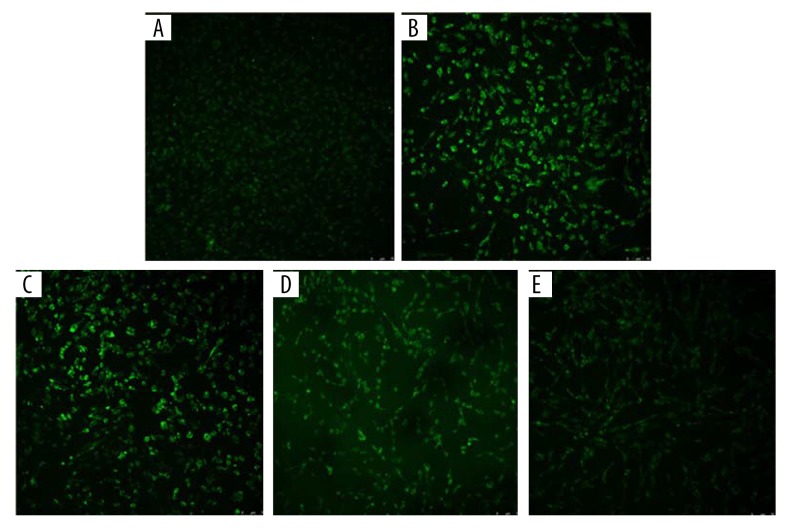
BACKGROUND This study aimed to explore the preventive effects of gallic acid (GA) on the toxicity induced by NiSO4 in Beas-2B cells. MATERIAL AND METHODS Beas-2B cell viability was measured by MTT assay. The degree of oxidative stress was detected by measuring the levels of reactive oxygen species (ROS) and lipid peroxide (LPO). The rate of apoptosis was measured by flow cytometry. Ras/ERK-related protein levels were analyzed by Western blot analysis, which including Ras, ERK, c-Myc, PARP, and PARP cleavage. RESULTS MTT assay showed that NiSO4 induced cytotoxicity, while GA had a protective role against toxicity. Additionally, GA could reduce the apoptotic cell number and the level of ROS in Beas-2B cells induced by NiSO4. Western blot analysis demonstrated that NiSO4 could up-regulate the related protein in the Ras/ERK signaling pathway. Furthermore, we observed that GA could alleviate the toxicity of NiSO4 through regulating protein changes in the Ras/ERK signaling pathway. CONCLUSIONS Preventive effects of GA on NiSO4-induced cytotoxicity in Beas-2B cells may be through the Ras/ERK signaling pathways.


Figures

Figure 1
Figure 1
Effect of GA on cell viability. Beas-2B cells were exposed to different concentrations of GA for 24 h, and cell viability was determined by MTT assay as described in the Material and Methods section. Data represent mean ±SD of three independent experiments made in three replicates. * P<0.05, compared with control group.
Figure 2
Figure 2
Effect of GA on NiSO4-induced apoptosis. Beas-2B cells were exposed to 500 μM NiSO4 for 24 h in the presence or advance of GA (0–150 μM), and apoptosis was determined by flow cytometry analysis as described in the Material and Methods section. (A) Control group. (B) 500 μM NiSO4. (C) 10 μM GA + 500 μM NiSO4. (D) 25 μM GA + 500 μM NiSO4. (E) 50 μM GA + 500 μM NiSO4. (F) 100 μM GA + 500 μM NiSO4. (G) 150 μM GA + 500 μM NiSO4. Data represent mean ±SD of three independent experiments made in three replicates. * Significant preventive effect of different concentrations of GA on reduction of oxidative stress caused by NiSO4 (P<0.05).
Figure 3
Figure 3
Effect of GA on NiSO4-induced oxidative stress. Beas-2B cells were exposed to 500 μM NiSO4 for 24 h in the presence or advance of GA (0–150 μM), and the cells’ oxidative stress parameters were determined according to the kit instructions as described in the Material and Methods section. (A) ROS, (B) LDH, (C) MDA, and (D) GSH. Data represent mean ±SD of three independent experiments made in three replicates. * Significant preventive effect of different concentrations of GA on reduction of oxidative stress caused by NiSO4 (P<0.05).
Figure 4
Figure 4
Effect of GA on NiSO4-induced reactive oxygen species fluorescence intensity. The expression of reactive oxygen species fluorescence intensity was observed by laser confocal microscopy. (A) Control group. (B) 500 μM NiSO4. (C) 10 μM GA+500 μM NiSO4. (D) 50 μM GA+500 μM NiSO4. (E) 150 μM GA+500 μM NiSO4. The original magnification is 20×.
Figure 5
Figure 5
Effect of GA on NiSO4-induced Ras/ERK signaling pathway protein expressions. Shown are expression levels of Ras/ERK signaling pathway proteins after Beas-2B cells were treated with NiSO4 alone or NiSO4 + GA. (A) NiSO4 regulated Ras/ERK signaling protein expressions in Beas-2B cells. Cells were exposed to different concentrations of NiSO4 for 24 h; effect of GA on NiSO4-induced Ras/ERK signaling pathway protein expressions. Cells were exposed to 500 μM NiSO4 for 24 h in the presence or advance of GA (0–150 μM). (B) Beas-2B cells were not treated or treated with Ras inhibitor manumycin A (10 μM) or ERK inhibitor PD98059 (20 μM) for 1 h, and then with 500 μM NiSO4 for 24 h. The protein expression levels were determined by Western blotting analysis as described in the Material and Methods section.

Similar articles

Cited by

References

    1. Grandjean P. Human exposure to nickel. IARC Sci Publ. 1984;53:469–85. - PubMed
    1. Ma L, Bai YN, Pu HQ, et al. A retrospective cohort mortality study in Jinchang, the largest nickel production enterprise in China. Biomed Environ Sci. 2014;27:567–71. - PubMed
    1. Li Q, Suen TC, Sun H, et al. Nickel compounds induce apoptosis in human bronchial epithelial Beas-2B cells by activation of c-Myc through ERK pathway. Toxical Appl Pharmacol. 2009;235:191–98. - PMC - PubMed
    1. Doll R, Morgan LG, Speizer FE. Cancers of the lung and nasal sinuses in nickel workers. Br J Cancer. 1970;24:623–32. - PMC - PubMed
    1. Chakrabati SK, Bai C, Subramanian KS. DNA-protein crosslinks induced by nickel compounds in isolated rat lymphocytes: Role of reactive oxygen species and specific amino acids. Tixicol Appl Pharmacol. 2001;170:153–65. - PubMed