. 2016 Dec;229(6):800-824.
doi: 10.1111/joa.12539. Epub 2016 Sep 28.

Vascular patterns in the heads of crocodilians: blood vessels and sites of thermal exchange

Affiliations

Vascular patterns in the heads of crocodilians: blood vessels and sites of thermal exchange

William Ruger Porter et al. J Anat. 2016 Dec.

Abstract

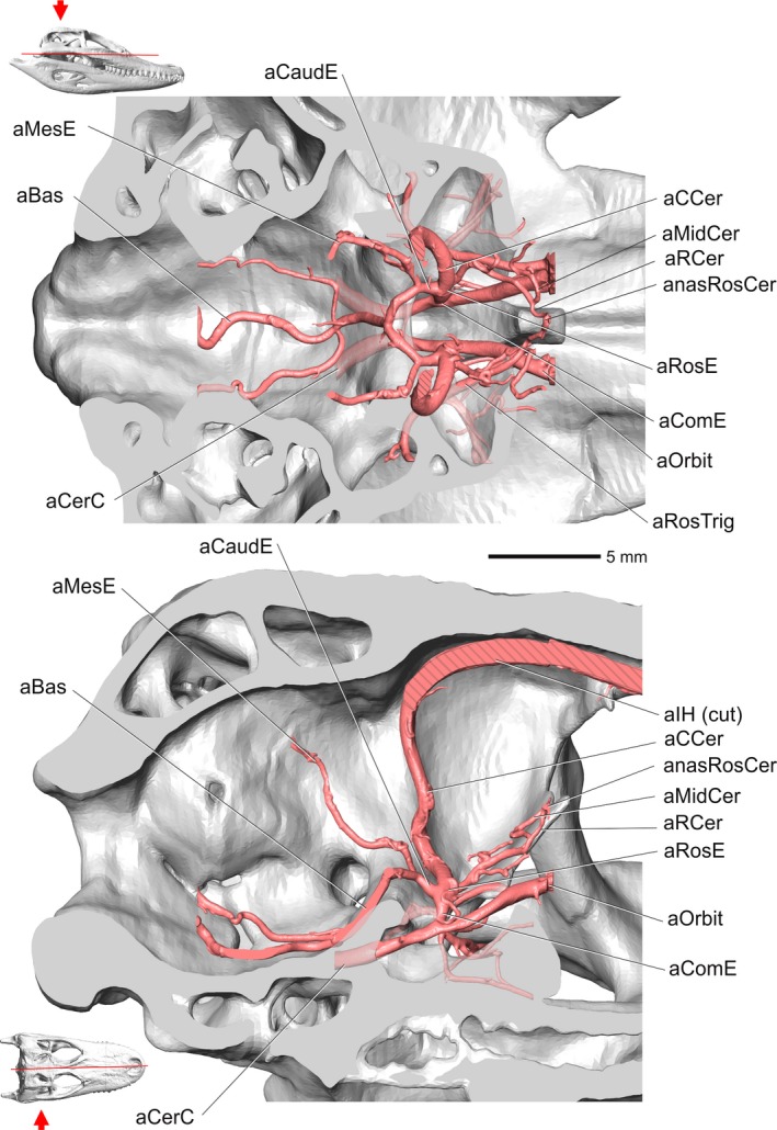
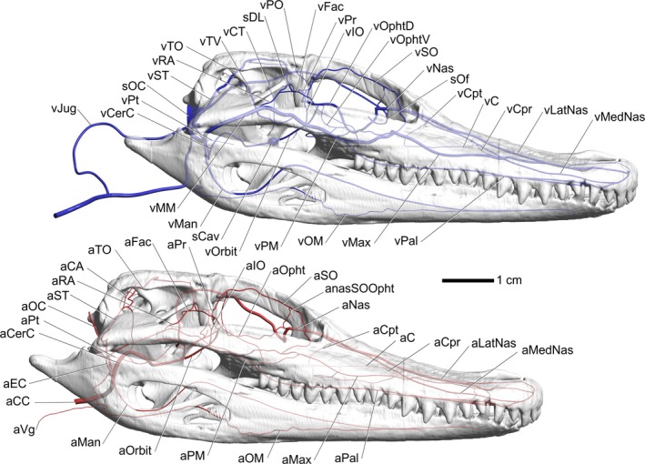
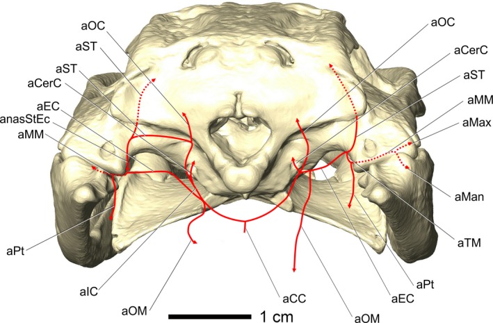
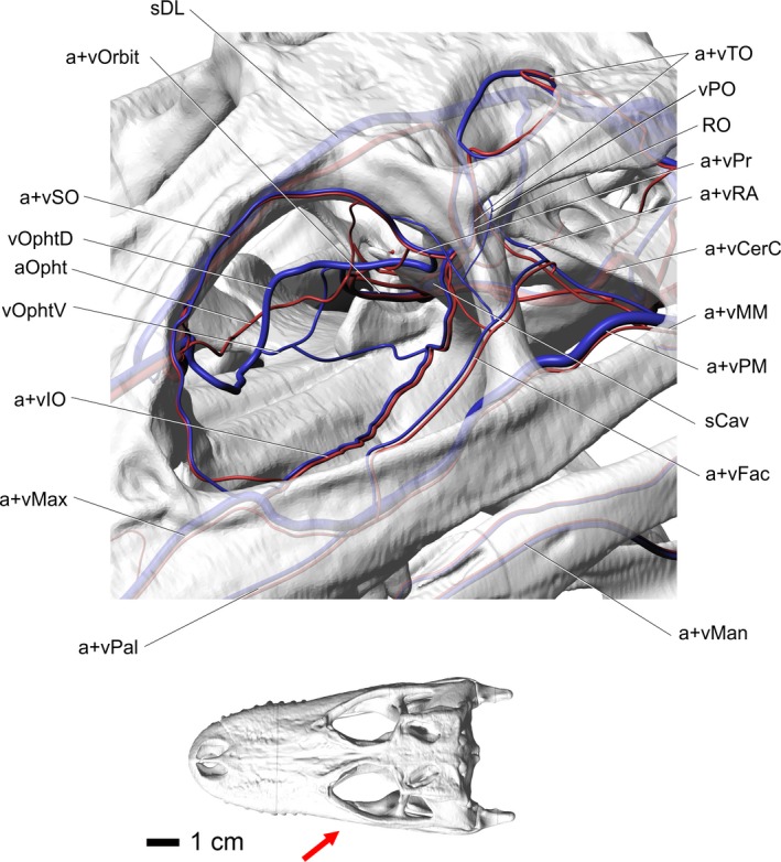
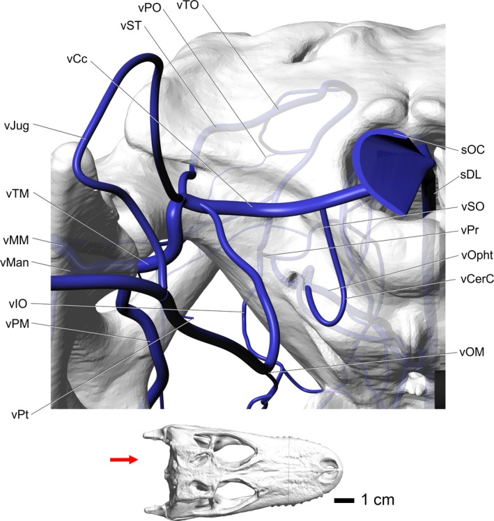
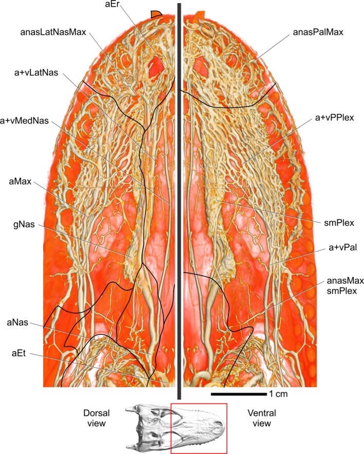
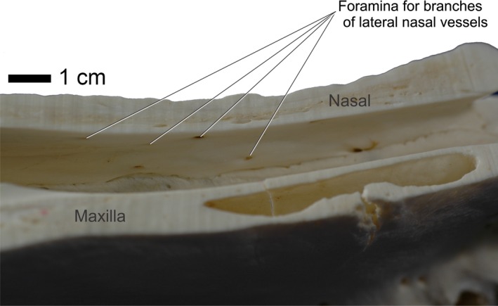
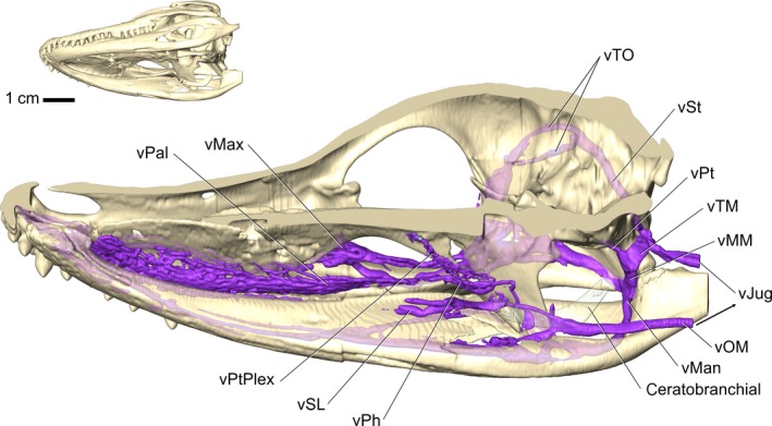
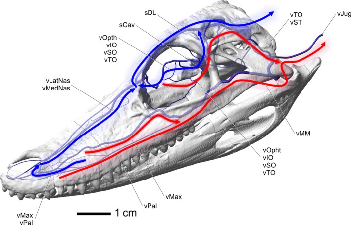
Extant crocodilians are a highly apomorphic archosaur clade that is ectothermic, yet often achieve large body sizes that can be subject to higher heat loads. Therefore, the anatomical and physiological roles that blood vessels play in crocodilian thermoregulation need further investigation to better understand how crocodilians establish and maintain cephalic temperatures and regulate neurosensory tissue temperatures during basking and normal activities. The cephalic vascular anatomy of extant crocodilians, particularly American alligator (Alligator mississippiensis) was investigated using a differential-contrast, dual-vascular injection technique and high resolution X-ray micro-computed tomography (μCT). Blood vessels were digitally isolated to create representations of vascular pathways. The specimens were then dissected to confirm CT results. Sites of thermal exchange, consisting of the oral, nasal, and orbital regions, were given special attention due to their role in evaporative cooling and cephalic thermoregulation in other diapsids. Blood vessels to and from sites of thermal exchange were studied to detect conserved vascular patterns and to assess their ability to deliver cooled blood to neurosensory tissues. Within the orbital region, both the arteries and veins demonstrated consistent branching patterns, with the supraorbital, infraorbital, and ophthalmotemporal vessels supplying and draining the orbit. The venous drainage of the orbital region showed connections to the dural sinuses via the orbital veins and cavernous sinus. The palatal region demonstrated a vast plexus that comprised both arteries and veins. The most direct route of venous drainage of the palatal plexus was through the palatomaxillary veins, essentially bypassing neurosensory tissues. Anastomotic connections with the nasal region, however, may provide an alternative route for palatal venous blood to reach neurosensory tissues. The nasal region in crocodilians is probably the most prominent site of thermal exchange, as it offers a substantial surface area and is completely surrounded by blood vessels. The venous drainage routes from the nasal region offer routes directly to the dural venous sinuses and the orbit, offering evidence of the potential to directly affect neurosensory tissue temperatures. The evolutionary history of crocodilians is complex, with large-bodied, terrestrial, and possibly endothermic taxa that may have had to deal with thermal loads that likely provided the anatomical building-blocks for such an extensive vascularization of sites of thermal exchange. A clear understanding of the physiological abilities and the role of blood vessels in the thermoregulation of crocodilians neurosensory tissues is not available but vascular anatomical patterns of crocodilian sites of thermal exchange indicate possible physiological abilities that may be more sophisticated than in other extant diapsids.

Keywords: blood vessels; cephalic; crocodilian; thermoregulation; vasculature.


Figures

Figure 1
Figure 1
Volume rendering of one crocodile (OUVC 10426) and eight alligator heads. Specimens are arranged by size, with OUVC accession number above the head and the skull length in mm below. Skulls were measured from the tip of the premaxilla to the dorsal aspect of the supraoccipital. OUVC 9601 and 9680 were not CT‐scanned, but were included in this study.
Figure 2
Figure 2
Volume renderings of four Alligator mississippiensis specimens in right rostrodorsolateral view showing blood vessels of the head. (A) OUVC 9757 venous and arterial injection. (B) OUVC 10389 arterial and venous injection. (C) OUVC 10395 arterial and venous injection. (D) OUVC 10597 arterial injection. Vessel names: aFac, facial artery; aLatNas, lateral nasal artery; aMax, maxillary artery; aMM, maxillomandibular artery; aPal, palatine artery; aRA, rostral auricular artery; aSO, supraorbital artery; aST, stapedial artery; aTO, temporoorbital artery; vJug, jugular vein; vLatNas, lateral nasal vein; vMM, maxillomandibular vein; vSO, supraorbital vein; vST, stapedial vein; vTO, temporoorbital vein.
Figure 3
Figure 3
Alligator mississippiensis, OUVC 9757. (A) CT slice of the nasal region showing arteries and veins. (B) Volume rendering of a dorsal view of OUVC 9757 showing arteries and veins. White line indicates the location of the CT slice and the white arrow indicates direction of view. Vessel names: vMax, maxillary vein; aMedNas, medial nasal artery; vLatNas, lateral nasal vein; vPPlex, venous palatal plexus; vMM, maxillomandibular vein; vTO, temporoorbital vein.
Figure 4
Figure 4
Alligator mississippiensis, caudal view, showing the modeled representations of the major arteries of the head. Skull is semitransparent. Arrow in inset at bottom shows the view in the image above. Vessel names: aTO, temporoorbital artery; aCA, caudal auricular artery; aST, stapedial artery; anasStEc, anastomosis between stapedial artery and external carotid vessels; aTM, temporomandibular artery; aMM, maxillomandibular artery; aMan, mandibular artery; aEC, external carotid artery; aPt, pterygoid artery; aIO, infraorbital artery; aVg, vagus artery; aOC, occipital artery; aCCer, caudal cerebral artery; aSO, supraorbital artery; aPr, profundus artery; aOpht, ophthalmotemporal artery; aIC, internal carotid artery; aCC, common carotid artery; aOM, oromandibular artery.
Figure 5
Figure 5
Alligator mississippiensis, OUVC 10395. Top, dorsal view showing the encephalic arteries. Hatching patterns represent cut arteries. Bottom, medial view showing the encephalic arteries. Inset with red line indicates the plane of section and the arrow indicates view. Vessel names: aCaudE, caudal encephalic artery; aMesE, mesencephalic artery; aBas, basilar artery; aCerC, cerebral carotid artery; aCCer, caudal cerebral artery; aMidCer, middle cerebral artery; aRCer, rostral cerebral artery; anasRosCer, anastomosis between rostral cerebral vessels; aRosE, rostral encephalic artery; aComE, common encephalic artery; aOrbit, orbital artery; aRosTrig, rostral branch of trigeminal artery; aIH, interhemispheric artery.
Figure 6
Figure 6
Alligator mississippiensis, right lateral view. Top, modeled representations of the cephalic veins. Bottom, modeled representations of the cephalic arteries. Skull is semitransparent. Vessel names: vPO, postorbital vein; sDL, dorsal longitudinal sinus; vCT, cerebrotectal vein; vTV, transverse sinus; vTO, temporoorbital vein; vRA, rostral auricular vein; vST, stapedial vein; sOC, occipital sinus; vPt, pterygoid vein; vCerC, cerebral carotid vein; vJug, jugular vein; vFac, facial vein; vPr, profundus vein; vIO, infraorbital vein; vOphtd, dorsal branch of ophthalmotemporal vein; vOphtv, ventral branch of ophthalmotemporal vein; vSO, supraorbital vein; vNas, common nasal vein; sOf, olfactory sinus; vCpt, postconchal vein; vC, conchal vein; vCpr, preconchal vein; vLatNas, lateral nasal vein; vMedNas, medial nasal vein; vMM, maxillomandibular vein; vMan, mandibular vein; sCav, cavernous sinus; vOrbit, orbital vein; vPM, palatomaxillary vein; vOM, oromandibular vein; vMax, maxillary vein; vPal, palatine vein; aFac, facial artery; aTO, temporoorbital artery; aCA, caudal auricular artery; aRA, rostral auricular artery; aST, stapedial artery; aOC, occipital artery; aPt, pterygoid artery; aCerC, cerebral carotid artery; aEC, external carotid artery; aCC, common carotid artery; aVg, vagus artery; aMan, mandibular artery; aOrbit, orbital artery; aPM, palatomaxillary artery; aOM, oromandibular artery; aMax, maxillary artery; aPal, palatine artery; aMedNas, medial nasal artery; aLatNas, lateral nasal artery; aCpr, preconchal artery; aC, conchal artery; aCpt, postconchal artery; aNas, common nasal artery; anasSOOpht, anastomosis between supraorbital and ophthalmotemporal vessels; aSO, supraorbital artery; aOpht, ophthalmotemporal artery; aIO, infraorbital artery; aPr, profundus artery.
Figure 7
Figure 7
Alligator mississippiensis, top view. Left, modeled representations of the cephalic arteries. Right, modeled representations of the cephalic veins. Skull is semitransparent. Vessel names: anasLatNasMax, anastomosis between lateral nasal and maxillary vessels; anasPalMax, anastomosis between palatine and maxillary vessels; aMedNas, medial nasal artery; aLatNas, lateral nasal artery; aPal, palatine artery; aMax, maxillary artery; aCpr, preconchal artery; aC, conchal artery; aCpt, postconchal artery; aNas, common nasal artery; aSO, supraorbital artery; aIO, infraorbital artery; aET, ethmoid artery; aOpht, ophthalmotemporal artery; aFac, facial artery; aPM, palatomaxillary artery; aRA, rostral auricular artery; aMan, mandibular artery; aOM, oromandibular artery; aTO, temporoorbital artery; aPt, pterygoid artery; aST, stapedial artery; aMM, maxillomandibular artery; aTM, temporomandibular artery; aOC, occipital artery; aVg, vagus artery; aCC, common carotid artery; sOC, occipital sinus; Anas, anastomosis; vMedNas, medial nasal vein; vLatNas, lateral nasal vein; vPal, palatine vein; vMax, maxillary vein; vCpr, preconchal vein; vC, conchal vein; vCpt, postconchal vein; sOf, olfactory sinus; vJug, jugular vein; vCc, caudal head vein; vTM, temporomandibular vein; vMM, maxillomandibular vein; vST, stapedial vein; vPt, pterygoid vein; vTO, temporoorbital vein; vOM, oromandibular vein; vRA, rostral auricular vein; vMan, mandibular vein; vPM, palatomaxillary vein; vFac, facial vein; sDL, dorsal longitudinal sinus; vOphtd, dorsal branch of ophthalmotemporal vein; vIO, infraorbital vein; vSO, supraorbital vein; vNas, common nasal vein.
Figure 8
Figure 8
Alligator mississippiensis, OUVC 10597. Caudal view of an alligator showing a diagram of the vascular variations in the caudal region of the head. Variations were present on the right side, whereas the left side had the more typical diapsid pattern. Vessel names: aCC, common carotid artery; aCerC, cerebral carotid artery; aEC, external carotid artery; aIC, internal carotid artery; aMan, mandibular artery; aMax, maxillary artery; aMM, maxillomandibular artery; anasStEc, anastomosis between stapedial artery and external carotid vessels; aOC, occipital artery; aOM, oromandibular artery; aPt, pterygoid artery; aST, stapedial artery; aTM, temporomandibular artery.
Figure 9
Figure 9
Alligator mississippiensis, rostrolateral oblique view of the blood vessels of the orbital region. Skull is semitransparent. Arrow in inset indicates orientation and view. Vessel names: sDL, dorsal longitudinal sinus; vOrbit, orbital vein; vSO, supraorbital vein; vOphtd, dorsal branch of ophthalmotemporal vein; aOpht, ophthalmotemporal artery; vOphtv, ventral branch of ophthalmotemporal vein; vIO, infraorbital vein; vMax, maxillary vein; vPal, palatine vein; vMan, mandibular vein; vFac, facial vein; sCav, cavernous sinus; vPM, palatomaxillary vein; vMM, maxillomandibular vein; vCerC, cerebral carotid vein; vRA, rostral auricular vein; vPr, profundus vein; RO, ophthalmic rete; vPO, postorbital vein; vTO, temporoorbital vein.
Figure 10
Figure 10
Alligator mississippiensis, caudal view, showing the modeled representations of the major veins of the head. Skull is semitransparent. Arrow in the inset indicates orientation and view. Vessel names: sDL, dorsal longitudinal sinus; vSO, supraorbital vein; vOphtv, ventral branch of ophthalmotemporal vein; vIO, infraorbital vein; vMan, mandibular vein; vPM, palatomaxillary vein; vMM, maxillomandibular vein; vCerC, cerebral carotid vein; vPr, profundus vein; vPO, postorbital vein; vTO, temporoorbital vein; vST, stapedial vein; vCc, caudal head vein; vJug, jugular vein; vTM, temporomandibular vein; vPt, pterygoid vein; vOM, oromandibular vein; sOC, occipital sinus.
Figure 11
Figure 11
Alligator mississippiensis, OUVC 9757. Volume rendering of both the nasal region in dorsal view (left) and palatal region in ventral view (right), indicating the extensive vascularization of these regions. Inset indicates area region. Black lines indicate the approximate location of sutures. Vessel names: aEr, erectile arteries; anasLatNasMax, anastomosis between lateral nasal and maxillary vessels; vLatNas, lateral nasal vein; vMedNas, medial nasal vein; aMax, maxillary artery; gNas, nasal gland; aNas, common nasal artery; aET, ethmoid artery; anasMaxsmPlex, anastomosis between maxillary artery and submucosal plexus; vPal, palatine vein; smPlex, submucosa plexus on ventral part of airway; vPPlex, venous palatal plexus; anasPalMax, anastomosis between palatine and maxillary vessels.
Figure 12
Figure 12
Alligator mississippiensis, OUVC 9601. Medial view of left side of sagittally sectioned skull demonstrating the foramina housing branches of the lateral nasal vessels and nerves.
Figure 13
Figure 13
Alligator mississippiensis, OUVC 10387. Right ventrolateral view of sagittally sectioned skull demonstrating the veins of the tongue. Perfusion into the distal aspects of the tongue was not sufficient, yet the veins draining the tongue are present. Blood from the ventral head drains into the jugular vein, bypassing neurosensory tissues like the brain and eyes. Vessel names: vTO, temporoorbital vein; vST, stapedial vein; vPt, pterygoid vein; vTM, temporomandibular vein; vMM, maxillomandibular vein; vJug, jugular vein; vMax, maxillary vein; vPal, palatine vein; vPtPlex, pterygoid plexus; vSL, sublingual vein; vPh, palatopharyngeal vein; vMan, mandibular vein; vOM, oromandibular vein.
Figure 14
Figure 14
Alligator mississippiensis, left rostrodorsolateral view, indicating possible pathways for blood to drain from sites of thermal exchange. Red arrows indicate pathways that likely have little influence on neurosensory tissues. Blue arrows indicate pathways that likely have an influence on neurosensory tissues. Labels indicate named blood vessels involved with indicated pathways. Vessel names: vMedNas, medial nasal vein; vLatNas, lateral nasal vein; sCav, cavernous sinus; sDL, dorsal longitudinal sinus; vJug, jugular vein; vMax, maxillary vein; vPal, palatine vein; vOphtd, dorsal branch of ophthalmotemporal vein; vOphtv, ventral branch of ophthalmotemporal vein; vIO, infraorbital vein; vSO, supraorbital vein; vTO, temporoorbital vein; vMM, maxillomandibular vein; vST, stapedial vein.

Similar articles

Cited by

References

    1. Almeida L, Campos R (2011) A systematic study of the brain base arteries in the broad‐snouted caiman (Caiman latirostris). J Morphol Sci 28, 62–68.
    1. Barbour HR, Archer MA, Hart NS, et al. (2002) Retinal characteristics of the ornate dragon lizard, Ctenophorus ornatus . J Comp Neurol 450, 334–344. - PubMed
    1. Baumel JJ (1975) Aves: heart and blood vessels In: Sisson and Grossman's the Anatomy of the Domestic Animals (ed. Getty R.), pp. 1968–2003. Philadelphia: W.B. Saunders.
    1. Baumel JJ (1993) Systema cardiovasculare In: Sisson and Grossman's the Anatomy of the Domestic Animals (ed. Getty R.), pp. 407–476. Philadelphia: W.B. Saunders.
    1. Bellairs AA, Shute CCD (1953) Observations on the narial musculature of Crocodilia and its innervation from the sympathetic system. J Anat 87, 367–380. - PMC - PubMed

Publication types