. 2017 Mar;21(3):422-431.
doi: 10.1111/jcmm.12986. Epub 2016 Sep 28.

Genistein suppresses leptin-induced proliferation and migration of vascular smooth muscle cells and neointima formation

Affiliations

Genistein suppresses leptin-induced proliferation and migration of vascular smooth muscle cells and neointima formation

Yung-Chieh Tsai et al. J Cell Mol Med. 2017 Mar.

Abstract

Obesity is a strong risk factor for the development of cardiovascular diseases and is associated with a marked increase in circulating leptin concentration. Leptin is a peptide hormone mainly produced by adipose tissue and is regulated by energy level, hormones and various inflammatory mediators. Genistein is an isoflavone that exhibits diverse health-promoting effects. Here, we investigated whether genistein suppressed the atherogenic effect induced by leptin. The A10 cells were treated with leptin and/or genistein, and then the cell proliferation and migration were analysed. The reactive oxygen species (ROS) and proteins levels were also measured, such as p44/42MAPK, cell cycle-related protein (cyclin D1 and p21) and matrix metalloproteinase-2 (MMP-2). Immunohistochemistry and morphometric analysis were used for the neointima formation in a rat carotid artery injury model. Genistein (5 μM) significantly inhibited both the proliferation and migration of leptin (10 ng/ml)-stimulated A10 cells. In accordance with these finding, genistein decreased the leptin-stimulated ROS production and phosphorylation of the p44/42MAPK signal transduction pathway. Meanwhile, genistein reversed the leptin-induced expression of cyclin D1, and cyclin-dependent kinase inhibitor, p21. Genistein attenuated leptin-induced A10 cell migration by inhibiting MMP-2 activity. Furthermore, the leptin (0.25 mg/kg)-augmented neointima formation in a rat carotid artery injury model was attenuated in the genistein (5 mg/kg body weight)-treated group when compared with the balloon injury plus leptin group. Genistein was capable of suppressing the atherogenic effects of leptin in vitro and in vivo, and may be a promising candidate drug in the clinical setting.

Keywords: Leptin; MMP2; carotid artery injury; genistein; reactive oxygen species.


Figures

Figure 1
Figure 1
Effect of genistein on leptin‐induced proliferation of A10 cells. (A and D) Growth‐arrested cells were stimulated with leptin (1–100 ng/ml) for 72 hrs in the presence or absence of genistein (1–10 μM). Cell proliferation was assayed by the CellTiter 96® AQueous One Solution kit. Relative proliferation activities were determined using untreated control cells as a standard. Data represent the mean ± S.E.M. of six independent observations with different cell passages and on different days. *P < 0.05 versus Control; # P < 0.05 versus Leptin (10 ng/ml) alone. (B and C) Cells were incubated with genistein at increasing concentrations (1–40 μM) for 24 hrs; the toxic effects of genistein were measured by the CellTiter 96® AQueous One Solution kit and LDH cytotoxicity assay kit, respectively. Data represent the mean ± S.E.M. of four independent observations with different cell passages and on different days. *P < 0.05 versus Control. (E) DNA synthesis was measured by the BrdU incorporation assay. Growth‐arrested cells were stimulated with leptin (1–100 ng/ml) for 72 hrs in the presence or absence of genistein (1 and 5 μM) or NAC (5 and 10 μM). Data represent the mean ± S.E.M. of six independent observations with different cell passages and on different days. *P < 0.05 versus Control; # P < 0.05 versus Leptin (10 ng/ml) alone.
Figure 2
Figure 2
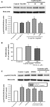
Effect of genistein on leptin‐induced p44/42MAPK phosphorylation in A10 cells. (A and C) Cells were stimulated with leptin (1–100 ng/ml) for 30 min. in the presence or absence of genistein (1 and 5 μM). The cells were lysed and proteins were analysed by Western blotting. β‐actin was used for normalization. Data represent the mean ± S.E.M. of five independent observations with different cell passages and on different days. *P < 0.05 versus Control; # P < 0.05 versus Leptin (10 ng/ml) alone. (B) Growth‐arrested cells were stimulated with leptin (10 ng/ml) for 72 hrs in the presence or absence of U0‐126 (1 μM). Cell proliferation was assayed by the CellTiter 96® AQueous One Solution kit. Relative proliferation activities were determined using untreated control cells as a standard. Data represent the mean ± S.E.M. of six independent observations with different cell passages and on different days. *P < 0.05 versus Control; # P < 0.05 versus Leptin (10 ng/ml) alone.
Figure 3
Figure 3
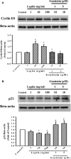
Effect of genistein on leptin‐induced cyclin D1 and p21 protein expression in A10 cells. (A and B) Cells were stimulated with leptin (1–100 ng/ml) for 3 hrs in the presence or absence of genistein (1 and 5 μM). The cells were lysed and both cyclin D1 and p21 proteins were analysed by Western blotting. β‐actin was used for normalization. Data represent the mean ± S.E.M. of three independent observations with different cell passages and on different days. *P < 0.05 versus Control; # P < 0.05 versus Leptin (10 ng/ml) alone.
Figure 4
Figure 4
Effects of genistein on leptin‐induced VSMC migration as well as MMP‐2 protein expression in A10 cells. (A and B) VSMC migration was examined using the Transwell® Permeable Support Culture Plate System. Genistein (1 and 5 μM) was added to both the upper and the lower compartments and was present throughout the duration of the experiment. Migration was induced by the addition of leptin (10 ng/ml) to the lower chamber. After incubation at 37°C for 48 hrs, the non‐migratory cells were removed from the upper surface of the membrane by scraping them with cotton swabs. The membrane was fixed with 90% ethanol and stained with 0.1% crystal. Migrated cells were counted at 200× magnification in five randomly chosen microscope fields per filter. The figure (B) indicates the fold value of cell migration. Data represent the mean ± S.E.M. of four independent observations with different cell passages and on different days. (C) Cells were stimulated with leptin (1–100 ng/ml) for 3 hrs in the presence or absence of genistein (1 and 5 μM). The cells were lysed and MMP‐2 protein was analysed by Western blotting. β‐actin was used for normalization. Data represent the mean ± S.E.M. of five independent observations with different cell passages and on different days. *P < 0.05 versus Control; # P < 0.05 versus Leptin (10 ng/ml) alone. (D) Gelatin zymography analysis was performed with conditioned media collected from A10 cells cultured in the presence or absence of genistein (1 and 5 μM) and leptin (10 ng/ml).
Figure 5
Figure 5
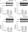
Effects of genistein alone on phosphorylated p44/42MAPK, cyclin D1, p21 and MMP2 protein expression in A10 cells. (AD) Cells were stimulated with genistein (1, 5 μM) for 24 hrs. The cells were lysed and proteins were analysed by Western blotting. β‐actin was used for normalization. Data represent the mean ± S.E.M. of three independent observations with different cell passages and on different days. *P < 0.05 versus Control.
Figure 6
Figure 6
Effects of genistein on leptin‐induced ROS production in A10 cells. Cells were stimulated with leptin (10 ng/ml) for 1 hr in the presence or absence of genistein (5 μM). Representative images showing that intracellular ROS production was detected using CellROX Green Reagent.
Figure 7
Figure 7
Typical examples of carotid arteries of rats treated with leptin alone or co‐treated with genistein after balloon injury. (A and E) Sham surgery, (B and F) arterial ballooning injury, (C and G) arterial ballooning injury then leptin (0.25 mg/kg body weight, i.p.) treatment, (D and H) arterial ballooning injury then leptin and genistein (5 mg/kg body weight, i.p.) treatment, showing significant attenuation of neointimal smooth muscle cell proliferation. AD (haematoxylin and eosin stain 400×) compared with EH (immuohistochemical stain of anti‐alpha smooth muscle actin, 400×).

References

    1. Chistiakov DA, Orekhov AN, Bobryshev YV. Vascular smooth muscle cell in atherosclerosis. Acta Physiol (Oxf). 2015; 214: 33–50. - PubMed
    1. Berg AH, Scherer PE. Adipose tissue, inflammation, and cardiovascular disease. Circ Res. 2005; 96: 939–49. - PubMed
    1. Sharma AM. Obesity and cardiovascular risk. Growth Horm IGF Res. 2003; 13 (Suppl A): S10–7. - PubMed
    1. Friedman JM, Halaas JL. Leptin and the regulation of body weight in mammals. Nature. 1998; 395: 763–70. - PubMed
    1. Fortuño A, Rodríguez A, Gómez‐Ambrosi J, et al Adipose tissue as an endocrine organ: role of leptin and adiponectin in the pathogenesis of cardiovascular diseases. J Physiol Biochem. 2003; 59: 51–60. - PubMed

Publication types

MeSH terms