Review
. 2016 Nov;13(11):633-642.
doi: 10.1038/nrgastro.2016.142. Epub 2016 Sep 28.

Human mini-guts: new insights into intestinal physiology and host-pathogen interactions

Affiliations
Review

Human mini-guts: new insights into intestinal physiology and host-pathogen interactions

Julie G In et al. Nat Rev Gastroenterol Hepatol. 2016 Nov.

Abstract

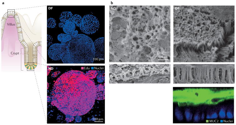
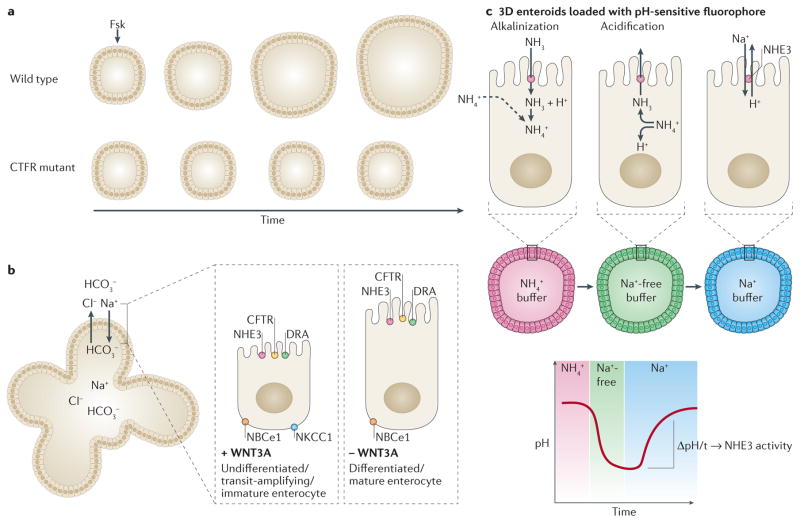
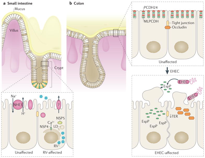
The development of indefinitely propagating human 'mini-guts' has led to a rapid advance in gastrointestinal research related to transport physiology, developmental biology, pharmacology, and pathophysiology. These mini-guts, also called enteroids or colonoids, are derived from LGR5+ intestinal stem cells isolated from the small intestine or colon. Addition of WNT3A and other growth factors promotes stemness and results in viable, physiologically functional human intestinal or colonic cultures that develop a crypt-villus axis and can be differentiated into all intestinal epithelial cell types. The success of research using human enteroids has highlighted the limitations of using animals or in vitro, cancer-derived cell lines to model transport physiology and pathophysiology. For example, curative or preventive therapies for acute enteric infections have been limited, mostly due to the lack of a physiological human intestinal model. However, the human enteroid model enables specific functional studies of secretion and absorption in each intestinal segment as well as observations of the earliest molecular events that occur during enteric infections. This Review describes studies characterizing these human mini-guts as a physiological model to investigate intestinal transport and host-pathogen interactions.


Conflict of interest statement

statement The authors declare no competing interests.

Figures

Figure 1
Figure 1. Human intestinal enteroids represent human intestinal tissue
a | In high WNT3A media (bottom panel), the enteroids represent the non-differentiated (ND) crypt and transit-amplifying regions of the intestine and are mostly proliferative, EdU+ cells. Withdrawal of WNT3A (top panel) leads to differentiation (DF), represented by the surface cells and lack of proliferation. b | Human colonoids develop a uniform brush border and thick MUC2 mucus layer only upon differentiation. The left-hand panel shows a non-differentiated (ND; maintained in high WNT3A media) colon monolayer. Note that the brush border (bottom strip) is short and disorganized. The right-hand panel shows a differentiated (DF; lacking WNT3A media) colon monolayer with a dense, uniform brush border, the presence of intermicrovillar bridges (middle strip), and a thick MUC2 mucus layer overlaying the cells (bottom strip). Permission for parta obtained from Elsevier © Foule-Abel, J. et al. Gastroenterology 150, 638–649 (2016). Permission for part b obtained from Elsevier © In, J. et al. Cell. Mol. Gastroenterol. Hepatol. 2, 48–62 (2016).
Figure 2
Figure 2. Enteroids can model transport physiology
a | Enteroids generated from healthy individuals swell over time in the presence of forskolin (Fsk), but enteroids generated from patients with cystic fibrosis do not respond to forskolin-induced cAMP. b | Nondifferentiated, or crypt-like, enteroids share many of the same transporters as differentiated, or surface-like enteroids, with the exception of NKCC1.c | To measure sodium absorption via the Na+/H+ exchanger 3 (NHE3), enteroids are loaded with a pH-sensitive fluorophore to monitor pH changes when exposed to various buffers. In the example shown, incubation in NH4+ buffer will lead to a basic pH and will be represented by the pH-dependent emission of a red fluorophore. Replacement of the buffer with a Na+-free buffer causes a sharp decrease to acidic pH and will be represented by emission of a green fluorophore. The pH is increased to basic levels by incubation in Na+ buffer. The change in pH over time indicates NHE3 activity of the enteroids.
Figure 3
Figure 3. Small intestinal and colonic enteroids can form a 2D monolayer and present a novel model to study intestinal–enteric pathogen interactions
a | Small intestinal enteroids are readily infected by rotavirus (RV), as noted by the presence of viral particles and lipid droplets (LD) in enteroids during RV infection. RV inhibits NHE3 (Na+/H+ exchanger 3), which might contribute to RV-induced diarrhoea. b | Colonoids infected with enterohemorrhagic Escherichia coli (EHEC) lead to a sharp decrease in transepithelial electrical resistance (TER), the degradation of brush border protein PCDH24, mislocalization of brush border protein MLPCDH (mucin-like protocadherin) and mislocalization of junctional protein occludin. The importance of EspP, an EHEC secreted serine protease, in advancing EHEC infection was first reported using human colonoid monolayers.

Similar articles

Cited by

References

    1. Seidler UE. Gastrointestinal HCO3− transport and epithelial protection in the gut: new techniques, transport pathways and regulatory pathways. Curr Opin Pharmacol. 2013;13:900–908. - PubMed
    1. Canny GO, McCormick BA. Bacteria in the intestine, helpful residents or enemies from within? Infect Immun. 2008;76:3360–33738. - PMC - PubMed
    1. Heath JP. Epithelial cell migration in the intestine. Cell Biol Int. 1996;20:139–146. - PubMed
    1. Cheng H, Bjerknes M. Whole population cell kinetics and postnatal development of the mouse intestinal epithelium. Anat Rec. 1985;211:420–426. - PubMed
    1. Barker N. Adult intestinal stem cells: critical drivers of epithelial homeostasis and regeneration. Nat Rev Mol Cell Biol. 2013;15:19–33. - PubMed