Review
. 2016 Oct;68(4):1074-1109.
doi: 10.1124/pr.115.012138.

Adolescent Alcohol Exposure Persistently Impacts Adult Neurobiology and Behavior

Affiliations
Review

Adolescent Alcohol Exposure Persistently Impacts Adult Neurobiology and Behavior

Fulton T Crews et al. Pharmacol Rev. 2016 Oct.

Abstract

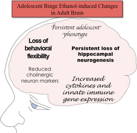
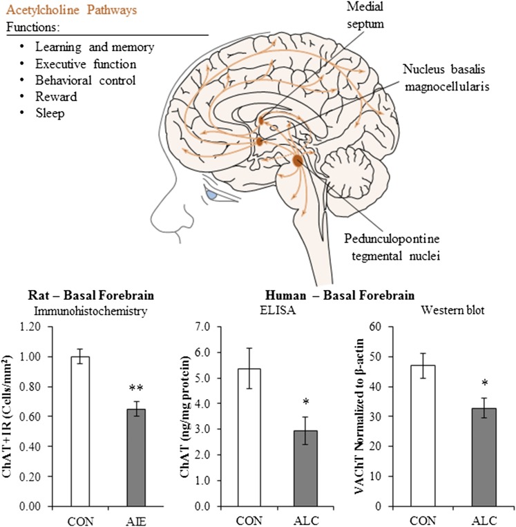
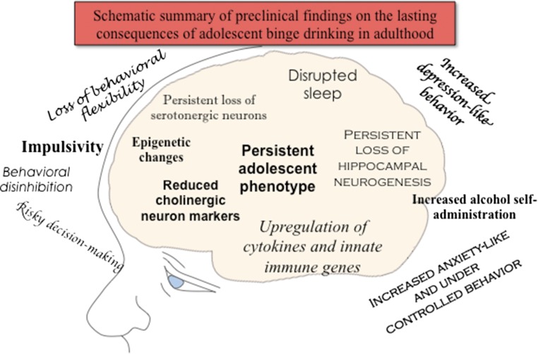
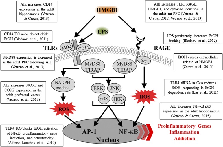
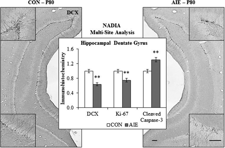
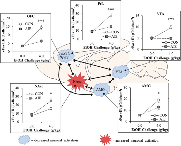
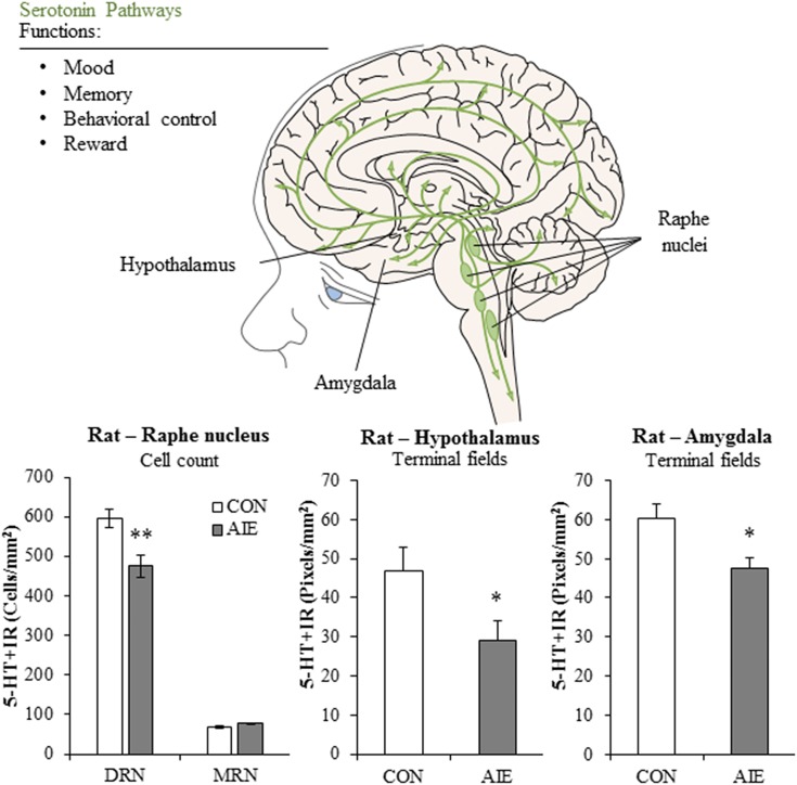
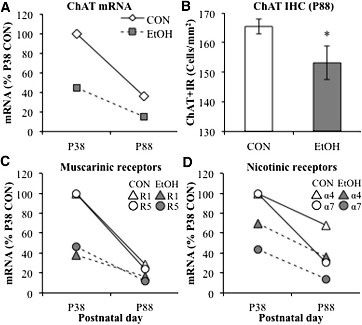
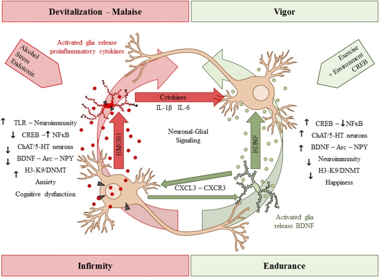
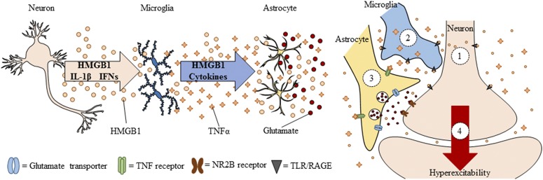
Adolescence is a developmental period when physical and cognitive abilities are optimized, when social skills are consolidated, and when sexuality, adolescent behaviors, and frontal cortical functions mature to adult levels. Adolescents also have unique responses to alcohol compared with adults, being less sensitive to ethanol sedative-motor responses that most likely contribute to binge drinking and blackouts. Population studies find that an early age of drinking onset correlates with increased lifetime risks for the development of alcohol dependence, violence, and injuries. Brain synapses, myelination, and neural circuits mature in adolescence to adult levels in parallel with increased reflection on the consequence of actions and reduced impulsivity and thrill seeking. Alcohol binge drinking could alter human development, but variations in genetics, peer groups, family structure, early life experiences, and the emergence of psychopathology in humans confound studies. As adolescence is common to mammalian species, preclinical models of binge drinking provide insight into the direct impact of alcohol on adolescent development. This review relates human findings to basic science studies, particularly the preclinical studies of the Neurobiology of Adolescent Drinking in Adulthood (NADIA) Consortium. These studies focus on persistent adult changes in neurobiology and behavior following adolescent intermittent ethanol (AIE), a model of underage drinking. NADIA studies and others find that AIE results in the following: increases in adult alcohol drinking, disinhibition, and social anxiety; altered adult synapses, cognition, and sleep; reduced adult neurogenesis, cholinergic, and serotonergic neurons; and increased neuroimmune gene expression and epigenetic modifiers of gene expression. Many of these effects are specific to adolescents and not found in parallel adult studies. AIE can cause a persistence of adolescent-like synaptic physiology, behavior, and sensitivity to alcohol into adulthood. Together, these findings support the hypothesis that adolescent binge drinking leads to long-lasting changes in the adult brain that increase risks of adult psychopathology, particularly for alcohol dependence.


Figures

None
Graphical abstract
Fig. 1.
Fig. 1.
AIE alters adult brain regional responses to an alcohol challenge in adulthood. Adult rats previously exposed to AIE exhibit altered neuronal responses to an ethanol challenge in adulthood as indexed by expression of the immediate early gene cFos, an indirect marker of neuronal activity. Comparison of cFos immunoreactivity (+IR) in adult Wistar rats that received an ethanol challenge (4.0 g/kg, i.g.) in adulthood (P80) revealed that prior AIE exposure (5.0 g/kg, i.g., 2 days on/2 days off from P25 to P55) significantly reduced cFos + IR in the orbitofrontal cortex (OFC; ↓57%), prelimbic cortex (PrL; ↓48%), ventral tegmental area (VTA; ↓50%), and basolateral amygdala (AMG; ↓33%), relative to ethanol-challenged control (CON) subjects. In contrast, previous AIE exposure increased neuronal activity in response to ethanol challenge in the nucleus accumbens core (NAcc; ↑43%) relative to CON subjects. These studies reveal that adolescent binge ethanol exposure causes long-lasting reductions in frontal cortical reactivity in areas involved in executive function and increased activation in reward circuitry in response to ethanol challenge in adulthood, indicative of an enduring alteration in the adult brain response to ethanol. Data are presented as mean ± S.E.M. *p < 0.05, ***p < 0.001, relative to CON. This figure is adapted from (Liu and Crews, 2015).
Fig. 2.
Fig. 2.
Spreading proinflammatory signals across neurons and glia contributes to innate immune gene induction and hyperexcitability following AIE exposure. Left: Alcohol, glutamate, and other inflammagens cause the nuclear release of HMGB1 from neurons that cause microglia to become hyper-ramified, resulting in further release of HMGB1 and other proinflammatory signals. As a consequence, astrocytes reduce glutamate reuptake (Zou and Crews, 2005), thereby increasing extracellular glutamate levels that induce neuronal excitability, and leading to further release of HMGB1 in a positive feedback cycle. Right: Simplified schematic depicting innate immune induction of hyperexcitability. (1) Ethanol administration leads to neuronal release of HMGB1 into the extracellular space. (2) Extracellular HMGB1 binds to TLRs on microglia and RAGE, leading to the release of TNF-α and other innate immune signals. (3) TNF-α binds to TNF receptors on astrocytes, leading to glutamate sensitivity and reduced reuptake through glutamate transporters. (4) Increased glutamate in the synapse activates N-methyl D-aspartate receptor subtype 2B (NR2B), culminating in hyperexcitability. Neuronal hyperexcitability can contribute to alterations in neuronal connectivity as well as causing excitotoxicity. Figure adapted from (Crews et al., 2011).
Fig. 3.
Fig. 3.
Innate immune-signaling cascades and evidence for upregulation in brain following AIE exposure. A simplified schematic of the TLR and RAGE signaling cascades. Stimulation of TLRs and RAGE with their endogenous agonist HMGB1 and other inflammagens [e.g., lipopolysaccharide (LPS)] leads to the generation of proinflammatory oxidases and reactive oxygen species (ROS) and downstream activation of NF-κB. Nuclear translocation of NF-κB leads to the secretion of proinflammatory gene expression, innate immune gene induction, cell death, and addiction-like behaviors. AP-1, activator protein-1; CD14, cluster of differentiation 14; ERK, extracellular signal-regulated kinase; IKK, inhibitor of nuclear factor κ-B; JNK, c-Jun N-terminal kinases; MyD88, myeloid differentiation primary response gene 88; Src, proto-oncogene tyrosine-protein kinase; TIRAP, Toll/interleukin-1 receptor domain-containing adaptor protein.
Fig. 4.
Fig. 4.
Alcohol disrupts the basal forebrain cholinergic system in rats and humans. Top: Simplified schematic of the cholinergic system of the brain. Animal studies have implicated the cholinergic system as important in a host of functions, including cognition and executive function, behavioral control, reward processes, and sleep. AIE exposure causes a reduction of ChAT + IR neurons, which synthesize acetylcholine, throughout cholinergic nuclei of the brain (Vetreno et al., 2014); this loss might contribute to persistent cognitive and emotive dysfunction in adulthood. Bottom left: Multi-site analysis of data from the NADIA Consortium reveals that AIE exposure causes a 35% reduction of ChAT + IR neurons in the adult basal forebrain. Bottom right: ChAT protein expression is reduced by 51% in the postmortem human alcoholic basal forebrain, relative to moderate drinking controls. Furthermore, protein expression of vesicular acetylcholine transporter, which packages acetylcholine into synaptic vesicles, is reduced by 30% in the postmortem human alcoholic basal forebrain, relative to moderate drinking controls. Multisite analysis was calculated by Dr. Margaret Burchinal from five independent data sets (unpublished data from Crews’ laboratory; Ehlers et al., 2011; Boutros et al., 2014; Vetreno et al., 2014). Data are presented as a mean ± S.E.M. *P < 0.05, **P < 0.01, relative to CON.
Fig. 5.
Fig. 5.
Adolescent binge ethanol exposure reduces cholinergic marker expression in the whole mouse brain. Adolescent mice received either water (CON) or ethanol (EtOH; 5.0 g/kg, i.g.) once per day for 10 consecutive days from P28 to P37. Alcohol treatment ended on P37. Shown in (A–D) are expression levels (mRNA) 1 day after the last AIE dose of ethanol and 50 days after the last dose in AIE animals (Coleman et al., 2011). Changes in controls represent maturation from adolescence to adulthood. (A) mRNA levels of ChAT, the acetylcholine-synthesizing enzyme, were reduced by 55% in adolescent mouse whole brain samples 24 hours after the conclusion of EtOH exposure (P38) as well as by 58% in adulthood (P88) compared with CON. (B) Comparison of ChAT immunohistochemistry revealed an 8% reduction of ChAT–immunopositive cells in the posterior basal forebrain of EtOH-treated adult mice, relative to CON. (C) mRNA expression of muscarinic acetylcholine receptor subtypes R1 and R5 was reduced 24 hours after the conclusion of EtOH exposure by 62% and 54%, respectively, which persisted into adulthood (R1: ↓45%; R5: ↓50%). (D) Similarly, mRNA expression of nicotinic acetylcholine receptor subtypes α4 and α7 was reduced by 30% and 56% at the conclusion of EtOH exposure, respectively, that persisted into adulthood (α4: ↓48%; α7: ↓54%). These data reveal that adolescent binge ethanol exposure leads to long-term alterations in the cholinergic system that might contribute to cognitive dysfunction in adulthood. Data are presented as mean ± S.E.M. *p < 0.05, relative to CON, and are adapted from Coleman et al. (2011).
Fig. 6.
Fig. 6.
AIE reduces 5-HT neurons in dorsal raphe nucleus, leading to alterations in terminal field projection densities. Top: Simplified schematic of the serotonergic system of the brain. The serotonergic system innervates the entire brain and plays a neuromodulatory role in mood regulation, memory, behavioral control, and reward processes. Dysregulation of this system has been identified as an etiological factor underlying several psychiatric disorders, including depression, impulsivity, and alcohol dependence (Michelsen et al., 2007; Donaldson et al., 2013; Muller and Homberg, 2015; Nautiyal et al., 2015). Bottom: Adult rats with a history of AIE exposure (5.0 g/kg, i.g., 2 days on/2 days off from P25 to P55) exhibit a 20% reduction of 5-HT–immunoreactive neurons in the adult (P80) dorsal raphe nucleus (DRN), whereas those in the median raphe nucleus (MRN) are spared. Quantification of serotonergic terminal field densities revealed reductions of 20% and 38% from control (CON) in both the amygdala and hypothalamus, respectively. The loss of 5-HT + IR neurons might contribute to AIE-induced cognitive and emotive dysfunction as well as increased alcohol self-administration in adulthood. Data are presented as a mean ± S.E.M. *p < 0.05, **p < 0.01, relative to CON.
Fig. 7.
Fig. 7.
Hippocampal neurogenesis is highly vulnerable to the neurodegenerative effects of adolescent binge ethanol exposure. Representative photomicrographs of doublecortin (DCX) immunoreactivity, a neuroprogenitor microtubule-associated protein expressed by immature neurons, in the adult dorsal and ventral hippocampal dentate gyrus following control (CON) and AIE (5.0 g.kg, i.g., 2 days on/2 days off from P25 to P55). Scale bars, 100 μm. The middle bar graph depicts multisite analyses of data from the NADIA Consortium. In adulthood (e.g., P80) following AIE, DCX + immunoreactive (+IR) cells are reduced by 36%, which is accompanied by a concomitant 25% reduction in Ki-67 + IR, which is an endogenous marker of progenitor cells (Vetreno and Crews, 2015). In parallel, cleaved caspase-3, which is a marker of cell death, was increased by 31% in the adult hippocampal dentate gyrus of AIE-exposed animals. These data reveal that AIE leads to long-term reductions of hippocampal neurogenesis that could contribute to cognitive deficits in adulthood. Multisite analysis was calculated by Dr. Margaret Burchinal from four independent data sets (Ehlers et al., 2013b; Broadwater et al., 2014b; Swartzwelder et al., 2015; Vetreno et al., 2015). Data are presented as a mean ± S.E.M. **p < 0.01, relative to CON.
Fig. 8.
Fig. 8.
Duality of stressors and enrichment on neuronal–glial signaling. Neuronal–glial communication lies along a continuum with devitalization-malaise on one end and vigor-endurance on the other end. Stimuli such as alcohol, stress, and endotoxins activate neurons and glia to release proinflammatory signals. As a consequence of innate immunity-system activation, neurotrophic support is reduced, leading to neurotransmitter system disruption as well as increased anxiety and cognitive dysfunction. In contrast, stimuli such as exercise and enrichment induce neuronal–glial communication to increase neurotrophin expression, such as BDNF, that blunts the innate immune system, creating an environment that facilitates neurotransmitter system survival and increases mood and cognition.
Fig. 9.
Fig. 9.
Schematic summary of preclinical findings on the lasting consequences of adolescent binge drinking in adulthood. Shown are the summarized, consensus findings of persistent adult pathologies from across the NADIA Consortium as well as other investigators, as described in this review. Multiple studies find that AIE leads to adult impairments in cognitive and executive functioning, increases depressive- and anxiety-like behaviors as well impulsivity, and increases alcohol self-administration in adulthood. Potential mechanisms of AIE disruption of maturation include increased expression of brain cytokines and other innate immune genes, loss of cholinergic and serotonergic neurons, and epigenetic changes that continue into adulthood following AIE treatment. Many of these changes are not found after similar treatment of adults, as outlined in the review. Additional studies are needed to clearly link mechanisms to adult behavioral pathologies; however, the persistent changes found are consistent with human studies, indicating that adolescent binge drinking is associated with lifelong risks for alcohol-related problems.

References

    1. Abrous DN, Koehl M, Le Moal M. (2005) Adult neurogenesis: from precursors to network and physiology. Physiol Rev 85:523–569. - PubMed
    1. Acheson SK, Bearison C, Risher ML, Abdelwahab SH, Wilson WA, Swartzwelder HS. (2013) Effects of acute or chronic ethanol exposure during adolescence on behavioral inhibition and efficiency in a modified water maze task. PLoS One 8:e77768. - PMC - PubMed
    1. Acheson SK, Stein RM, Swartzwelder HS. (1998) Impairment of semantic and figural memory by acute ethanol: age-dependent effects. Alcohol Clin Exp Res 22:1437–1442. - PubMed
    1. Agrawal A, Sartor CE, Lynskey MT, Grant JD, Pergadia ML, Grucza R, Bucholz KK, Nelson EC, Madden PA, Martin NG, et al. (2009) Evidence for an interaction between age at first drink and genetic influences on DSM-IV alcohol dependence symptoms. Alcohol Clin Exp Res 33:2047–2056. - PMC - PubMed
    1. Alaux-Cantin S, Warnault V, Legastelois R, Botia B, Pierrefiche O, Vilpoux C, Naassila M. (2013) Alcohol intoxications during adolescence increase motivation for alcohol in adult rats and induce neuroadaptations in the nucleus accumbens. Neuropharmacology 67:521–531. - PubMed