. 2016 Sep 27;7(5):e00796-16.
doi: 10.1128/mBio.00796-16.

Exposing the Three-Dimensional Biogeography and Metabolic States of Pathogens in Cystic Fibrosis Sputum via Hydrogel Embedding, Clearing, and rRNA Labeling

Affiliations

Exposing the Three-Dimensional Biogeography and Metabolic States of Pathogens in Cystic Fibrosis Sputum via Hydrogel Embedding, Clearing, and rRNA Labeling

William H DePas et al. mBio. .

Abstract

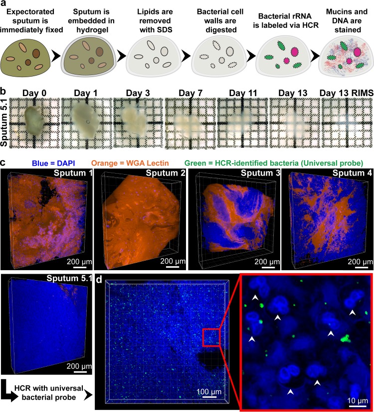
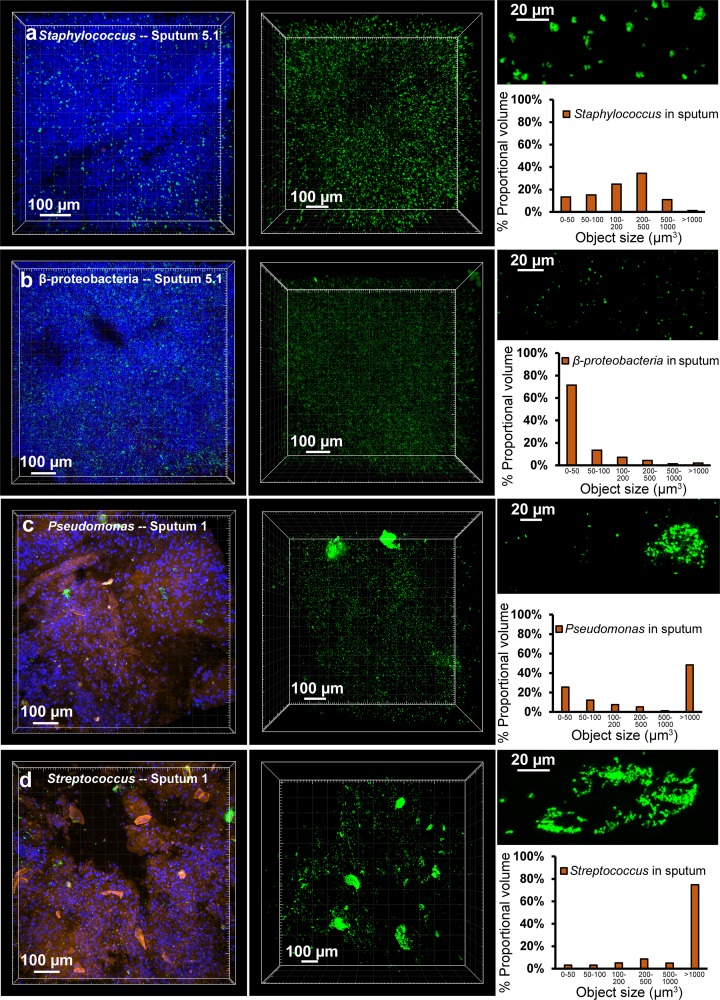
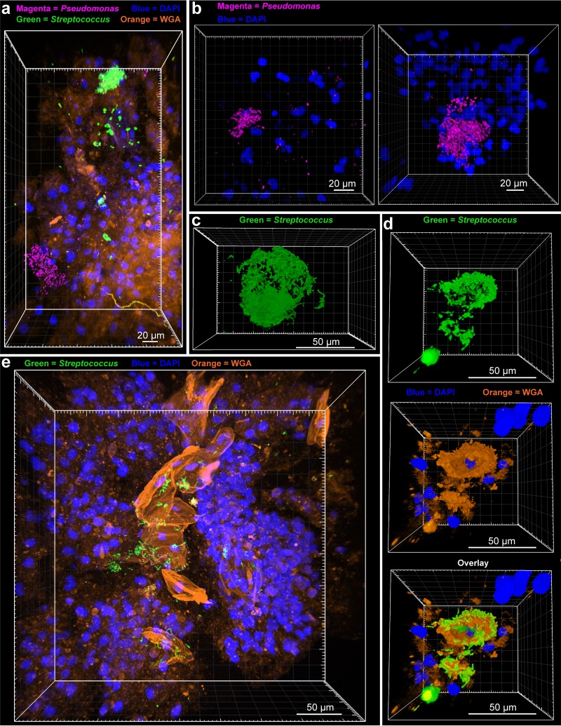
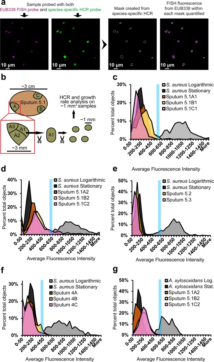
Physiological resistance to antibiotics confounds the treatment of many chronic bacterial infections, motivating researchers to identify novel therapeutic approaches. To do this effectively, an understanding of how microbes survive in vivo is needed. Though much can be inferred from bulk approaches to characterizing complex environments, essential information can be lost if spatial organization is not preserved. Here, we introduce a tissue-clearing technique, termed MiPACT, designed to retain and visualize bacteria with associated proteins and nucleic acids in situ on various spatial scales. By coupling MiPACT with hybridization chain reaction (HCR) to detect rRNA in sputum samples from cystic fibrosis (CF) patients, we demonstrate its ability to survey thousands of bacteria (or bacterial aggregates) over millimeter scales and quantify aggregation of individual species in polymicrobial communities. By analyzing aggregation patterns of four prominent CF pathogens, Staphylococcus aureus, Pseudomonas aeruginosa, Streptococcus sp., and Achromobacter xylosoxidans, we demonstrate a spectrum of aggregation states: from mostly single cells (A. xylosoxidans), to medium-sized clusters (S. aureus), to a mixture of single cells and large aggregates (P. aeruginosa and Streptococcus sp.). Furthermore, MiPACT-HCR revealed an intimate interaction between Streptococcus sp. and specific host cells. Lastly, by comparing standard rRNA fluorescence in situ hybridization signals to those from HCR, we found that different populations of S. aureus and A. xylosoxidans grow slowly overall yet exhibit growth rate heterogeneity over hundreds of microns. These results demonstrate the utility of MiPACT-HCR to directly capture the spatial organization and metabolic activity of bacteria in complex systems, such as human sputum.

Importance: The advent of metagenomic and metatranscriptomic analyses has improved our understanding of microbial communities by empowering us to identify bacteria, calculate their abundance, and profile gene expression patterns in complex environments. We are still technologically limited, however, in regards to the many questions that bulk measurements cannot answer, specifically in assessing the spatial organization of microbe-microbe and microbe-host interactions. Here, we demonstrate the power of an enhanced optical clearing method, MiPACT, to survey important aspects of bacterial physiology (aggregation, host interactions, and growth rate), in situ, with preserved spatial information when coupled to rRNA detection by HCR. Our application of MiPACT-HCR to cystic fibrosis patient sputum revealed species-specific aggregation patterns, yet slow growth characterized the vast majority of bacterial cells regardless of their cell type. More broadly, MiPACT, coupled with fluorescent labeling, promises to advance the direct study of microbial communities in diverse environments, including microbial habitats within mammalian systems.


Figures

FIG 1
FIG 1
MiPACT-HCR allows visualization of bacteria in cleared sputum samples. (a) Cartoon depicting the process of embedding and clearing sputum for visualization of bacteria via HCR. (b) The clearing process for sputum sample 5.1. Each grid square represents 1 mm2. (c) Blend projections of five sputum samples after staining with DAPI (blue) and WGA (orange) from Z-stacks acquired with a 10× objective. (d) HCR with a universal bacterial probe (green; EUB338 with B1 hairpins conjugated to AlexaFluor 647) in sputum sample 5.1. The middle panel is a maximum intensity projection acquired with a 10× objective, and the right panel is a single-plane image acquired with a 25× objective. White arrows indicate PMNs.
FIG 2
FIG 2
Aggregation patterns vary between species. (a) HCR with a Staphylococcus-specific probe in sputum sample 5.1 (green). The first panel is a maximum intensity projection of a Z-stack after HCR and staining with DAPI (blue) and WGA (orange), acquired with a 10× objective. The second panel is a maximum intensity projection of a separate Z-stack acquired with a 10× objective while only collecting HCR signal (Staphylococcus-specific probe mix with B4 amplifier and AlexaFluor 488-conjugated B4 hairpins) (7,910 objects analyzed). Each object identified in the second panel’s Z-stack was binned according to proportional object volume (each object’s fluorescent volume relative to the total fluorescent volume for that Z-stack; shown in the graph on the right). The top right panel is a maximum intensity projection of a Z-stack acquired with a 25× objective, highlighting a representative region from the same sputum sample. (b to d) The same analysis was applied to sputum 5.1 using a Betaproteobacteria-specific probe with B4 amplifier and AlexaFluor 488-conjugated B4 hairpin (21,255 objects analyzed) (b), to sputum 1 with a Pseudomonas-specific probe mixture with B4 amplifier and AlexaFluor 647-conjugated B4 hairpins (9,520 objects analyzed) (c), or a Streptococcus-specific probe mixture with AlexaFluor 488-conjugated B4 hairpins (4,603 objects analyzed) (d).
FIG 3
FIG 3
Pseudomonas and Streptococcus biofilm structure. (a) A maximum intensity projection was generated after HCR was performed on sputum 1 with a Pseudomonas-specific probe mixture (with B1 hairpins conjugated to AlexaFluor 647) and a Streptococcus-specific probe mixture (with B4 hairpins conjugated to AlexaFluor 488). (b) Maximum intensity projections showing Pseudomonas aggregates from sputum 1 after HCR with a Pseudomonas probe mixture and B4 hairpins conjugated to AlexaFluor 488 and DAPI staining. (c) Blend projection of a Streptococcus biofilm from sputum 1 (HCR with Streptococcus probe mixture with B4 hairpins conjugated to AlexaFluor 488). (d) Blend projections showing, stepwise, a Streptococcus aggregate (top; green), DAPI (blue), and WGA (orange) staining of host cells (middle), and an overlay of the two showing the arrangement of the Streptococcus biofilm around WGA-stained host cells (bottom). (e) Maximum intensity projection of HCR-identified Streptococcus (green), DAPI (blue), and WGA (orange) staining in sputum 1.
FIG 4
FIG 4
Growth rate estimates of CF pathogens in situ. (a) Diagram showing the process of estimating growth rates in situ. Samples were first stained with a species-specific B4 amplifier HCR probe, using B4 hairpins conjugated to AlexaFluor 488. Samples were then stained with the universal bacterial FISH probe EUB338, conjugated to two Cy5 fluorophores. Masks were made based upon HCR signal, and fluorescence intensity from FISH was quantified within each mask. (b) The basic sputum sampling technique. (c) For this and subsequent panels, Z-stacks of cultured cells and sputum samples were acquired with a 25× objective in parallel. The average fluorescence intensity of the FISH channel of each object is plotted on the x axis as a histogram. The blue line denotes the bin above which 90% of the logarithmic objects fell (for each particular experimental set). Growth rate analysis was performed on three distinct regions of sputum sample 5.1: 5.1A1 (409 objects analyzed), 5.1B1 (697 objects analyzed), and 5.1C1 (575 objects analyzed) (c), and on 5.1A2 (418 objects analyzed) 5.1B2 (1,087 objects analyzed), and 5.1C2 (419 objects analyzed) (d). (e) Analysis of temporal samples 5.2 (520 objects analyzed) and 5.3 (893 objects analyzed). (f) Analysis of three distinct regions of sputum 4: 4A (1,067 objects analyzed), 4B (73 objects analyzed), and 4C (599 objects analyzed). (g) Analysis of Betaproteobacteria from samples 5.1A2 (1,919 objects analyzed), 5.1B2 (2,351 objects analyzed), and 5.1C2 (1,523 objects analyzed).

Similar articles

Cited by

References

    1. Hall-Stoodley L, Stoodley P. 2009. Evolving concepts in biofilm infections. Cell Microbiol 11:1034–1043. doi:10.1111/j.1462-5822.2009.01323.x. - DOI - PubMed
    1. Cho I, Blaser MJ. 2012. The human microbiome: at the interface of health and disease. Nat Rev Genet 13:260–270. doi:10.1038/nrg3182. - DOI - PMC - PubMed
    1. Parsek MR, Singh PK. 2003. Bacterial biofilms: an emerging link to disease pathogenesis. Annu Rev Microbiol 57:677–701. doi:10.1146/annurev.micro.57.030502.090720. - DOI - PubMed
    1. Stacy A, McNally L, Darch SE, Brown SP, Whiteley M. 2016. The biogeography of polymicrobial infection. Nat Rev Microbiol 14:93–105. doi:10.1038/nrmicro.2015.8. - DOI - PMC - PubMed
    1. Jorth P, Staudinger BJ, Wu X, Hisert KB, Hayden H, Garudathri J, Harding CL, Radey MC, Rezayat A, Bautista G, Berrington WR, Goddard AF, Zheng C, Angermeyer A, Brittnacher MJ, Kitzman J, Shendure J, Fligner CL, Mittler J, Aitken ML, Manoil C, Bruce JE, Yahr TL, Singh PK. 2015. Regional isolation drives bacterial diversification within cystic fibrosis lungs. Cell Host Microbe 18:307–319. doi:10.1016/j.chom.2015.07.006. - DOI - PMC - PubMed