

Static and dynamic functional connectivity in patients with chronic fatigue syndrome: use of arterial spin labelling fMRI
- PMID: 27678090
- PMCID: PMC5373941
- DOI: 10.1111/cpf.12393
Static and dynamic functional connectivity in patients with chronic fatigue syndrome: use of arterial spin labelling fMRI
Abstract
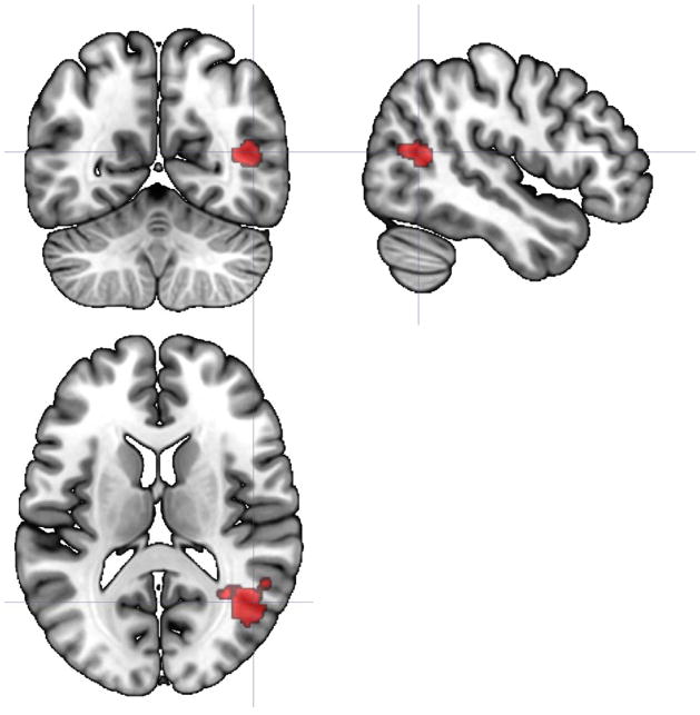
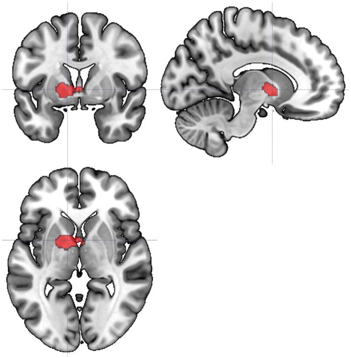
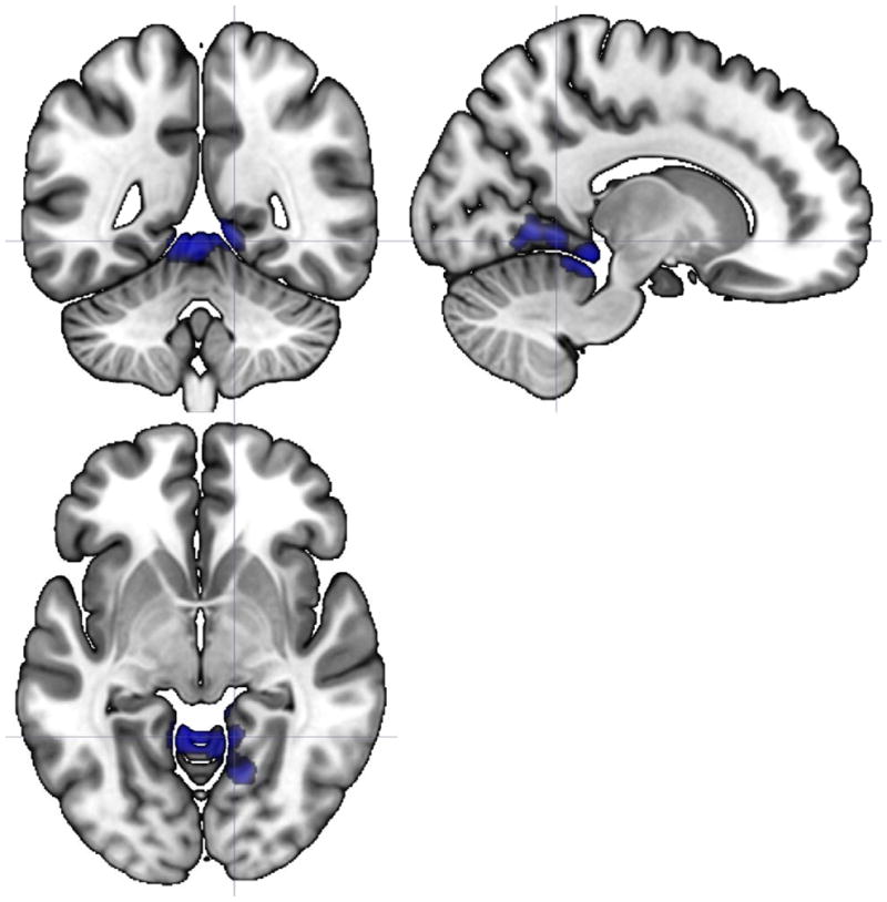
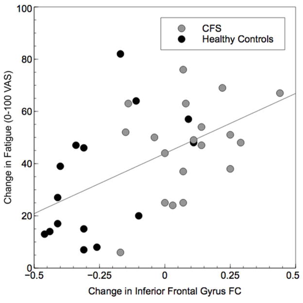
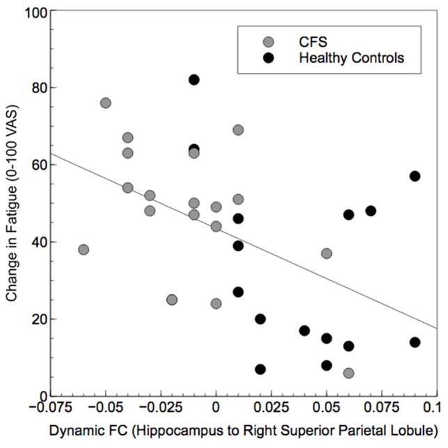
Studies using arterial spin labelling (ASL) have shown that individuals with chronic fatigue syndrome (CFS) have decreased regional cerebral blood flow, which may be associated with changes in functional neural networks. Indeed, recent studies indicate disruptions in functional connectivity (FC) at rest in chronically fatigued patients including perturbations in static FC (sFC), that is average FC at rest between several brain regions subserving neurocognitive, motor and affect-related networks. Whereas sFC often provides information of functional network reorganization in chronic illnesses, investigations of temporal changes in functional connectivity between multiple brain areas may shed light on the dynamic characteristics of brain network activation associated with such maladies. We used ASL fMRI in 19 patients with CFS and 15 healthy controls (HC) to examine both static and dynamic changes in FC among several a priori selected brain regions during a fatiguing cognitive task. HC showed greater increases than CFS in static FC (sFC) between insula and temporo-occipital structures and between precuneus and thalamus/striatum. Furthermore, inferior frontal gyrus connectivity to cerebellum, occipital and temporal structures declined in HC but increased in CFS. Patients also showed lower dynamic FC (dFC) between hippocampus and right superior parietal lobule. Both sFC and dFC correlated with task-related fatigue increases. These data provide the first evidence that perturbations in static and dynamic FC may underlie chronically fatigued patients' report of task-induced fatigue. Further research will determine whether such changes in sFC and dFC are also characteristic for other fatigued individuals, including patients with chronic pain, cancer and multiple sclerosis.
Keywords: MRI; arterial spin labelling; chronic fatigue; dynamic functional connectivity.
© 2016 Scandinavian Society of Clinical Physiology and Nuclear Medicine. Published by John Wiley & Sons Ltd.
Conflict of interest statement
Disclosure: None of the authors have any financial or other relationships that might result in a conflict of interest.
Figures
References
-
- Behrmann M, Geng JJ, Shomstein S. Parietal cortex and attention. Curr Opin Neurobiol. 2004;14:212–217. - PubMed
-
- Benjamini Y, Hochberg Y. Controlling the False Discovery Rate - a Practical and Powerful Approach to Multiple Testing. Journal of the Royal Statistical Society Series B-Methodological. 1995;57:289–300.
MeSH terms
Substances
Grants and funding
LinkOut - more resources
Full Text Sources
Other Literature Sources
Medical
