. 2016 Sep 28:6:34251.
doi: 10.1038/srep34251.

13C and 15N natural isotope abundance reflects breast cancer cell metabolism

Affiliations

13C and 15N natural isotope abundance reflects breast cancer cell metabolism

Illa Tea et al. Sci Rep. .

Abstract

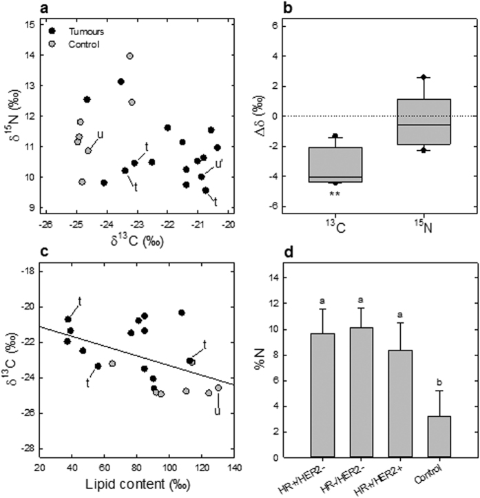
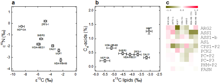
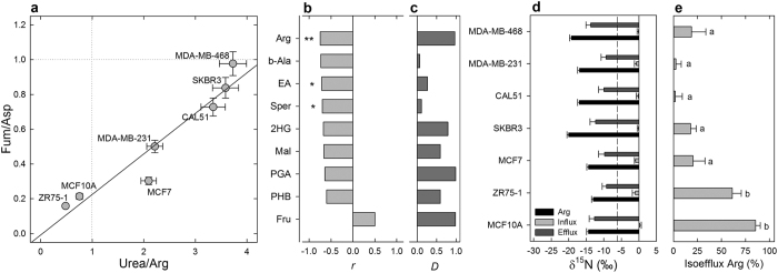
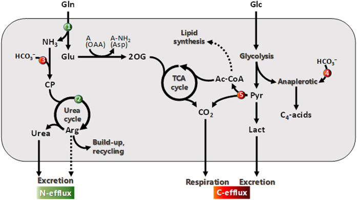
Breast cancer is the most common cancer in women worldwide. Despite the information provided by anatomopathological assessment and molecular markers (such as receptor expression ER, PR, HER2), breast cancer therapies and prognostics depend on the metabolic properties of tumor cells. However, metabolomics have not provided a robust and congruent biomarker yet, likely because individual metabolite contents are insufficient to encapsulate all of the alterations in metabolic fluxes. Here, we took advantage of natural 13C and 15N isotope abundance to show there are isotopic differences between healthy and cancer biopsy tissues or between healthy and malignant cultured cell lines. Isotope mass balance further suggests that these differences are mostly related to lipid metabolism, anaplerosis and urea cycle, three pathways known to be impacted in malignant cells. Our results demonstrate that the isotope signature is a good descriptor of metabolism since it integrates modifications in C partitioning and N excretion altogether. Our present study is thus a starting point to possible clinical applications such as patient screening and biopsy characterization in every cancer that is associated with metabolic changes.


Figures

Figure 1
Figure 1. Breast cancer biopsies are naturally 13C-enriched and nitrogen-rich.
(a) The natural abundance in 13C (δ13C, vs. V-PDB) and 15N (δ15N, vs. air N2) differentiates control from tumor patient tissue samples (n = 23). One sample pair of healthy (adjacent) and cancerous tissues not characterized for receptors (u and u’, respectively) and three triple negative tumor samples (t) are also shown. (b) Taken as a whole, the change in the 13C-abundance (Δδ) in cancerous tissues in tumor/adjacent tissue pairs was significant (P < 0.01, n = 5). (c) 13C-abundance (Δδ) was inversely correlated with the total lipid content (r2 = 0.48, P < 0.03). (d) Regardless of receptor expression, cancerous tissues had a higher elemental content in N (P < 0.05). In (a,c), each datum is the average of 3 sub-samples. In (d), lower case letters stand for statistical classes, P < 0.05.
Figure 2
Figure 2. Cancerous cell lines are naturally 13C-enriched and 15N-depleted.
When compared to source C and N used for growth, the natural isotopic enrichment (ε13C and ε15N) differentiates the control immortalized line (MCF10A) and the breast tumor ascites-derived line (ZR75-1) from other adenocarcinoma lines (a). The isotope enrichment in lipids parallels that in total organic matter, and cancerous lines contain more (13C-enriched) C4-acids (b). Despite some variations, transcript quantitation of key enzymes shows a generally lower expression in cancer cells compared to MCF10A cells of enzymes of the urea cycle, lipid synthesis or the anaplerotic pathway (gene abbreviations in Supplementary Table S5), suggesting that metabolite excretion in addition to biosynthesis is involved in the isotopic differences (c).
Figure 3
Figure 3. Cancerous cell lines excrete less 15N-depleted arginine (Arg).
The role of Arg metabolism (urea cycle) is further demonstrated by the differences in ratios of endometabolites since Arg appears to be more retained relative to urea in cancerous cell lines (a). The extracellular-to-intracellular relative difference (D) in Arg was found to (i) be the best D value that anti-correlates to δ15N (b) and (ii) have the largest average D value across cell lines (c). In all cases, the δ15N value of the influx (substrate consumption) was close to 0‰ while the efflux (excreted N) was 15N-depleted and Arg was even more 15N-depleted in cancerous cell lines by up to ≈7‰ (d). Therefore, the relative isotopic efflux represented by Arg excretion (i.e., the quotient of Arg isoefflux to total isoefflux) was large in ZR75-1 and MCF10A and low in other cell lines (e). Asterisks: significant differences between cell lines (*P < 0.05; **P < 0.01); dashed line: δ15N of source Gln (d); lower case letters stand for statistical classes (P < 0.05) (e). EA, ethanolamine, 2HG, 2-hydroxyglutarate; PGA, phosphoglycerate; PHB, parahydroxybenzoate.
Figure 4
Figure 4. Major metabolic pathways are responsible for changes in the natural 13C and 15N abundance in cancerous cultured cells.
Glutamine (Gln) is a major N and C source from which N is removed via hydrolysis (glutaminase ❶) and the urea cycle (❷). These reactions are both fractionating against 15N thereby yielding 15N-depleted urea and arginine (Arg). Therefore, build-up and recycling rather than excretion of Arg (dotted arrow) tend to deplete cancer cells of 15N. Cells are also 13C-enriched due to the fixation of bicarbonate by carbamoyl-phosphate (CP) synthesis (to feed the urea cycle ❸) and the anaplerotic pathway (❹), as well as a lower 13C content in non-structural lipids (dotted arrow). Lipids that are 13C-depleted come from the natural 13C-depletion in acetyl-CoA (Ac-CoA) (inherited from naturally 13C-depleted C-atom positions in glucose) and the isotope effect of pyruvate dehydrogenase (❺). Amine acceptors are denoted as ‘A’ and oxaloacetate (OAA) converted to aspartate (Asp) is provided as an example. 2OG, 2-oxoglutarate; Pyr, pyruvate; Lact, lactate. The potential contribution of respiration (CO2 loss) and lactate excretion to the natural 13C-abundance is described in Supplementary Table S4.

References

    1. Graham D. Y. et al.. Campylobacter pylori detected noninvasively by the 13C-urea breath test. The Lancet 1, 1174–1177 (1987). - PubMed
    1. Schmidt H.-L., Robins R. J. & Werner R. A. Multi-factorial in vivo stable isotope fractionation: causes, correlations, consequences and applications. Isotopes Environ Health Stud 51, 155–199 (2015). - PubMed
    1. Bartstow T. J., Cooper D. M., Epstein S. & Wasserman K. Changes in 13CO2/12CO2 consequent to exercise and hypoxia. J Appl Physiol 66, 936–942 (1989). - PubMed
    1. Perkins S. E. & Speakman J. R. Measuring natural abundance of 13C in respired CO2: variability and implications for non-invasive dietary analysis. Funct Ecol 15, 791–797 (2001).
    1. Balter V. et al.. Natural variations of copper and sulfur stable isotopes in blood of hepatocellular carcinoma patients. Proc Natl Acad Sci USA 4, 982–985 (2015). - PMC - PubMed