. 2016 Sep 27;17(1):198.
doi: 10.1186/s13059-016-1054-5.

Time-resolved dual RNA-seq reveals extensive rewiring of lung epithelial and pneumococcal transcriptomes during early infection

Affiliations

Time-resolved dual RNA-seq reveals extensive rewiring of lung epithelial and pneumococcal transcriptomes during early infection

Rieza Aprianto et al. Genome Biol. .

Abstract

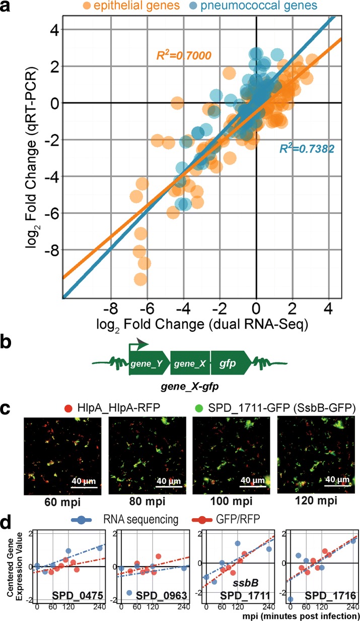
Background: Streptococcus pneumoniae, the pneumococcus, is the main etiological agent of pneumonia. Pneumococcal infection is initiated by bacterial adherence to lung epithelial cells. The exact transcriptional changes occurring in both host and microbe during infection are unknown. Here, we developed a time-resolved infection model of human lung alveolar epithelial cells by S. pneumoniae and assess the resulting transcriptome changes in both organisms simultaneously by using dual RNA-seq.

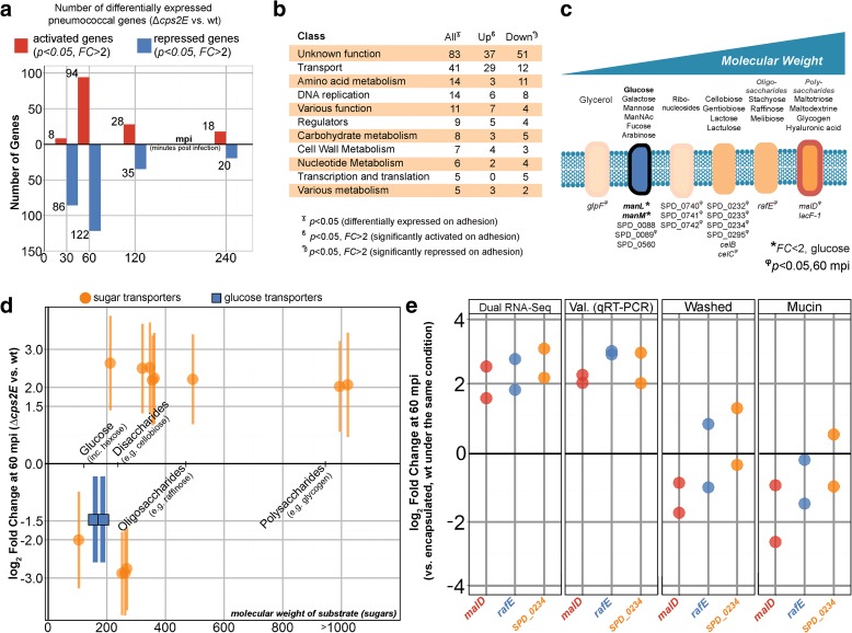
Results: Functional analysis of the time-resolved dual RNA-seq data identifies several features of pneumococcal infection. For instance, we show that the glutathione-dependent reactive oxygen detoxification pathway in epithelial cells is activated by reactive oxygen species produced by S. pneumoniae. Addition of the antioxidant resveratrol during infection abates this response. At the same time, pneumococci activate the competence regulon during co-incubation with lung epithelial cells. By comparing transcriptional changes between wild-type encapsulated and mutant unencapsulated pneumococci, we demonstrate that adherent pneumococci, but not free-floating bacteria, repress innate immune responses in epithelial cells including expression of the chemokine IL-8 and the production of antimicrobial peptides. We also show that pneumococci activate several sugar transporters in response to adherence to epithelial cells and demonstrate that this activation depends on host-derived mucins.

Conclusions: We provide a dual-transcriptomics overview of early pneumococcal infection in a time-resolved manner, providing new insights into host-microbe interactions. To allow easy access to the data by the community, a web-based platform was developed ( http://dualrnaseq.molgenrug.nl ). Further database exploration may expand our understanding of epithelial-pneumococcal interaction, leading to novel antimicrobial strategies.

Keywords: Adherence; Competence; Dual RNA-seq; Host-microbe interaction; Lung epithelial cells; Streptococcus pneumoniae; Systems biology; Transcriptomics.


Figures

Fig. 1
Fig. 1
The early infection model. A confluent monolayer of alveolar epithelial cells (A549) was co-incubated with S. pneumoniae strain D39 at an MOI of 10 (ten pneumococci per epithelial cell). a Five infection time points were interrogated: 0, 30, 60, 120, and 240 minutes post-infection (mpi). b Since adherence is a hallmark of infection, we used an unencapsulated S. pneumoniae strain (∆cps2E), which adheres more readily to epithelial cells than its encapsulated parental strain. c At 30 mpi, ∆cps2E (orange bar) showed significantly (p < 0.001) more adherent cells than the wild-type (wt) parental strain (cyan bar). Data are presented as mean ± standard error of the mean. d At 240 mpi, both strains multiplied significantly (p < 0.01) with no significant difference between them. e The setup with the encapsulated strain contains more free-floating than adherent bacteria while ∆cps2E has a higher fraction of adherent bacteria. f After quality control (QC), low-quality reads were trimmed and aligned to a synthetic chimeric genome. Aligned reads were counted and classified as epithelial or pneumococcal counts. We removed three gene fractions and performed clustering and functional enrichment of the working libraries
Fig. 2
Fig. 2
Dual RNA-seq generates high-quality datasets suitable for probing host–pathogen transcriptomes. a On average, there are 70 million reads per library: 42 % of the reads aligned to the human genome and 58 % to the S. pneumoniae D39 genome. b In order to simplify downstream analysis, we excluded three gene fractions: unexpressed genes, i.e., those without counts in any libraries; genes that were differentially expressed at 0 mpi (p < 0.05) between ∆cps2E and wild-type (wt) libraries; and genes with no statistical significance (p > 0.05) and fold changes (FC) < 2 in all comparisons. After exclusion, the epithelial working libraries contained 4337 genes (7 % of all human genes) while the pneumococcal working libraries contained 860 genes (41 % of all pneumococcal genes). c Gene expression in epithelial working libraries was normalized, centered, and clustered. The left panel shows epithelial genes in response to the encapsulated strain while the right panel shows the epithelial response to ∆cps2E S. pneumoniae at different time points. Clear clusters of co-expressed epithelial genes can be observed in the heat map. Blue indicates relatively lower expression while red indicates a higher value. d Pneumococcal expression: the left panel shows the wild-type pneumococcal response to epithelial cells, while the right panel shows the response of the ∆cps2E strain
Fig. 3
Fig. 3
Validation of dual RNA-seq. a We confirmed dual RNA-seq gene expression values by qRT-PCR. The infection study was repeated in duplicates and total RNA was isolated as previously described. Ten human and 19 pneumococcal genes were chosen as validation targets. We plotted fold changes from qRT-PCR against dual RNA-seq fold changes and observed a high degree of correlation for both species (R 2 > 0.7, Pearson). b We also confirmed pneumococcal gene expression at the protein level by quantitative fluorescence microscopy. Four target genes (SPD_0475, SPD_0963, SPD_1711, and SPD_1716) were C-terminally tagged with green fluorescent protein (GFP) at their own locus. GFP fusions were introduced in the ∆cps2E strain expressing red fluorescent protein (RFP) fused to HlpA. c Non-deconvolved image of SPD_1711-GFP up to 120 mpi. While RFP emitted a relatively constant signal, the GFP signal increased. d Dual RNA-seq expression values superimposed on the GFP/RFP ratio. To some extent, transcriptional changes corresponded to protein expression
Fig. 4
Fig. 4
Epithelial glutathione-associated genes are activated in response to pneumococcal ROS. a We clustered epithelial working libraries exposed to wild-type pneumococci and found a cluster of 242 co-expressed genes showing sustained upregulation (p < 0.05, FC > 2) at 60 mpi compared with 30 mpi. Gene ontology (GO) analysis showed that “oxidation reduction” was enriched in 17 genes. b GPX2, encoding glutathione peroxidase-2, is one of the enriched genes. Eight genes are associated with glutathione (GSH), an important antioxidant. The main glutathione-associated processes are biosynthesis of glutathione and detoxification of ROS assisted by ligand and glutathione recycling. c Between 30 and 60 mpi, expression of GCLC increased 2.7 ± 1.1 times and of GCLM 2.3 ± 1.2 times. Expression of GPX2, the main detoxification gene, increased 18.1 ± 1.3 times while that of the gene encoding its ligand, MGST2, increased 3.1 ± 1.2 times. Genes involved in the recycling of glutathione were activated: IDH1, 4.5 ± 1.2; IDH2, 2.3 ± 1.2; PGD, 2.9 ± 1.2; and G6PD, 6.5 ± 1.2. d We validated GPX2, GSR, IDH1, and PGD expression using qRT-PCR. Epithelial incubation with pneumococcal supernatant showed similar upregulation of glutathione-associated genes. Addition of resveratrol (100 μM) into the model diminished the upregulation (FC < 2) altogether
Fig. 5
Fig. 5
S. pneumoniae transcriptional adaptation in response to co-incubation with epithelial cells. We clustered pneumococcal genes based on centered normalized expression and recovered clusters of genes with shared function. a A subset of 20 genes encoding carbohydrate transporters displayed early activation at 30 mpi and returned to basal expression levels at later time points. The unencapsulated strain (∆cps2E, orange box) exhibited a more sustained expression up to 60 mpi than the wild type (wt). b Similarly, 13 genes encoding adherence factors showed the same profile of early activation at 30 mpi and a more sustained expression at 60 mpi in the unencapsulated strain. c Seventeen genes encoding non-carbohydrate transporters were repressed at 30 mpi. d And 45 genes that are part of the competence regulon were activated beginning at 60 mpi until the end of the experiment. e Interestingly, subsets of genes of unknown function showed mixed profiles, including early activation and repression followed by de-repression and late activation
Fig. 6
Fig. 6
Adherent S. pneumoniae repress epithelial innate immune responses. a At 60 mpi, 124 epithelial genes were significantly repressed upon exposure to ∆cps2E bacteria compared with wild-type pneumococci. b GO term enrichment analysis of 60-mpi repressed genes showed enrichment of “oxidation reduction” (11 genes, p = 4.2 × 10–2), “humoral immune response” (six genes, p = 2.0 × 10–2) and “quinone metabolic process” (six genes, p = 6.0 × 10–7) among others. c ∆cps2E-exposed epithelial cells expressed 2.8 ± 1.3-fold less CXCL8 and 3.0 ± 1.2-fold less DEFB1 than epithelial cells exposed to wild type pneumococci. d We validated CXCL8 and DEFB1 repression by qRT-PCR. Heat-inactivated encapsulated bacteria showed no repression of CXCL8 and DEFB1, i.e., no difference (p > 0.05) compared to viable encapsulated S. pneumoniae (Dead Encap.). While infection with heat-inactivated ∆cps2E repressed CXCL8 to the level of viable ∆cps2E, DEFB1 was more repressed (p < 0.05) by dead ∆cps2E than by viable unencapsulated pneumococci
Fig. 7
Fig. 7
Adherent pneumococci gain access to host-derived carbohydrates and activate non-glucose sugar importers. a There were 295 genes differentially expressed between pneumococcal strains exposed to epithelial cells: 118 unique genes of the 295 genes were activated in ∆cps2E compared to wild type (wt) pneumococci while 185 unique genes were repressed. Note that eight genes showed activation and repression at different time points. b Most of the differentially expressed genes are of unknown function (83 genes, 28 % of 295), followed by cellular transport (41 genes, 14 %), amino acid metabolism (14 genes, 5 %), and DNA replication, repair, and recombination (14 genes, 5 %). Note that an individual gene can be part of multiple classes. c Of the 41 transporter genes, 16 are described to transport carbohydrates. The carbohydrate importers transport a wide range of carbohydrates, from simple monosaccharides to complex polysaccharides. d At 60 mpi, the expression of glucose transporters (manLM, blue boxes) was repressed (p < 0.05, FC = 1.5) in ∆cps2E compared to encapsulated S. pneumoniae. Eight non-glucose transporters were activated (p < 0.05, FC > 2) in the ∆cps2E strain: SPD_0089, celC, SPD_0295, SPD_0232/33/34, rafE, and malD. e We validated the data by qRT-PCR for three sugar importers: malD (polysaccharides), rafE (oligosaccharide), and SPD_0234 (non-glucose disaccharide). By removing epithelial mucus prior to infection, the importers were no longer activated in ∆cps2E compared to wild type (FC < 2, Washed). Incubation with type III porcine mucin (5 g/L) did not activate the genes in ∆cps2E compared to encapsulated pneumococci (FC < 2)

References

    1. Prina E, Ranzani OT, Torres A. Community-acquired pneumonia. Lancet. 2015;386(9998):1097–108. doi: 10.1016/S0140-6736(15)60733-4. - DOI - PMC - PubMed
    1. Kadioglu A, Weiser JN, Paton JC, Andrew PW. The role of Streptococcus pneumoniae virulence factors in host respiratory colonization and disease. Nat Rev Microbiol. 2008;6(4):288–301. doi: 10.1038/nrmicro1871. - DOI - PubMed
    1. Hammerschmidt S, Bergmann S, Paterson GK, Mitchell TJ. Community-acquired pneumonia, Birkhäuser advances in infectious diseases. Basel: Birkhäuser; 2007. Pathogenesis of Streptococcus pneumoniae infections: adaptive immunity, innate immunity, cell biology, virulence factors; pp. 139–81.
    1. Lee H-Y, Andalibi A, Webster P, Moon S-K, Teufert K, Kang S-H, et al. Antimicrobial activity of innate immune molecules against Streptococcus pneumoniae, Moraxella catarrhalis and nontypeable Haemophilus influenzae. BMC Infect Dis. 2004;4(1):12. doi: 10.1186/1471-2334-4-12. - DOI - PMC - PubMed
    1. Bootsma HJ, Egmont-Petersen M, Hermans PWM. Analysis of the in vitro transcriptional response of human pharyngeal epithelial cells to adherent Streptococcus pneumoniae: evidence for a distinct response to encapsulated strains. Infect Immun. 2007;75(11):5489–99. doi: 10.1128/IAI.01823-06. - DOI - PMC - PubMed