Review
. 2016 Oct;94(10):1111-1127.
doi: 10.1007/s00109-016-1465-5. Epub 2016 Sep 27.

Insights from Mendelian Interferonopathies: Comparison of CANDLE, SAVI with AGS, Monogenic Lupus

Affiliations
Review

Insights from Mendelian Interferonopathies: Comparison of CANDLE, SAVI with AGS, Monogenic Lupus

Hanna Kim et al. J Mol Med (Berl). 2016 Oct.

Abstract

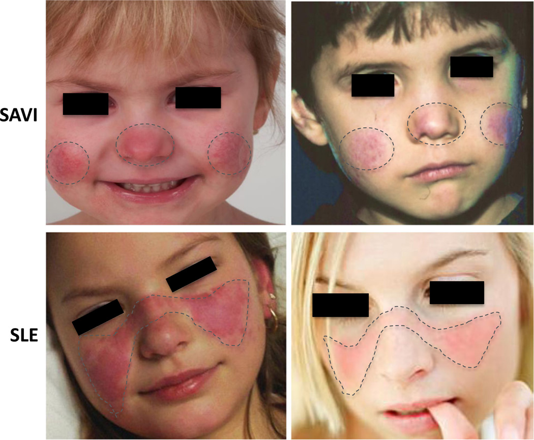
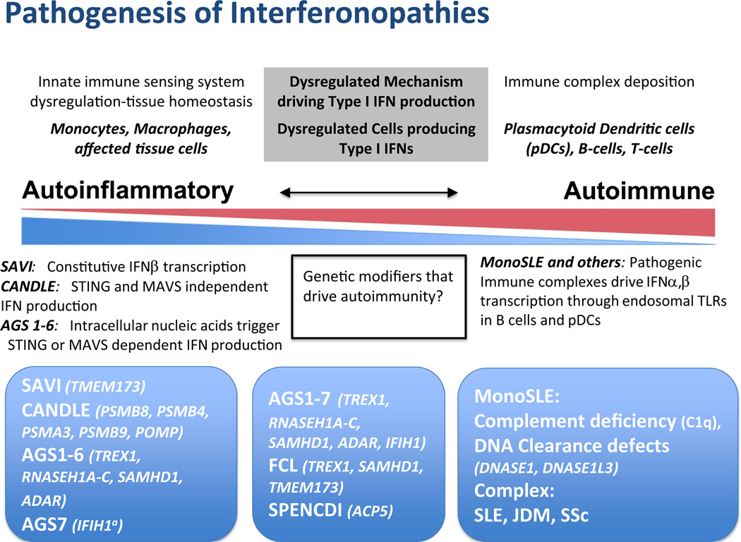
Autoinflammatory disorders are sterile inflammatory conditions characterized by episodes of early-onset fever and disease-specific patterns of organ inflammation. Recently, the discoveries of monogenic disorders with strong type I interferon (IFN) signatures caused by mutations in proteasome degradation and cytoplasmic RNA and DNA sensing pathways suggest a pathogenic role of IFNs in causing autoinflammatory phenotypes. The IFN response gene signature (IGS) has been associated with systemic lupus erythematosus (SLE) and other autoimmune diseases. In this review, we compare the clinical presentations and pathogenesis of two IFN-mediated autoinflammatory diseases, CANDLE and SAVI, with Aicardi Goutières syndrome (AGS) and monogenic forms of SLE (monoSLE) caused by loss-of-function mutations in complement 1 (C1q) or the DNA nucleases, DNASE1 and DNASE1L3. We outline differences in intracellular signaling pathways that fuel a pathologic type I IFN amplification cycle. While IFN amplification is caused by predominantly innate immune cell dysfunction in SAVI, CANDLE, and AGS, autoantibodies to modified RNA and DNA antigens interact with tissues and immune cells including neutrophils and contribute to IFN upregulation in some SLE patients including monoSLE, thus justifying a grouping of "autoinflammatory" and "autoimmune" interferonopathies. Understanding of the differences in the cellular sources and signaling pathways will guide new drug development and the use of emerging targeted therapies.

Keywords: Autoimmune; Autoinflammatory; Candle; Interferonopathies; SAVI; Type I IFN.


Figures

Fig. 1
Fig. 1
Different patterns in facial rash of SAVI and SLE. In SAVI patients (upper panels), the rashes are aggravated by cold exposure and are prominent in cold-sensitive areas including the tip of the nose and the lower part of the cheeks and the ears (not shown). The characteristic malar (facial) rash with a butterfly distribution on pictures from genetically undefined SLE patients (lower panels) show a photosensitive rash that is often induced by exposure to sunlight and associated with immune complex deposition at the dermal-epidermal junction (LE band) on biopsy. References: Lower left photo: © 2016 American College of Rheumatology. Used with permission. Lower right photo: www.mollysfund.org. Used with permission.
Fig. 2
Fig. 2
Comparative pathogenic considerations in autoinflammatory and autoimmune interfernopathies. On the left end of the spectrum, in monogenic conditions CANDLE/PRAAS and SAVI, the IFN signature is derived from proteasome suppression leading to IFNA and IFNB1 transcription and constitutively increased IFNB1 transcription, respectively, and are not triggered by immune complex deposition and can thus be called autoinflammatory. In AGS 1–6, sterile pyrexia and CNS damage are likely driven STING- or MAVS-dependent type I IFN production, suggesting innate (autoinflammatory) immune activation triggered by intracellular nucleic acid accumulation. IFIH1a (AGS7) mutations directly activate MAVS-dependent upregulation of IFN production without nucleic acid accumulation. The incomplete penetrance and variable clinical phenotype with IFIH1 and TREX1 mutations suggests a possible role for modifier genes. FCL and SMS have clinical features not seen in other interfernopathies. Though upregulated interferon-regulated genes are reported in SMS, little information regarding pathogenesis is available. Both autoimmune and autoinflammatory features are seen in the middle box. On the right end of the spectrum, clinical features of monoSLE (C1q deficiency, DNA clearance defects due to deficiency in DNASE1 or DNASE1L3) are likely driven by immune complex deposition (see text). The immune complexes can drive IFN production through activation of interferogenic dendritic and B cell responses or interferogenic release of oxidized mitochondrial DNA, suggesting involvement of adaptive “autoimmune” pathways in disease pathogenesis. Pathogenic antibodies and immune complexes are seen in SLE, and likely in JDM and SSc

References

    1. Kastner DL, Aksentijevich I, Goldbach-Mansky R. Autoinflammatory disease reloaded: a clinical perspective. Cell. 2010;140:784–790. - PMC - PubMed
    1. de Jesus AA, Canna SW, Liu Y, Goldbach-Mansky R. Molecular mechanisms in genetically defined autoinflammatory diseases: disorders of amplified danger signaling. Annu Rev Immunol. 2015;33:823–874. - PMC - PubMed
    1. Liu Y, Ramot Y, Torrelo A, Paller AS, Si N, Babay S, Kim PW, Sheikh A, Lee CC, Chen Y, Vera A, et al. Mutations in proteasome subunit beta type 8 cause chronic atypical neutrophilic dermatosis with lipodystrophy and elevated temperature with evidence of genetic and phenotypic heterogeneity. Arthritis Rheumatism. 2012;64:895–907. - PMC - PubMed
    1. Liu Y, Jesus AA, Marrero B, Yang D, Ramsey SE, Montealegre Sanchez GA, Tenbrock K, Wittkowski H, Jones OY, Kuehn HS, et al. Activated STING in a vascular and pulmonary syndrome. N Engl J Med. 2014;371:507–518. - PMC - PubMed
    1. Rice GI, Forte GM, Szynkiewicz M, Chase DS, Aeby A, Abdel-Hamid MS, Ackroyd S, Allcock R, Bailey KM, Balottin U, et al. Assessment of interferon-related biomarkers in Aicardi-Goutieres syndrome associated with mutations in TREX1, RNASEH2A, RNASEH2B, RNASEH2C, SAMHD1, and ADAR: a case-control study. Lancet Neurol. 2013;12:1159–1169. - PMC - PubMed

Publication types