. 2017 Apr;45(4):990-1002.
doi: 10.1007/s10439-016-1735-y. Epub 2016 Sep 27.

Development of a Smart Pump for Monitoring and Controlling Intraocular Pressure

Affiliations

Development of a Smart Pump for Monitoring and Controlling Intraocular Pressure

Simon A Bello et al. Ann Biomed Eng. 2017 Apr.

Abstract

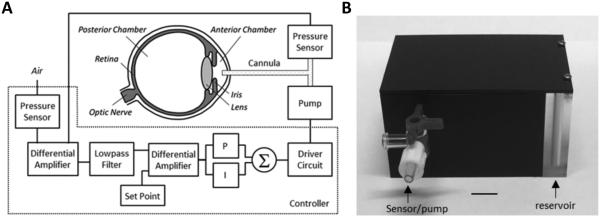
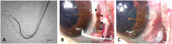
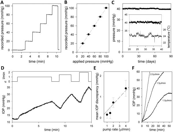
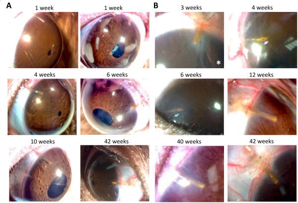
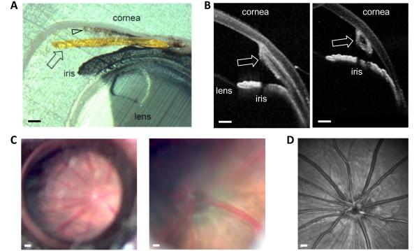
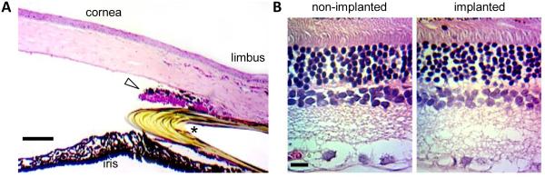
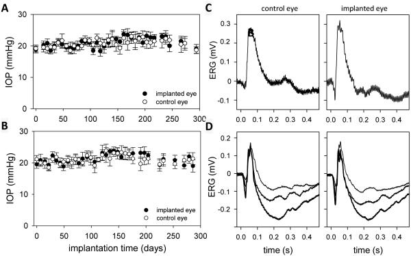
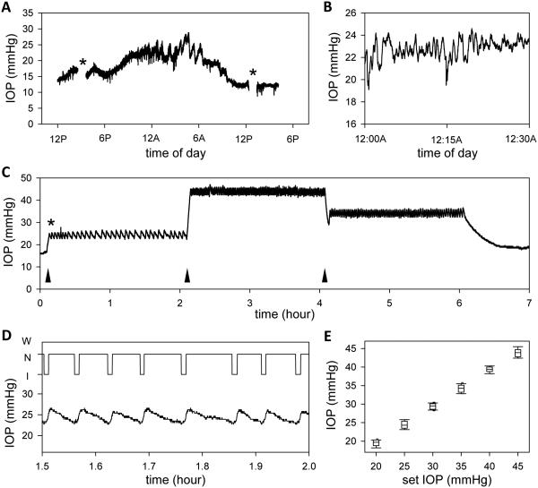
Animal models of ocular hypertension are important for glaucoma research but come with experimental costs. Available methods of intraocular pressure (IOP) elevation are not always successful, the amplitude and time course of IOP changes are unpredictable and irreversible, and IOP measurement by tonometry is laborious. Here we present a novel system for monitoring and controlling IOP without these limitations. It consists of a cannula implanted in the anterior chamber of the eye, a pressure sensor that continually measures IOP, and a bidirectional pump driven by control circuitry that can infuse or withdraw fluid to hold IOP at user-desired levels. A portable version was developed for tethered use on rats. We show that rat eyes can be cannulated for months without causing significant anatomical or physiological damage although the animal and its eyes freely move. We show that the system measures IOP with <0.7 mmHg resolution and <0.3 mmHg/month drift and can maintain IOP within a user-specified window of desired levels for any duration necessary. We conclude that the system is ready for cage- or bench-side applications. The results lay the foundation for an implantable version that would give glaucoma researchers unprecedented knowledge and control of IOP in rats and potentially larger animals.

Keywords: Closed loop control; Eye; Glaucoma; Implant; Rat; Telemetry.


Figures

Figure 1
Figure 1
IOP control system. (A) Block diagram of the system. A pressure sensor measures IOP via a fine cannula implanted in the anterior chamber of the eye and a second sensor measures atmospheric pressure. A controller circuit amplifies and filters the difference in pressure signals and compares the result against a user-specified set point. If IOP deviates from the desired level, proportional (P) and integrator (I) circuit elements combine to produce a command signal that drives a small pump to inject or withdraw fluid through the cannula until IOP returns to the set point. (B) Picture of portable iPump system. All electronic components are housed in a small plastic box (8 × 5 × 4 cm) that contains a fluid reservoir which can be filled or drained. The box tethers to the animal via tubing that runs inside a protective spring to a plastic head mount which connects subdermally to the implanted cannula. Bar: 1 cm.
Figure 2
Figure 2
Eye implantation procedure. (A) Cannula pre-shaped for rat eyes with heat. Bar: 500 μm. (B) Cannula implanted in the anterior chamber of a rat eye and secured with half-thickness sutures (arrowheads) to the sclera. (C) Rat eye after cannula implantation
Figure 3
Figure 3
iPump system properties. (A) System output as applied pressure was raised in steps of 20 mmHg every 1-2 minutes and released. (B) Mean (symbols) and standard deviation (bars) of pressure readings for each step in applied pressure. (C) Signal recorded over 90 days with applied pressure set at 40 mmHg. Data were fit by the regression line: f(x) = 40.7 − 0.0077x. Inset, segment of the pressure record during which room temperature fluctuated greatest. (D) Top: Time course of pump rates applied by the system to a dead rat eye in situ. The rate was stepped on and off to 1, 1.5 and 2uL/min over a 15-min period. Bottom: Pump-induced IOP changes recorded simultaneously by the iPump (thick line) and a second pressure sensor (thin line) independently connected to the eye. (E) Mean and standard deviation of the difference record obtained by subtracting the simultaneously-recorded IOP signals for pump rates of 1 to 4uL/min. Dashed line estimates the system resistance based on Pouiselle’s law . (F) IOP dynamics following the perfusion rate steps.
Figure 4
Figure 4
Rat eyes with chronically implanted cannulas. (A) Time series of images of two rat eyes implanted with a cannula for over 3 months (I4: left) and 10 months (I10: right). (B) Magnified images of the cannula tip in these (I4: right middle, I10: right bottom) and four other animals at experiment end. Asterisk indicates an eye that exhibited a local corneal response to the implant.
Figure 5
Figure 5
Internal structure of an implanted rat eye (I4). (A) Iridocorneal angle after fixation and paraffin embedding. Arrows mark the cannula. Arrowhead points to scar tissue growth between the cornea and cannula. Bars: 200μm. (B) Iridocorneal angle viewed along the longitudinal (left) and transverse (right) plane of the cannula. Images were acquired at experiment end with a Heidelberg-Spectralis II OCT scanner with the rat under anesthesia. (C) Fundus viewed at low (left) and high (right) magnification via a digital stereoscope, ring light, and coverslip on the cornea. Bars: 500μm and 200μm. (D) Fundus image acquired at experiment end with the OCT scanner. Bar: 500μm.
Figure 6
Figure 6
Histology of an implanted rat eye (I4). (A) HE stained section of the iridocorneal angle. Asterisk marks the cannula. Triangle points at an outgrowth of scar tissue. Bar: 100μm. (B) HE stained section of the retina of the non-implanted and implanted eyes. Inspection of serial retinal sections all presented similar cell density and layer thickness. Bar: 25μm.
Figure 7
Figure 7
(A,B) IOP history of implanted rat eyes (A: I10, B: I14). Each data point is the average of 6 tonometer readings. Bars are standard deviation. (C,D) Flash ERG records of implanted and non-implanted eyes of two rats (C: I4, D: I10) after 6 weeks. Each record is the average of 30 responses to full-field flashes with an interstimulus interval of 3 s. Flash intensity: 4.3 (thin trace), 8.6 (thicker trace), and 17.2 kcd/m2 (thickest trace).
Figure 8
Figure 8
IOP monitoring and controlling with the iPump system. (A) IOP record of the implanted eye of an anesthetized rat. Asterisks mark data periods that were excluded because IOP was manipulated to examine system behavior. (B) Short (30-min) segment of the IOP record at midnight. (C) IOP record of the implanted eye of an anesthetized rat during which the system was programmed to hold pressure at 25, 45, and 35 mmHg for 2 hours each. Asterisk marks the start of IOP control. Arrowheads mark the start of each IOP step. (D) Top, record of pump action (I: injection, N: neutral, W: withdrawal) for a 30-min period. Bottom, IOP dynamics over the same period. (E) Box-and-whiskers plot of the distribution of IOP readings for each IOP step. Center line: mean IOP, lower and upper edges: 25% to 75% range, lower and upper whiskers: 10th and 90th data percentiles.

References

    1. Abrams LS, Vitale S, Jampel HD. Comparison of three tonometers for measuring intraocular pressure in rabbits. Invest. Ophthalmol. Vis. Sci. 1996;37:940–944. - PubMed
    1. Akaishi T, Ishida N, Shimazaki A, Hara H, Kuwayama Y. Continuous monitoring of circadian variations in intraocular pressure by telemetry system throughout a 12-week treatment with timolol maleate in rabbits. J. Ocul. Pharmacol. Therap. 2005;21:436–444. - PubMed
    1. Akula JD, Favazza TL, Mocko JA, Benador IY, Asturias AL, Kleinman MS, Hansen RM, Fulton AB. The anatomy of the rat eye with oxygen-induced retinopathy. Doc. Ophthalmol. 2010;120:41–50. - PubMed
    1. Asrani S, Zeimer R, Wilensky J, Gieser D, Vitale S, Lindenmuth K. Large diurnal fluctuations in intraocular pressure are an independent risk factor in patients with glaucoma. J. Glaucoma. 2000;9:134–142. - PubMed
    1. Bengtsson B, Leske MC, Hyman L, Heijl A. Fluctuation of intraocular pressure and glaucoma progression in the early manifest glaucoma trial. Ophthalmol. 2007;114:205–209. - PubMed

Publication types