. 2016 Sep 13:8:213.
doi: 10.3389/fnagi.2016.00213. eCollection 2016.

Regional and Gender Study of Neuronal Density in Brain during Aging and in Alzheimer's Disease

Affiliations

Regional and Gender Study of Neuronal Density in Brain during Aging and in Alzheimer's Disease

Eva Martínez-Pinilla et al. Front Aging Neurosci. .

Abstract

Background: Learning processes or language development are only some of the cognitive functions that differ qualitatively between men and women. Gender differences in the brain structure seem to be behind these variations. Indeed, this sexual dimorphism at neuroanatomical level is accompanied unequivocally by differences in the way that aging and neurodegenerative diseases affect men and women brains.

Objective: The aim of this study is the analysis of neuronal density in four areas of the hippocampus, and entorhinal and frontal cortices to analyze the possible gender influence during normal aging and in Alzheimer's disease (AD).

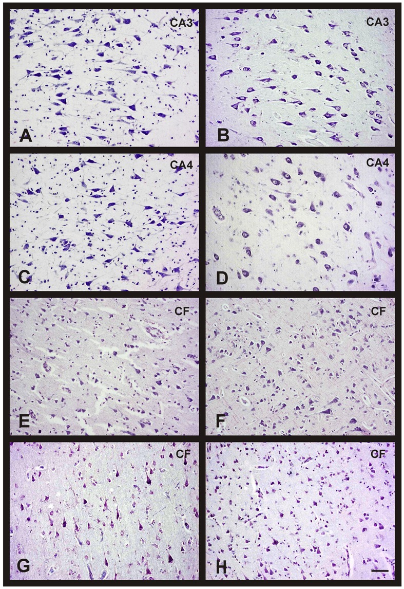
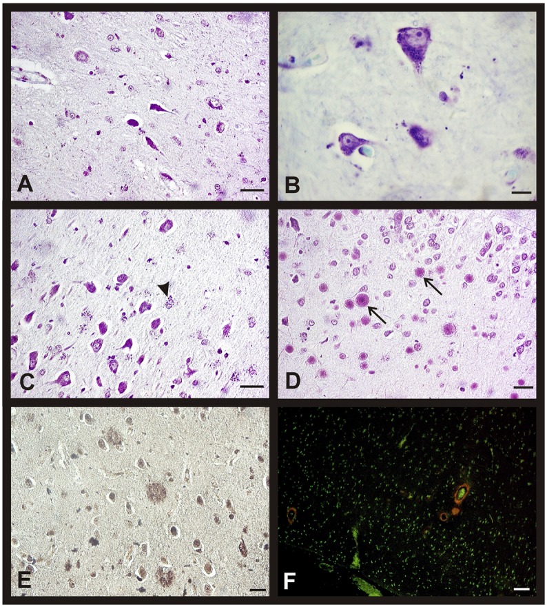
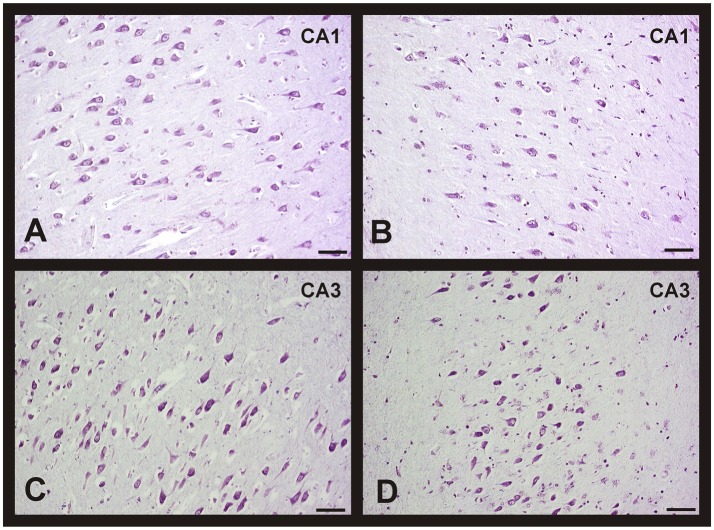
Methods: Human brain tissues of different age and from both sexes, without neurological pathology and with different Braak's stages of AD, were studied. Neuronal density was quantified using the optical dissector.

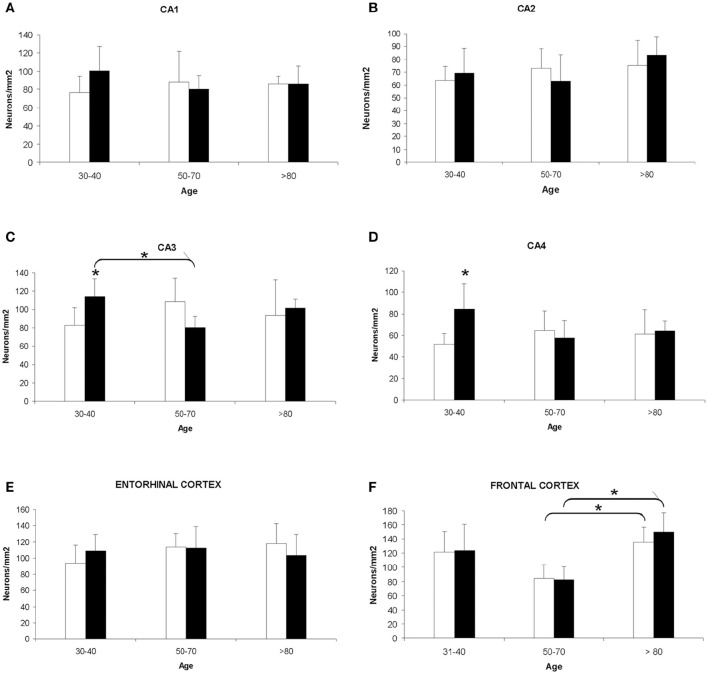
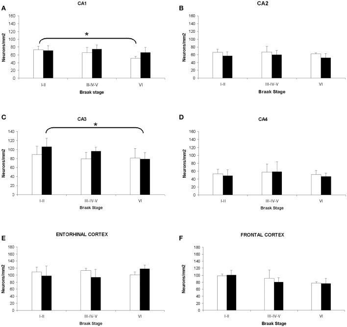
Results: Our results showed the absence of a significant neuronal loss during aging in non-pathological brains in both sexes. However, we have demonstrated specific punctual significant variations in neuronal density related with the age and gender in some regions of these brains. In fact, we observed a higher neuronal density in CA3 and CA4 hippocampal areas of non-pathological brains of young men compared to women. During AD, we observed a negative correlation between Braak's stages and neuronal density in hippocampus, specifically in CA1 for women and CA3 for men, and in frontal cortex for both, men and women.

Conclusion: Our data demonstrated a sexual dimorphism in the neuronal vulnerability to degeneration suggesting the need to consider the gender of the individuals in future studies, regarding neuronal loss in aging and AD, in order to avoid problems in interpreting data.

Keywords: Alzheimer's disease; age; entorhinal cortex; frontal cortex; hippocampus; human; sexual dimorphism.


Figures

Figure 1
Figure 1
Quantitative changes in neuronal density in certain hippocampal fields (CA1–4) and in frontal and entorhinal cortices between men and women during aging. (A) CA1 hippocampal area. (B) CA2 hippocampal area. (C) CA3 hippocampal area. (D) CA4 hippocampal area. (E) Entorhinal cortex. (F) Frontal cortex. Bars represent mean density in a 20x field ± SD (n = 6). *Statistically significant differences, p ≤ 0.05. Women, white bars; Men, black bars.
Figure 2
Figure 2
Changes in neuronal density in hippocampus and frontal cortex between men and women during aging. Representative microphotographs of human brain sections of non-pathological individuals contrasted with a Nissl method modification. (A) Hippocampus (CA3) of a 37 years old man. (B) Hippocampus (CA3) of a 35 years old woman. (C) Hippocampus (CA4) of a 37 years old man. (D) Hippocampus (CA4) of a 35 years old woman. (E) Frontal cortex of a 40 years old man. (F) Frontal cortex of a 65 years old man. (G) Frontal cortex of a 37 years old woman. (H) Frontal cortex of a 69 years old woman. Bar, 60 μm.
Figure 3
Figure 3
Morphological changes in hippocampus and entorhinal cortex during aging in both sexes. Representative microphotographs of human brain sections of non-pathological individuals contrasted with a Nissl method modification (A–D), silver technique of Reusche (E) and a modification of Congo Red method (F). (A) Shrinking neurons in the hippocampus of an 80 years old man. (B) Frontal cortex of a 75 years old woman, lipofuscin in neurons can be observed. (C) Hippocampus (CA3) of an 80 years old man, several astrocytes with lipofuscin granules are showed (arrowhead). (D) Hippocampus (CA4) of a 75 years old woman, several corpora amylacea can be observed (arrows). (E) Diffuse senile plaques in the entorhinal cortex of an 85 years old man. (F) Vessels with amyloid (red fluorescence) in frontal cortex of an 80 years old woman. Bars: (A,C), 60 μm; (B,D), 10 μm; (E), 40 μm; (F), 50 μm.
Figure 4
Figure 4
Quantitative changes in neuronal density in certain hippocampal fields (CA1–4) and in frontal and entorhinal cortices between men and women during AD progression (I–VI Braak's stages). (A) CA1 hippocampal area. (B) CA2 hippocampal area. (C) CA3 hippocampal area. (D) CA4 hippocampal area. (E) Entorhinal cortex. (F) Frontal cortex. Bars represent mean density in a 20x field ± SD (n = 6). *Statistically significant differences, p ≤ 0.05. Women, white bars; Men, black bars.
Figure 5
Figure 5
Changes in neuronal density in hippocampus between men and women during AD progression (I-VI Braak's stages). Representative microphotographs of human brain sections of individuals with AD pathology, contrasted with a Nissl method modification. (A) Hippocampus (CA1) of a 70 years old woman (Braak's stage I). (B) Hippocampus (CA1) of a 75 years old woman (Braak's stage V). (C) Hippocampus (CA3) of a 75 years old man (Braak's stage I). (D) Hippocampus (CA3) of an 80 years old man (Braak's stage VI). Bar: 20 μm.
Figure 6
Figure 6
Morphological brain changes in men and women during AD progression. Representative microphotographs of human brain sections of individuals with AD pathology, contrasted with a silver technique of Reusche (A,B) and a modification of Congo Red method (C,D). (A) Diffuse and mature plaques in the entorhinal cortex of an 80 years old man (Braak's stage II). (B) Senile plaques and neurofibrillary tangles in the hippocampus (CA2) of an 85 years old woman (Braak's stage VI). (C) Diffuse and mature plaques (red fluorescence) in the entorhinal cortex of an 80 years old man (Braak's stage II). (D) Senile plaques, in different maturation stages, and neurofibrillary tangles (red fluorescence) in the entorhinal cortex of an 80 years old man (Braak's stage VI). Bars: (A,C), 60 μm; (B), 10 μm; (D), 60 μm.

Similar articles

Cited by

References

    1. Adachi M., Kawakatsu S., Hosoya T., Otani K., Honma T., Shibata A., et al. . (2003). Morphology of the inner structure of the hippocampal formation in Alzheimer disease. Am. J. Neuroradiol. 24, 1575–1581. - PMC - PubMed
    1. Allen L. S., Gorski R. A. (1991). Sexual dimorphism of the anterior commissure and massa intermedia of the human brain. J. Comp. Neurol. 312, 97–104. 10.1002/cne.903120108 - DOI - PubMed
    1. Aloisi A. M., Zimmermann M., Herdegen T. (1997). Sex-dependent effects of formalin and restraint on c-Fos expression in the septum and hippocampus of the rat. Neuroscience 81, 951–958. 10.1016/S0306-4522(97)00270-4 - DOI - PubMed
    1. Alvarez J. C., Díaz C., Suárez C., Fernández J. A., González del Rey C., Navarro A., et al. . (1998). Neuronal loss in human medial vestibular nucleus. Anat. Rec. 251, 431–438. 10.1002/(SICI)1097-0185(199808)251:4<431::AID-AR2>3.0.CO;2-V - DOI - PubMed
    1. Alvarez J. C., Díaz C., Suárez C., Fernández J. A., González del Rey C., Navarro A., et al. . (2000). Aging and the human vestibular nuclei: morphometric analysis. Mech. Ageing Dev. 114, 149–172. 10.1016/S0047-6374(00)00098-1 - DOI - PubMed

LinkOut - more resources