

Magnetic Resonance Angiography in the Diagnosis of Cerebral Arteriovenous Malformation and Dural Arteriovenous Fistulas: Comparison of Time-Resolved Magnetic Resonance Angiography and Three Dimensional Time-of-Flight Magnetic Resonance Angiography
- PMID: 27679690
- PMCID: PMC5036458
- DOI: 10.5812/iranjradiol.19814
Magnetic Resonance Angiography in the Diagnosis of Cerebral Arteriovenous Malformation and Dural Arteriovenous Fistulas: Comparison of Time-Resolved Magnetic Resonance Angiography and Three Dimensional Time-of-Flight Magnetic Resonance Angiography
Abstract
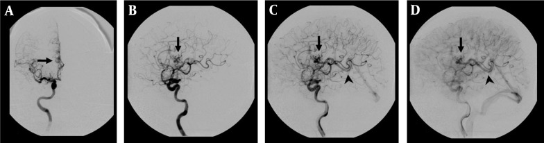
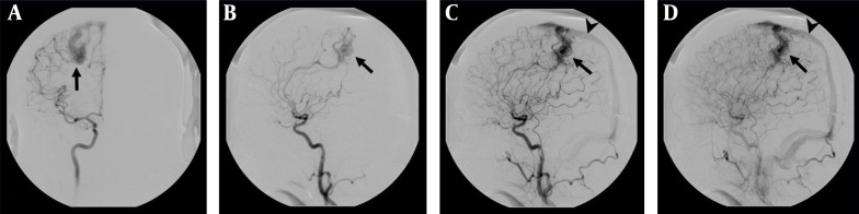
Background: Traditional digital subtraction angiography (DSA) is currently the gold standard diagnostic method for the diagnosis and evaluation of cerebral arteriovenous malformation (AVM) and dural arteriovenous fistulas (dAVF).
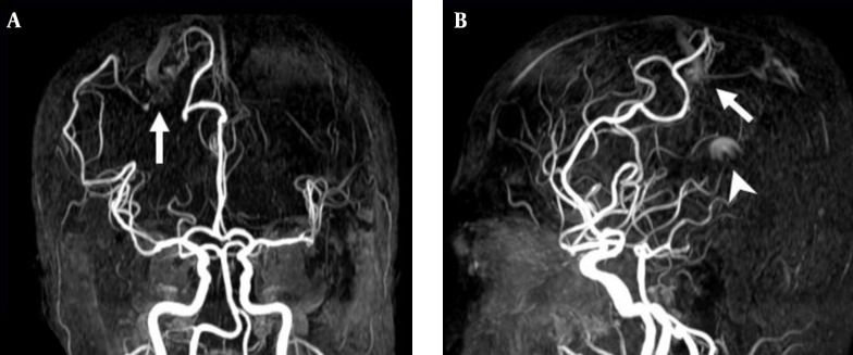
Objectives: The aim of this study was to analyze different less invasive magnetic resonance angiography (MRA) images, time-resolved MRA (TR-MRA) and three-dimensional time-of-flight MRA (3D TOF MRA) to identify their diagnostic accuracy and to determine which approach is most similar to DSA.
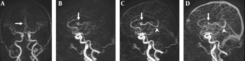
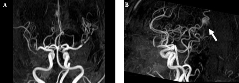
Patients and methods: A total of 41 patients with AVM and dAVF at their initial evaluation or follow-up after treatment were recruited in this study. We applied time-resolved angiography using keyhole (4D-TRAK) MRA to perform TR-MRA and 3D TOF MRA examinations simultaneously followed by DSA, which was considered as a standard reference. Two experienced neuroradiologists reviewed the images to compare the diagnostic accuracy, arterial feeder and venous drainage between these two MRA images. Inter-observer agreement for different MRA images was assessed by Kappa coefficient and the differences of diagnostic accuracy between MRA images were evaluated by the Wilcoxon rank sum test.
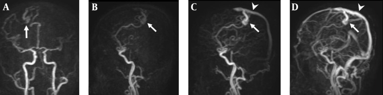
Results: Almost all vascular lesions (92.68%) were correctly diagnosed using 4D-TRAK MRA. However, 3D TOF MRA only diagnosed 26 patients (63.41%) accurately. There were statistically significant differences regarding lesion diagnostic accuracy (P = 0.008) and venous drainage identification (P < 0.0001) between 4D-TRAK MRA and 3D TOF MRA. The results indicate that 4D-TRAK MRA is superior to 3D TOF MRA in the assessment of lesions.
Conclusion: Compared with 3D TOF MRA, 4D-TRAK MRA proved to be a more reliable screening modality and follow-up method for the diagnosis of cerebral AVM and dAVF.
Keywords: 3D TOF MRA; 4D-TRAK MRA; Cerebral Arteriovenous Malformation; Cerebral Dural Arteriovenous Fistulas.
Figures
Similar articles
-
A comparison of 4D time-resolved MRA with keyhole and 3D time-of-flight MRA at 3.0 T for the evaluation of cerebral aneurysms.BMC Neurol. 2012 Jul 6;12:50. doi: 10.1186/1471-2377-12-50. BMC Neurol. 2012. PMID: 22784396 Free PMC article.
-
MR angiography of dural arteriovenous fistulas: diagnosis and follow-up after treatment using a time-resolved 3D contrast-enhanced technique.AJNR Am J Neuroradiol. 2007 May;28(5):877-84. AJNR Am J Neuroradiol. 2007. PMID: 17494662 Free PMC article. Clinical Trial.
-
Contrast-enhanced MR 3D angiography in the assessment of brain AVMs.Eur J Radiol. 2006 Dec;60(3):367-78. doi: 10.1016/j.ejrad.2006.08.007. Epub 2006 Sep 11. Eur J Radiol. 2006. PMID: 16965882
-
The use of contrast-enhanced, time-resolved magnetic resonance angiography in cerebrovascular pathology.Neurosurg Focus. 2019 Dec 1;47(6):E3. doi: 10.3171/2019.9.FOCUS19627. Neurosurg Focus. 2019. PMID: 31786556 Review.
-
A Systematic Review Comparing Digital Subtraction Angiogram With Magnetic Resonance Angiogram Studies in Demonstrating the Angioarchitecture of Cerebral Arteriovenous Malformations.Cureus. 2022 Jun 9;14(6):e25803. doi: 10.7759/cureus.25803. eCollection 2022 Jun. Cureus. 2022. PMID: 35706438 Free PMC article. Review.
Cited by
-
Dural Arteriovenous Fistula Mimicking a Brain Tumor on Methionine-positron Emission Tomography: A Case Report.NMC Case Rep J. 2022 Sep 15;9:289-294. doi: 10.2176/jns-nmc.2022-0055. eCollection 2022. NMC Case Rep J. 2022. PMID: 36263190 Free PMC article.
-
Quantitative evaluation of arteriovenous malformation hemodynamic changes after endovascular treatment using parametric color coding: A case series study.Interv Neuroradiol. 2017 Dec;23(6):650-655. doi: 10.1177/1591019917721867. Epub 2017 Aug 1. Interv Neuroradiol. 2017. PMID: 28764614 Free PMC article.
-
A Review of the Current State and Future Directions for Management of Scalp and Facial Vascular Malformations.J Korean Neurosurg Soc. 2024 May;67(3):315-325. doi: 10.3340/jkns.2024.0032. Epub 2024 May 1. J Korean Neurosurg Soc. 2024. PMID: 38720545 Free PMC article.
References
-
- Lim RP, Shapiro M, Wang EY, Law M, Babb JS, Rueff LE, et al. 3D time-resolved MR angiography (MRA) of the carotid arteries with time-resolved imaging with stochastic trajectories: comparison with 3D contrast-enhanced Bolus-Chase MRA and 3D time-of-flight MRA. AJNR Am J Neuroradiol. 2008;29(10):1847–54. doi: 10.3174/ajnr.A1252. - DOI - PMC - PubMed
-
- Nussel F, Wegmuller H, Huber P. Comparison of magnetic resonance angiography, magnetic resonance imaging and conventional angiography in cerebral arteriovenous malformation. Neuroradiology. 1991;33(1):56–61. - PubMed
LinkOut - more resources
Full Text Sources
Other Literature Sources