. 2016 Oct 11;113(41):E6036-E6044.
doi: 10.1073/pnas.1609571113. Epub 2016 Sep 27.

Intracellular mechanisms of molecular recognition and sorting for transport of large extracellular matrix molecules

Affiliations

Intracellular mechanisms of molecular recognition and sorting for transport of large extracellular matrix molecules

Yoshihiro Ishikawa et al. Proc Natl Acad Sci U S A. .

Abstract

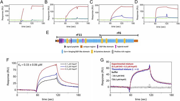
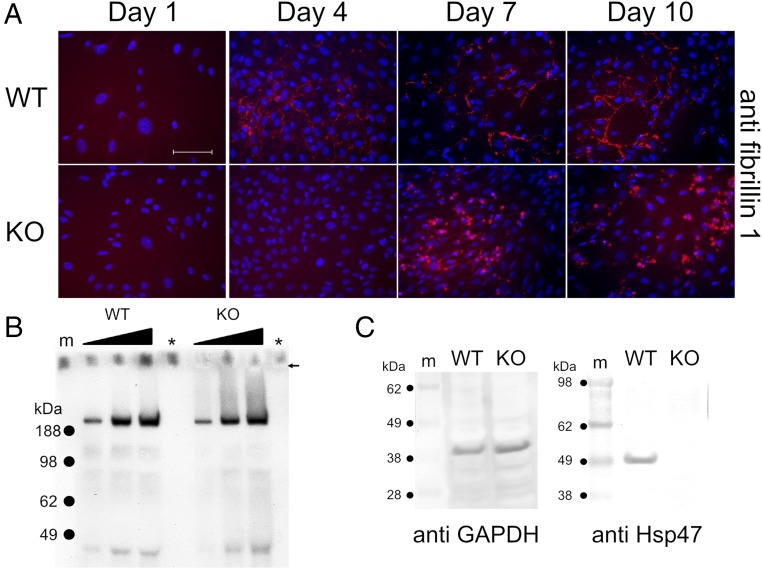
Extracellular matrix (ECM) proteins are biosynthesized in the rough endoplasmic reticulum (rER) and transported via the Golgi apparatus to the extracellular space. The coat protein complex II (COPII) transport vesicles are approximately 60-90 nm in diameter. However, several ECM molecules are much larger, up to several hundreds of nanometers. Therefore, special COPII vesicles are required to coat and transport these molecules. Transmembrane Protein Transport and Golgi Organization 1 (TANGO1) facilitates loading of collagens into special vesicles. The Src homology 3 (SH3) domain of TANGO1 was proposed to recognize collagen molecules, but how the SH3 domain recognizes various types of collagen is not understood. Moreover, how are large noncollagenous ECM molecules transported from the rER to the Golgi? Here we identify heat shock protein (Hsp) 47 as a guide molecule directing collagens to special vesicles by interacting with the SH3 domain of TANGO1. We also consider whether the collagen secretory model applies to other large ECM molecules.

Keywords: COPII vesicles; Hsp47; TANGO1; collagen; secretion.


Conflict of interest statement

The authors declare no conflict of interest.

Figures

Fig. 1.
Fig. 1.
Characterization of recombinant SH3 domain of human TANGO1. (A) SDS/PAGE analysis of purified recombinant SH3 domain of human TANGO1. The SH3 domain was purified from an E. coli expression system, and the figure shows the final purified material in the presence and absence of DTT in lanes 1 and 2, respectively. The purified SH3 domain was run on NuPAGE Novex Bis-Tris 4–12% gel (Life Technology) and stained with GelCode Blue Stain Reagent. Asterisk indicates blank lane. (B) CD spectra of the SH3 domain. The CD spectrum was measured at 4 °C in 10 mM phosphate, pH 7.5. The concentration of the SH3 domain of human TANGO1 was 8 µM. (C) Thermal stability of the SH3 domain of human TANGO1 was monitored by CD at 210 nm, and the rate of heating was 10 °C/h. The concentration of the SH3 domain of human TANGO1 was 8 µM in PBS solution.
Fig. 2.
Fig. 2.
Direct binding kinetics of the SH3 domain of human TANGO1 to collagens. Direct binding kinetics were measured by SPR analysis using a BIAcore X instrument. The SH3 domain of human TANGO1 (10 µM; red) was injected over CM5 chips immobilized with eight different types of collagen: bovine type I collagen (A), bovine type II collagen (B), bovine type III collagen (C), mouse type IV collagen (D), bovine type V collagen (E), human type VI collagen (F), human recombinant type X collagen (G), and bovine type XI collagen (H). Hsp47 (0.05 µM; blue) was used as a positive control.
Fig. 3.
Fig. 3.
Determination of the direct binding between the SH3 domain of human TANGO1 and Hsp47. (A) SPR analysis was carried out by using a BIAcore X instrument. Hsp47 (0.1 µM; black), FKBP22 (1.0 µM; red), and FKBP65 (1.0 µM; blue) were run over the CM5 chip with immobilized SH3 domain of human TANGO1. (B) Various concentrations of Hsp47 were run over the SH3 domain of human TANGO1 chip. The following binding curves are shown: 0.05 µM (black), 0.1 µM (red), and 0.2 µM (blue) Hsp47. The calculated Kd value is also shown. (C) CD spectra were measured at 4 °C in 10 mM phosphate, pH 7.5 The filled and open circles are the SH3 domain of human TANGO1 (4.0 µM) and Hsp47 (1.0 µM), respectively. Red and blue curves indicate the theoretical signal derived from addition of individual SH3 domain of human TANGO1 and Hsp47 signals, and the experimental signal from a mixture of the SH3 domain of human TANGO1 and Hsp47, respectively. (D) SPR analysis was carried out by using a BIAcore X instrument to determine the binding orientation. The open squares and circles are the SH3 domain of human TANGO1 (0.5 µM) and Hsp47 (0.05 µM), respectively. HBS-P buffer (10 mM Hepes buffer, pH 7.4, containing 150 mM NaCl, 1 mM CaCl2, and 0.005% surfactant P20) is shown as a black curve. Red and blue curves indicate the theoretical signal derived from addition of individual SH3 domain of human TANGO1 and Hsp47 signals and the experimental signal from a mixture of both SH3 domain of human TANGO1 and Hsp47, respectively.
Fig. 4.
Fig. 4.
Direct binding kinetics of Hsp47 to ECM molecules. Direct binding kinetics were measured by SPR analysis using a BIAcore X instrument. Antibodies against each ECM protein (red) were used as a positive control. Hsp47 (blue) and the SH3 domain of human TANGO1 (green) were injected over CM5 chips with immobilized ECM proteins. (A) Human fibronectin chip with 20 µg/mL anti-fibronectin, 0.8 µM Hsp47, and 10 µM SH3 domain of human TANGO1. (B) Human COMP chip with 40 µg/mL anti-COMP, 0.8 µM Hsp47, and 10 µM SH3 domain of human TANGO1. (C) Recombinant amino terminal fragment of human fibrillin-1 rF11 chip with 86 µg/mL anti-rF11, 0.2 µM Hsp47, and 10 µM SH3 domain of human TANGO1. (D) Recombinant carboxyl terminal fragment of human fibrillin-1 rF6 chip with 0.36 mg/mL anti-rF6, 0.8 µM Hsp47, and 10 µM SH3 domain of human TANGO1. (E) Domain structure of fibrillin-1 describing the fragments rF11 and rF6. (F) Various concentrations of Hsp47 were run over the recombinant amino-terminal fragment of human fibrillin-1 rF11 chip. The following binding curves are shown: 0.1 µM (black), 0.2 µM (red), and 0.3 µM (blue) Hsp47. The calculated Kd value is also shown. (G) SPR analysis was carried out by using a BIAcore X instrument to determine the binding orientation. The open squares and circles are the SH3 domain of human TANGO1 (0.5 µM) and Hsp47 (0.2 µM), respectively. HBS-P buffer (10 mM Hepes buffer, pH 7.4, containing 150 mM NaCl, 1 mM CaCl2, and 0.005% Surfactant P20) is shown as a black curve. Red and blue curves indicate the theoretical signal derived from the addition of individual SH3 domain of human TANGO1 and Hsp47 signals and the experimental signal from a mixture of SH3 domain of human TANGO1 and Hsp47, respectively.
Fig. 5.
Fig. 5.
Immunofluorescence staining in Hsp47+/+ and Hsp47−/− MEFs. Hsp47+/+ and Hsp47−/− MEFs were stained against anti-Hsp47 (A), anti-fibronectin (B), and anti-type I collagen (C). Cells were cultured with ascorbic acid phosphate (150 µg /mL) continuously, and fixed/permeabilized with cold methanol before staining was performed. The red and blue staining is derived from specific primary antibody and DAPI, respectively. Confocal images are included for day 4 of anti-fibronectin and day 10 of anti-type I collagen. These images are labeled as “confocal MS.” (Scale bars: 100 µm.)
Fig. 6.
Fig. 6.
Analysis of fibrillin-1 in Hsp47+/+ and Hsp47−/− MEFs. (A) Hsp47+/+ and Hsp47−/− MEFs were stained with anti–fibrillin-1. Cells were cultured with ascorbic acid phosphate (150 µg/mL) continuously and fixed/permeabilized with cold methanol before staining. The red and blue stainings are derived from specific primary antibody and DAPI, respectively. (Scale bar: 100 µm.) (B) Secreted amounts of fibrillin-1 in the serum-free medium from equal cell numbers were analyzed by Western blotting against fibrillin-1. The filled triangle and arrow indicate loading amounts on the well and well bottom, respectively. Blank and marker lanes are shown as asterisk and “m”, respectively. (C) Total soluble proteins from equal cell numbers in Hsp47+/+ and Hsp47−/− MEFs were extracted and blotted by using anti-GAPDH and anti-Hsp47. Marker is indicated as “m.”
Fig. 7.
Fig. 7.
Schematic diagram of the model and hypotheses for secretion of large ECM molecules. Type VII collagen is directed to special COPII vesicles by the SH3 domain of TANGO1 and by the SH3 domain–Hsp47 complex. All collagen types (type I collagen is shown in the model) are sorted and loaded into special COPII vesicles by the SH3 domain–Hsp47 complex. Type IX collagen may traffic to the extracellular space with COMP. Fibronectin and fibrillin-1 must be sorted into special COPII vesicles, but the SH3 domain–Hsp47 complex is not the mechanism of molecular recognition. The function of Hsp47 interaction with fibrillin-1 in the rER is unknown. Further investigations should be directed toward molecular mechanisms by which fibrillin-1 and fibronectin are sorted into special COPII vesicles and to the effects of mutations in Hsp47 (or loss of Hsp47) on matrix organization.

References

    1. Bateman JF, Boot-Handford RP, Lamandé SR. Genetic diseases of connective tissues: Cellular and extracellular effects of ECM mutations. Nat Rev Genet. 2009;10(3):173–183. - PubMed
    1. D’Arcangelo JG, Stahmer KR, Miller EA. Vesicle-mediated export from the ER: COPII coat function and regulation. Biochim Biophys Acta. 2013;1833(11):2464–2472. - PMC - PubMed
    1. Miller EA, Schekman R. COPII - A flexible vesicle formation system. Curr Opin Cell Biol. 2013;25(4):420–427. - PMC - PubMed
    1. Unlu G, Levic DS, Melville DB, Knapik EW. Trafficking mechanisms of extracellular matrix macromolecules: Insights from vertebrate development and human diseases. Int J Biochem Cell Biol. 2014;47:57–67. - PMC - PubMed
    1. Bächinger HP, Mizuno K, Vranka J, Boudko S. Collagen formation and structure. In: Mander L, Liu HW, editors. Comprehensive Natural Products II: Chemistry and Biology. Elsevier; Amsterdam: 2010. pp. 469–530.

Publication types

MeSH terms