. 2016 Nov;4(11):936-947.
doi: 10.1158/2326-6066.CIR-16-0177. Epub 2016 Sep 28.

Kinase Regulation of Human MHC Class I Molecule Expression on Cancer Cells

Affiliations

Kinase Regulation of Human MHC Class I Molecule Expression on Cancer Cells

Elliott J Brea et al. Cancer Immunol Res. 2016 Nov.

Abstract

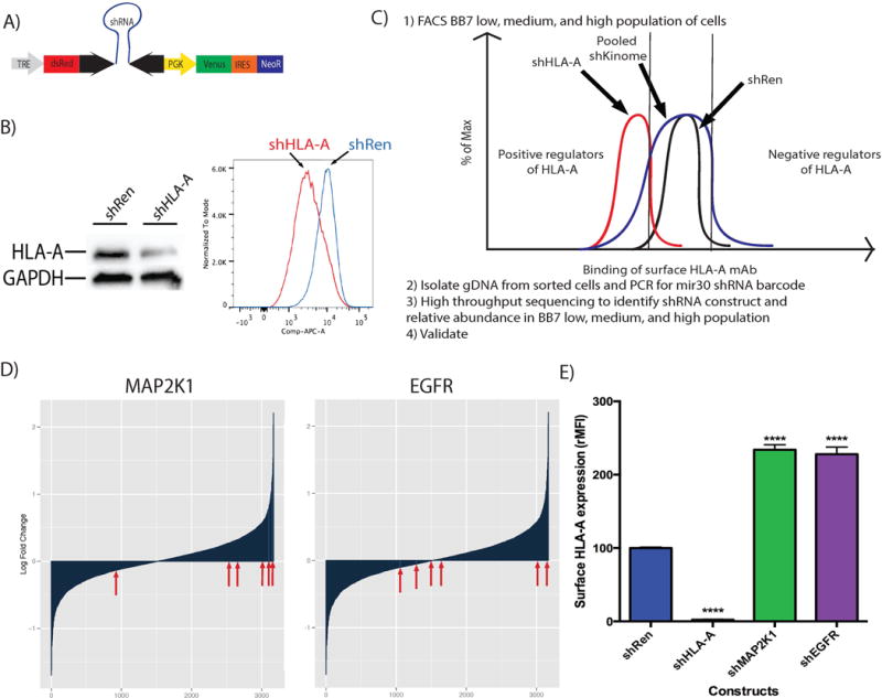
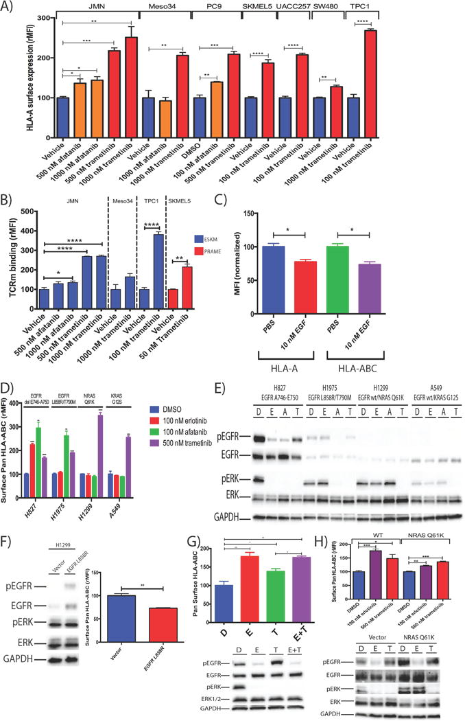
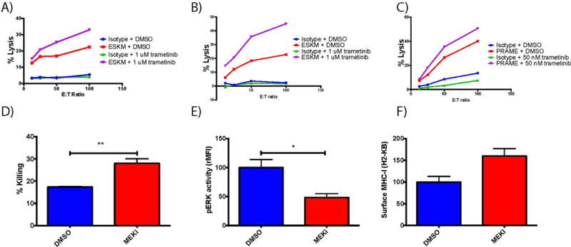
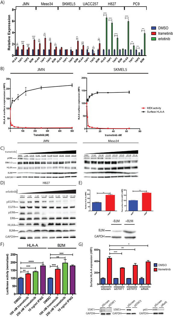
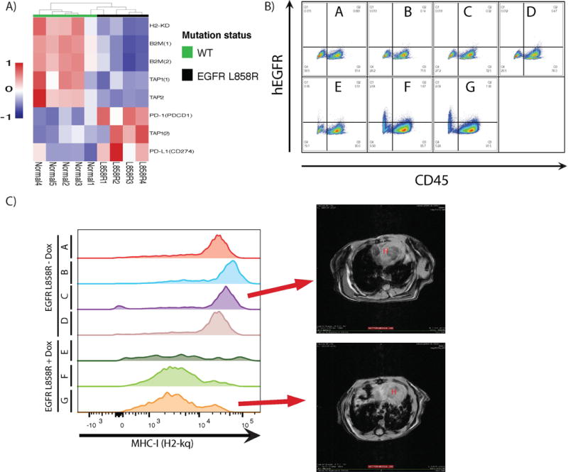
The major histocompatibility complex I (MHC-1) presents antigenic peptides to tumor-specific CD8+ T cells. The regulation of MHC-I by kinases is largely unstudied, even though many patients with cancer are receiving therapeutic kinase inhibitors. Regulators of cell-surface HLA amounts were discovered using a pooled human kinome shRNA interference-based approach. Hits scoring highly were subsequently validated by additional RNAi and pharmacologic inhibitors. MAP2K1 (MEK), EGFR, and RET were validated as negative regulators of MHC-I expression and antigen presentation machinery in multiple cancer types, acting through an ERK output-dependent mechanism; the pathways responsible for increased MHC-I upon kinase inhibition were mapped. Activated MAPK signaling in mouse tumors in vivo suppressed components of MHC-I and the antigen presentation machinery. Pharmacologic inhibition of MAPK signaling also led to improved peptide/MHC target recognition and killing by T cells and TCR-mimic antibodies. Druggable kinases may thus serve as immediately applicable targets for modulating immunotherapy for many diseases. Cancer Immunol Res; 4(11); 936-47. ©2016 AACR.


Conflict of interest statement

D.A.S. is an inventor of the ESKM technology described in this paper and licensed by Memorial Sloan Kettering Cancer Center to Novartis.

Figures

Figure 1
Figure 1
Screen for kinase regulators of surface HLA. A. A TRMPV inducible shRNA retroviral vector was used for transducing JMN (HLA-A*02:01 positive human mesothelioma line). TRE is the Tet responsive element, which drives expression of the fluorophore dsRed and the shRNA hairpin. The constitutive PGK promoter drives the Venus fluorophore along with Neomycin resistance cassette. B. Western blot and flow cytometry data showing knockdown of HLA-A using TRMPV retroviral system with a positive control shRNA to HLA-A02. The shRen is a negative control shRNA designed against the Renilla gene C. Schema depicting the workflow pipeline for the screen of regulators of surface HLA-A. D. Waterfall plot showing distribution of shRNA constructs against MAP2K1 and EGFR as log fold difference between BB7 high sorted population and BB7 low sorted population. E. shRNA knockdown of MAP2K1 and EGFR in JMN cells validates them as a negative regulator of surface HLA-A. BB7.2 is a mAb specific for HLA-A02. shRNA against Renilla was used as a negative control, whereas an shRNA against HLA-A was used as a positive control. The screen was done in triplicate. Inhibition experiments were performed at least twice with similar results, and data shown are representative. Student’s t-test was done to compare each shRNA gene knockdown MFI to the shRen control. (*≤0.05, **≤0.01, ***≤0.001, ****≤0.0001)
Figure 2
Figure 2
Use of selective EGFRi and MEKi increased cell surface HLA-A expression and tumor antigen presentation, whereas activation of EGFR caused downregulation of MHC-I. A. MEK inhibition and EGFR inhibition for 72 h with indicated inhibitors increased HLA-A (BB7 binding) by flow cytometry in JMN, Meso34, PC-9, UACC257, SK-MEL-5, SW480, and TPC1 cell lines. 1% DMSO was used as a vehicle control. B. Binding of TCRm antibodies to peptide/MHC epitopes. In blue, use of ESK antibody to a peptide derived from the oncoprotein WT1 that is presented on HLA-A0201. Binding increased after inhibition of EGFR and MEK for 72 h in JMN, Meso34, and TPC1. In red, the PRAME TCRm antibody to an epitope of PRAME tumor antigen presented on HLA-A0201 on SKMEL5 cells. Experimental setup was similar to A. C. Treatment of JMN with 10 nM EGF for 72 h, causing activation of the downstream MAPK pathway, led to decreased surface HLA-A and total HLA-ABC. D. Use of EGFRi erlotinib, along with MEKi trametinib, on H827 (EGFR E746del-A750 mutation), H1975 (L858R/T790M), H1299 (EGFR wt, NRAS Q61K), and A549 (EGFR wt/KRAS G12S) to alter surface HLA-ABC expression. Student t-test was done to compare each treatment to vehicle control. *P values annotated as in figure 1. E. Western blot analysis showing degree of inhibition of the MAP kinase pathway on a panel of NSCLC cell lines using 1% DMSO (D), 100 nM erlotinib (E), 100 nM afatinib (A), or 500 nM trametinib (T). F. H1299 cells were transduced with retroviral vectors expressing EGFR L858R and were analyzed for surface pan HLA-ABC using W6/32. Activation of EGFR is demonstrated by western blot G. EGFR inhibition upregulated surface HLA-ABC more than MEKi, despite equivalent inhibition of pERK output. H. EGFRi upregulated MHC-I despite downstream mutations causing constitutive MAPK activation. The NRAS Q61K mutation was introduced into H827 and cells were treated with EGFRi or MEKi as done in 2G. Experiments were performed 2–4 times with similar results, and data shown are representative.
Figure 3
Figure 3
Improving cytolysis efficacy by up-regulating cell surface MHC-I. A. Antibody dependent cellular cytotoxicity assay was performed on JMN human mesothelioma cell line. Cells were incubated for 72 h with either vehicle control or trametinib and subsequently exposed to either isotype antibody or ESKM in ADCC assay B. ADCC assay on Meso34 (human mesothelioma). Experimental setup was similar to 3A. C. ADCC assay on SKMEL5 (human melanoma) using TCRm mAb PRAME against the PRAME epitope, experimental setup similar to 3A. D. B16F10 cells were exposed to pmel-1 (gp100)–specific TCR T cells for 24 h, then killing was assessed using a clonogenic assay described previously. E. B16F10 pERK protein, as measured by pERK intracellular staining, in cells treated with vehicle or 1 μM trametinib. F. B16F10 MHC-I expression assessed by flow cytometry after treatment with 1 μM trametinib for 72 h. Experiments were performed 2–4 times with similar results, and data shown are representative.
Figure 4
Figure 4
MAPK signaling suppresses antigen presentation machinery and MAPK inhibition broadly up-regulates antigen presentation machinery. A. MEK and EGFR inhibition for 48 h led to increased HLA-A, along with TAP1, TAP2, and β2M in JMN, Meso34, SK-MEL-5 and UACC257, H827, and PC9 B. Dose dependent increase in surface HLA-A with increasing MEKi in JMN and SKMEL5. Cells were analyzed by flow cytometry at 72 h C. MEK inhibition leads to increasing amounts of HLA-A and β2M protein. Cells were treated with indicating amounts of trametinib (MEKi) for 72 h and specific antibodies to the indicated proteins were blotted. D. EGFR inhibition led to increasing HLA-A and β2M protein. Experimental setup similar to Fig 4C. E. Overexpression of β2M led to increased surface HLA-A and HLA-ABC. F. Treatment of JMN with trametinib for 72 h led to increased activity on the HLA-A and B2M promoters. The HLA-A and B2M promoter was cloned upstream of the Gaussian Luciferase gene. SEAP under the CMV promoter was used as a normalization factor. G. Knockdown of STAT1, on JMN cells treated with MEKi demonstrates role in mediating surface HLA-A up-regulation. JMN cells were transfected with siRNA against the genes shown and treated with either DMSO or 1 μM trametinib 24 h after siRNA transfection, then assayed by flow cytometry for surface HLA-A expression 72 h after treatment. Experiments were performed 2–4 times with similar results, and data shown are representative.
Figure 5
Figure 5
Activation of MAPK pathway via activating EGFR mutations causes in vivo suppression of MHC-I in addition to upregulation of checkpoint blockade. A. Unsupervised hierarchical clustering microarray expression profiling analysis of lung tumors from CC10/L858R mice with EGFR L858R tumor bearing lungs (right side, black) or normal lungs (left side, green) focusing on H2-KD, B2M, TAP1, TAP2, PD-L1 (PDCD1), and PD-1 (CD274) gene expression. B. Flow cytometry data of FVB CC10-rtTA/TetO EGFR L858R expressing mice. Mice were induced with doxycycline for >6 weeks before sacrificed (mice E–G). Control mice were kept on normal diet, but genotypically identical (A–D). Lungs were isolated and stained with markers for CD45 (pan leukocyte), hEGFR, and H2-Kq (MHC-I). C. The CD45- lung population was stained with mouse H2-kq specific mAb. CD45hEGR population shows higher MHC-I expression than CD45hEGFR+ population. Representative MRI images of mouse lungs are shown for two samples.

References

    1. Agrawal S, Kishore MC. MHC Class I Gene Expression and Regulation. J Hematother Stem Cell Res. 2000;9:795–812. - PubMed
    1. Morgan RA, Dudley ME, Wunderlich JR, Hughes MS, Yang JC, Sherry RM, et al. Cancer regression in patients after transfer of genetically engineered lymphocytes. Science. 2006;314:126–9. - PMC - PubMed
    1. Dao T, Yan S, Veomett N, Pankov D, Zhou L, Korontsvit T, et al. Targeting the intracellular WT1 oncogene product with a therapeutic human antibody. Sci Transl Med. 2013;5:176ra33. - PMC - PubMed
    1. Yee C, Thompson JA, Byrd D, Riddell SR, Roche P, Celis E, et al. Adoptive T cell therapy using antigen-specific CD8+ T cell clones for the treatment of patients with metastatic melanoma: In vivo persistence, migration, and antitumor effect of transferred T cells. Proc Natl Acad Sci. 2002;99:16168–73. - PMC - PubMed
    1. Chapuis AG, Ragnarsson GB, Nguyen HN, Chaney CN, Pufnock JS, Schmitt TM, et al. Transferred WT1-reactive CD8+ T cells can mediate antileukemic activity and persist in post-transplant patients. Sci Transl Med. 2013;5:174ra27. - PMC - PubMed

Publication types

MeSH terms