. 2016 Sep 29:7:12949.
doi: 10.1038/ncomms12949.

Structural and quantum chemical analysis of exciton coupling in homo- and heteroaggregate stacks of merocyanines

Affiliations

Structural and quantum chemical analysis of exciton coupling in homo- and heteroaggregate stacks of merocyanines

David Bialas et al. Nat Commun. .

Abstract

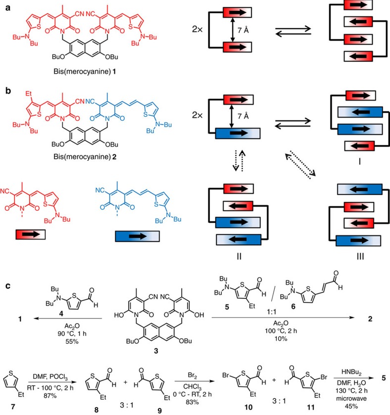
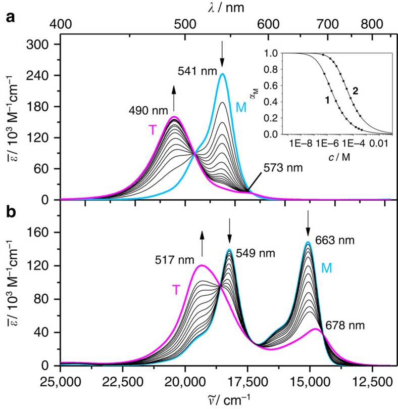
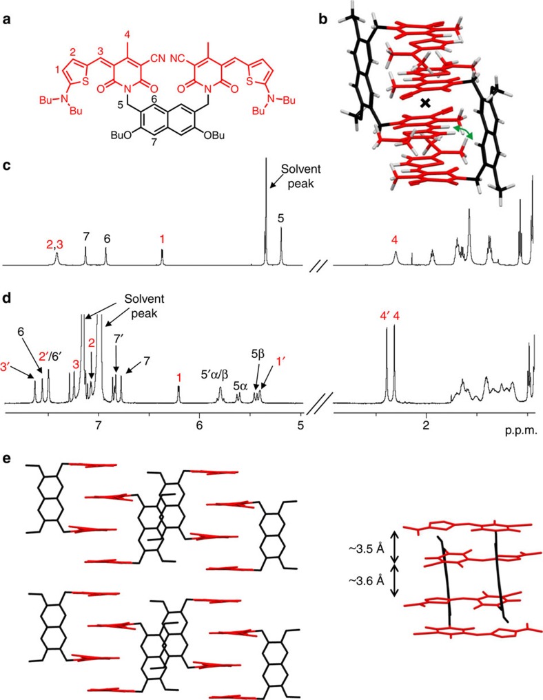
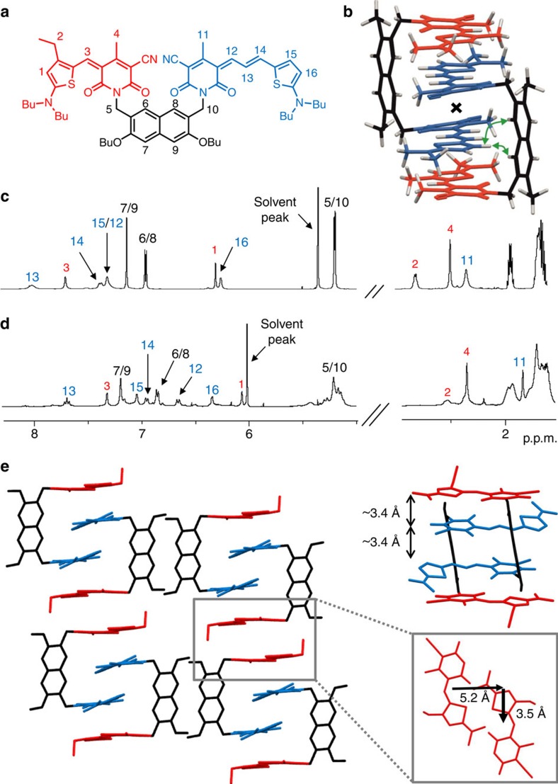
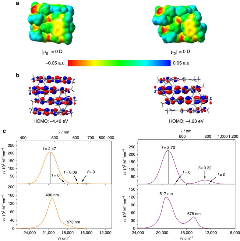
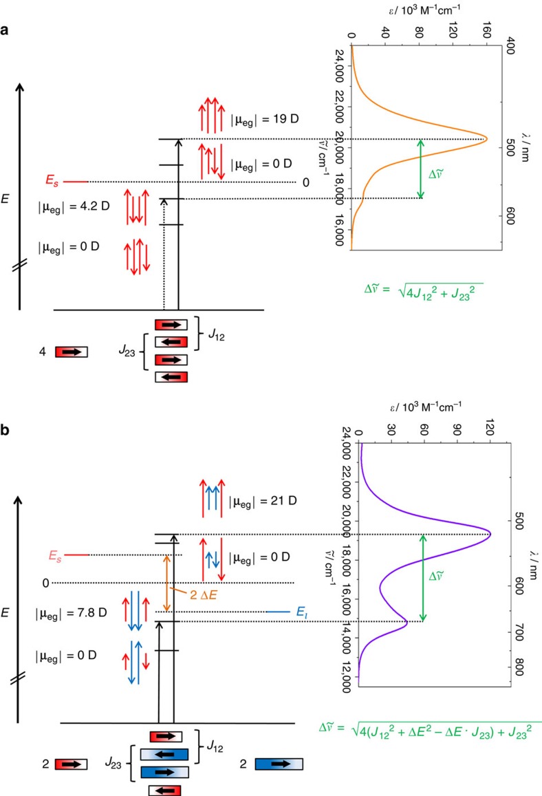
Exciton coupling is of fundamental importance and determines functional properties of organic dyes in (opto-)electronic and photovoltaic devices. Here we show that strong exciton coupling is not limited to the situation of equal chromophores as often assumed. Quadruple dye stacks were obtained from two bis(merocyanine) dyes with same or different chromophores, respectively, which dimerize in less-polar solvents resulting in the respective homo- and heteroaggregates. The structures of the quadruple dye stacks were assigned by NMR techniques and unambiguously confirmed by single-crystal X-ray analysis. The heteroaggregate stack formed from the bis(merocyanine) bearing two different chromophores exhibits remarkably different ultraviolet/vis absorption bands compared with those of the homoaggregate of the bis(merocyanine) comprising two identical chromophores. Quantum chemical analysis based on an extension of Kasha's exciton theory appropriately describes the absorption properties of both types of stacks revealing strong exciton coupling also between different chromophores within the heteroaggregate.


Figures

Figure 1
Figure 1. Chemical structures, synthesis and schematic representation of the self-assembly of bis(merocyanine) dyes.
(a) Chemical structure of bis(merocyanine) 1 with two identical chromophores and its self-aggregation into homodimer stack. (b) Chemical structure of bis(merocyanine) 2 with two different chromophores and its self-aggregation into three possible dimer structures. The bold arrows illustrate ground state dipole moments of the chromophore units. (c) Synthesis of symmetric bis(merocyanine) 1 and unsysmmetric bis(merocyanine) 2, and the literature unknown precursor aminothiophene 5.
Figure 2
Figure 2. UV/vis spectroscopic studies.
Concentration-dependent UV/vis spectra of (a) bis(merocyanine) 1 (c=4.4 × 10⁻⁷–6.0 × 10⁻⁴ M) and (b) bis(merocyanine) 2 (c=5.0 × 10⁻⁷–3.4 × 10⁻⁴ M) in chlorobenzene at 298 K. The arrows indicate the spectral changes upon increasing the concentration. The turquoise (denoted with M) and the magenta lines (denoted with T) represent the calculated spectra of the monomers and the tetrachromophoric (dimer) stacks, respectively, by extrapolation of the spectral data towards most diluted and most concentrated solutions. Inset in a: Plot of fractions of monomeric species αM against concentration and non-linear regression analysis of the data based on the dimerization model for bis(merocyanine) dyes 1 and 2 in chlorobenzene.
Figure 3
Figure 3. NMR studies and single-crystal X-ray analysis of bis(merocyanine) 1.
(a) Chemical structure of 1 with the significant protons numbered. (b) Geometry-optimized structure (B97D3/def2-SVP, butyl chains were replaced by methyl groups) of dimer aggregate. The inversion center of the dimer is indicated with a cross, and the green curved arrow indicates the close proximity of chromophore and backbone protons in the dimer. (c) Relevant sections of 1H NMR (600 MHz) spectrum of 1 monomer in CD2Cl2 (c=1 × 10⁻³ M) at 295 K. (d) Spectrum of aggregate in C6D5Cl (c=1 × 10⁻³ M) at 233 K with the assignment of significant proton signals. The two sets of signals for the aggregate chromophore protons are denoted with and without prime of the respective signals. (e) Molecular packing of bis(merocyanine) 1 in the solid state (side view) with enlargement of the dimer structure motif (front view). Solvent molecules and hydrogen atoms are omitted for clarity and butyl chains were replaced by methyl groups.
Figure 4
Figure 4. NMR studies and single-crystal X-ray analysis of bis(merocyanine) 2.
(a) Chemical structure of 2 with the significant protons numbered. (b) Geometry-optimized structure (B97D3/def2-SVP, alkyl chains were replaced by methyl groups) of dimer aggregate. The inversion center of the dimer is indicated with a cross, and the green curved arrows indicate the close proximity of chromophore and backbone protons in the dimer. (c) Relevant sections of 1H NMR (600 MHz) spectrum of 2 monomer in CD2Cl2 (c=5 × 10⁻³ M) at 295 K. (d) Spectrum of aggregate in a 1:1 mixture of 1,1,2,2-tetrachloroethane-d2/tetrachloromethane (c=8 × 10⁻³ M) at 253 K with the assignment of significant proton signals. (e) Molecular packing of 2 in the solid state (side view) with enlargement of the dimer structure motif (front view). Solvent molecules and hydrogen atoms are omitted for clarity and butyl chains were replaced by methyl groups. A section of the crystal structure (top view) of 2 is shown (e, right) illustrating the lateral and transversal shift of two chromophores of next-neighboured quadruple dye stacks.
Figure 5
Figure 5. Quantum chemical calculation studies.
(a) Electrostatic potential surface (isovalue=0.001 a.u.) and (b) HOMO distribution (isovalue=0.02 a.u.) of the geometry-optimized structures (B97D3, def2-SVP) of the dimers of bis(merocyanine) dye 1 (left) and 2 (right). (c) time-dependent DFT (ωB97/def2-SVP) calculated (top) and experimental dimer spectra (bottom) of bis(merocyanine) 1 (left) and 2 (right). The calculated spectra were shifted by 0.6 eV towards lower energies. The experimental dimer spectra were calculated from the data obtained by concentration-dependent UV/vis studies in chlorobenzene.
Figure 6
Figure 6. Exciton state diagram and UV/vis absorption spectra.
State diagram and spectra of the quadruple dye stacks of (a) bis(merocyanine) 1 and (b) bis(merocyanine) 2. The arrows (red for the short chromophore and blue for the long chromophore, respectively) indicate the phase relations between the corresponding transition dipole moments. The lengths of the arrows reflect the magnitude of the coefficients of the oscillating transition dipole moments. The corresponding experimental dimer spectrum obtained from concentration-dependent UV/vis spectroscopy in chlorobenzene is depicted to indicate the allowed transitions. J12 and J23 denote the exciton coupling energies for the chromophore pairs in the exterior and in the interior of the dye stack, respectively, while Es and El are the excited state energies of the short and long chromophore, respectively.

References

    1. Kasha M., Rawls H. R. & El-Bayoumi M. A. The exciton model in molecular spectroscopy. Pure Appl. Chem. 11, 371–392 (1965).
    1. Davydov A. S. The theory of molecular excitons. Sov. Phys. Usp. 7, 145–178 (1964).
    1. Bhosale R. et al.. Zipper assembly of SHJ photosystems: focus on red naphthalenediimides, optoelectronic finetuning and topological matching. Chem. Sci. 1, 357–368 (2010).
    1. Wasielewski M. R. Self-assembly strategies for integrating light harvesting and charge separation in artificial photosynthetic systems. Acc. Chem. Res. 42, 1910–1921 (2009). - PubMed
    1. Praveen V. K., Ranjith C., Bandini E., Ajayaghosh A. & Armaroli N. Oligo(phenylenevinylene) hybrids and self-assemblies: versatile materials for excitation energy transfer. Chem. Soc. Rev. 43, 4222–4242 (2014). - PubMed

LinkOut - more resources