. 2016 Sep 29:7:12979.
doi: 10.1038/ncomms12979.

Aromatic thiol-mediated cleavage of N-O bonds enables chemical ubiquitylation of folded proteins

Affiliations

Aromatic thiol-mediated cleavage of N-O bonds enables chemical ubiquitylation of folded proteins

Caroline E Weller et al. Nat Commun. .

Abstract

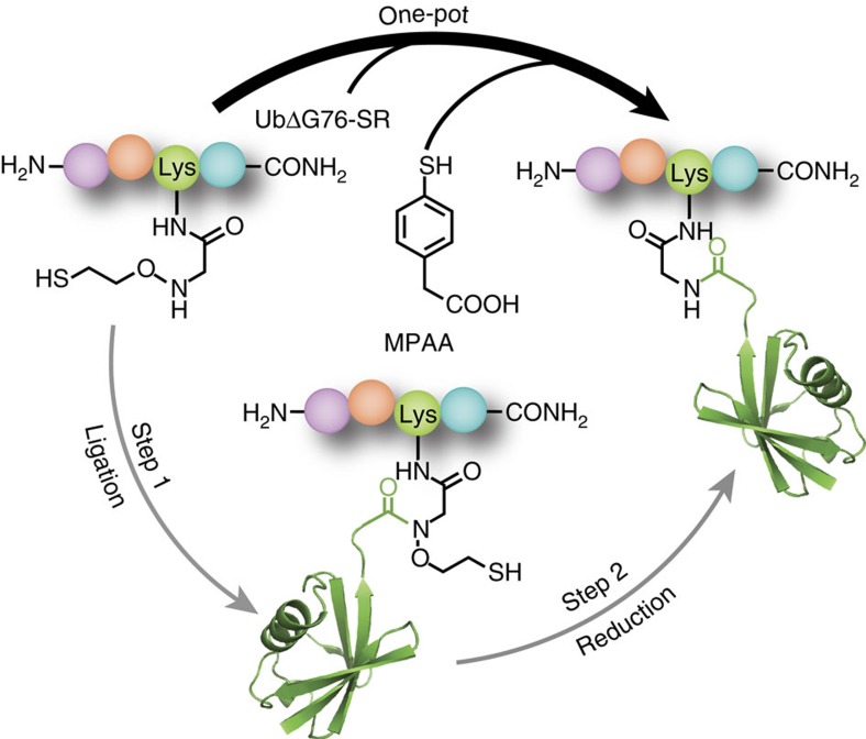
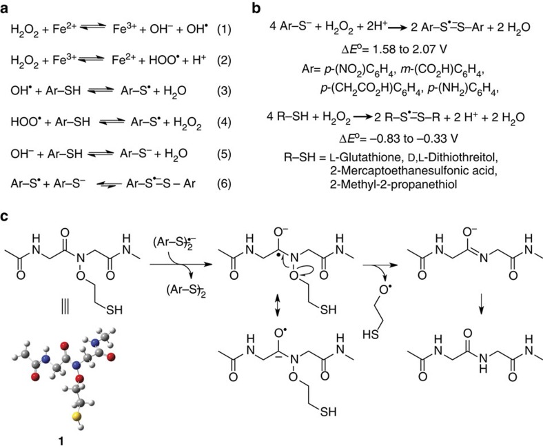
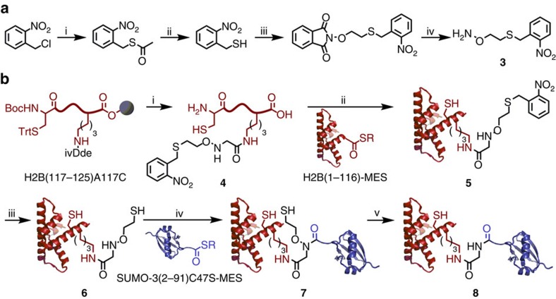
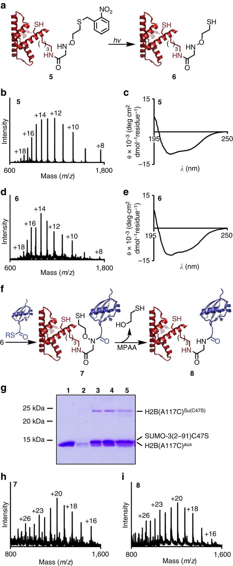
Access to protein substrates homogenously modified by ubiquitin (Ub) is critical for biophysical and biochemical investigations aimed at deconvoluting the myriad biological roles for Ub. Current chemical strategies for protein ubiquitylation, however, employ temporary ligation auxiliaries that are removed under harsh denaturing conditions and have limited applicability. We report an unprecedented aromatic thiol-mediated N-O bond cleavage and its application towards native chemical ubiquitylation with the ligation auxiliary 2-aminooxyethanethiol. Our interrogation of the reaction mechanism suggests a disulfide radical anion as the active species capable of cleaving the N-O bond. The successful semisynthesis of full-length histone H2B modified by the small ubiquitin-like modifier-3 (SUMO-3) protein further demonstrates the generalizability and compatibility of our strategy with folded proteins.


Figures

Figure 1
Figure 1. Aromatic thiol-mediated one-pot traceless native chemical ubiquitylation.
MPAA, 4-mercaptophenylacetic acid; UbΔG76-SR, ubiquitin(1–75)-α-thioester with 2-mercaptoethanesulfonic acid. PDB code 1UBQ (ubiquitin).
Figure 2
Figure 2. N–O bond cleavage in the native chemical ligation product KAKUb(aux)I.
(a) ESI-MS spectrum of the final ligation product of ubiquitin(1–75)-α-thioester with KAKauxI. Calculated for KAKUb(aux)I, 9,081.1 Da. Observed KAKUbI, 9,004.8±2.7 Da. (b) Time-course of N–O bond cleavage and KAKUbI formation from the auxiliary-containing test substrate KAKUb(aux)I in a buffer consisting of 200 mM MPAA, 100 mM NaH2PO4 at pH 7.3 under the indicated conditions. Error bars represent the s.d. from three independent measurements.
Figure 3
Figure 3. EPR spectra of DMPO/·S-Ar adduct.
(a) Spectrum obtained upon incubating 50 mM MPAA and 100 mM DMPO in a 1:1 water-DMF mixture at 25 °C. (b) Computer simulation of the spectrum observed in a with hyperfine splitting constants aN=14.22 G and aH=16.16 G. (c) Spectrum obtained upon incubating 50 mM MPAA and 100 mM DMPO in 50 mM Na2HPO4 at pH 7.5, in a 1:1 water-DMF mixture at 25 °C. (d) ESI-MS spectrum obtained by LC-ESI-MS analysis of the reaction components in c. Inset shows the proposed radical adduct. (e) EPR spectrum obtained upon pre-incubating 50 mM MPAA with 70 mM 2-iodoacetamide for 1.5 h followed by 100 mM DMPO in 50 mM Na2HPO4 at pH 7.5, in a 1:1 water-DMF mixture at 25 °C. Incubation of 50 mM MPAA and 100 mM DMPO in 50 mM Na2HPO4 at pH 7.5, in a 1:1 water-DMF mixture at 25 °C for 1.5 h without the addition of 2-iodoacetamide resulted in a spectrum similar to that seen in c. Spectrometer settings: microwave power, 20 mW; modulation amplitude, 1.0 G; time constant, 163 ms; scan rate, 0.6 G/s.
Figure 4
Figure 4. Formation of disulfide radical anions and their role in N–O bond cleavage.
(a) Production of aromatic thiyl radicals mediated by trace-metal-catalysed Fenton chemistry (1–4) and their combination with aromatic thiolates to form disulfide radical anions (5–6). (b) Net chemical equations for the formation of disulfide radical anions from aromatic thiolates and aliphatic thiols at pH 7.3. The calculated range of standard redox potentials is indicated for compounds from each class of molecules. (c) Proposed mechanism for disulfide radical anion-mediated N–O bond cleavage in the model compound 1.
Figure 5
Figure 5. Semisynthesis of full-length sumoylated histone H2B(A117C).
(a) Synthesis of photoprotected auxiliary 3. (i) CH3C(O)SH, K2CO3, THF, 8 h, 25 °C. (ii) HCl, CH3OH, 6 h, 60 °C, 75% (2 steps). (iii) N-(2-bromoethoxy)phthalimide, Et3N, DMSO, 4 h, 25 °C, 74%. (iv) H2NNH2, CHCl3, 1 h, 25 °C, 98%. (b) (i) Site-specific coupling of 3 to H2B(117–125)A117C Lys120 followed by acidolytic release of the unprotected peptide, 4, from the solid-phase. (ii) Expressed protein ligation of 4 with H2B(1–116)-α-thioester to generate full-length H2B(A117C) with protected auxiliary at Lys120, 5. (iii) photolytic removal of the auxiliary protecting group to give H2B(A117C) with unprotected auxiliary at Lys120, 6. (iv) Expressed protein ligation of 6 with SUMO-3(2–91)C47S-α-thioester to generate sumoylated H2B(A117C) 7, with retention of the ligation auxiliary. (v) Selective removal of the ligation auxiliary with 150 mM MPAA under non-denaturing conditions to yield sumoylated H2B(A117C) 8. ivDde=1-(4,4-Dimethyl-2,6-dioxocyclohexylidene)-3-methylbutyl group. PDB codes, 1KX5 (H2B) and 1U4A (SUMO-3).
Figure 6
Figure 6. Photodeprotection and sumoylation of folded histone H2B.
(a) Scheme depicting photolytic cleavage of the auxiliary protecting group from H2B(A117C)photoaux (5) to generate H2B(A117C)aux (6). (b) ESI-MS spectrum of 5. Calculated for 5, 14,059.2 Da. Observed for 5, 14,062.7±2.8 Da. (c) Circular dichroism spectrum of 5 in 50 mM Na2HPO4, pH 7.5. (d) ESI-MS spectrum of 6. Calculated for 6, 13,924.1 Da. Observed for 6, 13,925.8±2.6 Da. (e) Circular dichroism spectrum of 6 in 50 mM Na2HPO4, pH 7.5. (f) Scheme depicting ligation of 6 to SUMO-3(2–91)C47S-α-thioester to yield H2B(A117C)Su(C47S)aux (7) and subsequent MPAA-mediated auxiliary removal to yield H2B(A117C)Su(C47S) (8) under folded conditions. (g) Coomassie-stained 15% SDS–polyacrylamide gel electrophoresis gel of ligation between H2B(A117C)aux (6) and SUMO3(2–91)C47S-α-thioester under non-denaturing conditions. Lane 1=SUMO-3(2–91)C47S-MES, 2=H2B(A117C)aux, 3=24 h ligation, 4=48 h ligation, 5=24 h MPAA incubation. (h) ESI-MS spectrum of the ligation product, H2B(A117C)Su(C47S)aux (7). Calculated for 7, 24,225.6 Da. Observed for 7, 24,230.9±4.0 Da. (i) ESI-MS spectrum of the final product, H2B(A117C)Su(C47S) (8). Calculated for 8, 24,149.6 Da. Observed for 8, 24,153.1±3.2 Da.

References

    1. Sharp P. M. & Li W. H. Molecular evolution of ubiquitin genes. Trends Ecol. Evol. 2, 328–332 (1987). - PubMed
    1. Van der Veen A. G. & Ploegh H. L. Ubiquitin-like proteins. Annu. Rev. Biochem. 81, 323–357 (2012). - PubMed
    1. Li W. et al.. Genome-wide and functional annotation of human E3 ubiquitin ligases identifies MULAN, a mitochondrial E3 that regulates the organelle's dynamics and signaling. PLoS ONE 3, e1487 (2008). - PMC - PubMed
    1. Komander D. & Rape M. The ubiquitin code. Annu. Rev. Biochem. 81, 203–229 (2012). - PubMed
    1. Danielsen J. M. R. et al.. Mass spectrometric analysis of lysine ubiquitylation reveals promiscuity at site level. Mol. Cell Proteomics 10, M110.003590 (2011). - PMC - PubMed

LinkOut - more resources