. 2016 Sep 29;16(1):760.
doi: 10.1186/s12885-016-2792-1.

Novel biomarkers that assist in accurate discrimination of squamous cell carcinoma from adenocarcinoma of the lung

Affiliations

Novel biomarkers that assist in accurate discrimination of squamous cell carcinoma from adenocarcinoma of the lung

Kazuya Takamochi et al. BMC Cancer. .

Abstract

Background: Targeted therapies based on the molecular and histological features of cancer types are becoming standard practice. The most effective regimen in lung cancers is different between squamous cell carcinoma (SCC) and adenocarcinoma (AD). Therefore a precise diagnosis is crucial, but this has been difficult, particularly for poorly differentiated SCC (PDSCC) and AD without a lepidic growth component (non-lepidic AD). Biomarkers enabling a precise diagnosis are therefore urgently needed.

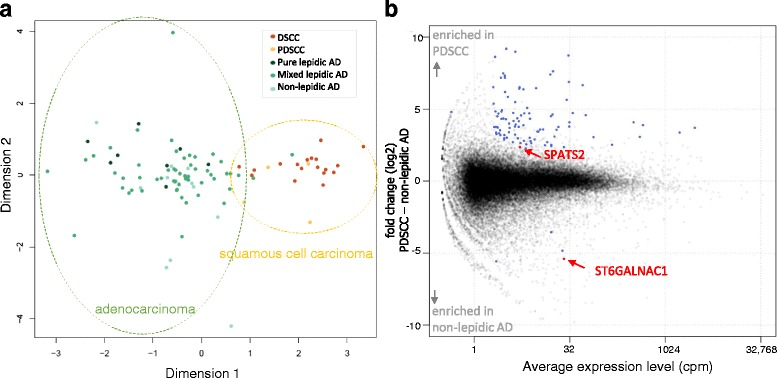
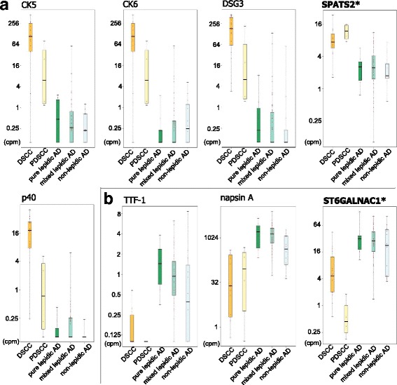
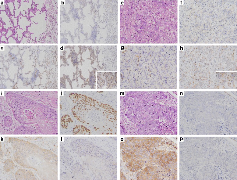
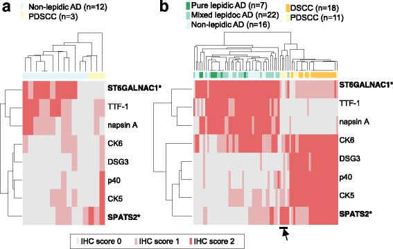
Methods: Cap Analysis of Gene Expression (CAGE) is a method used to quantify promoter activities across the whole genome by determining the 5' ends of capped RNA molecules with next-generation sequencing. We performed CAGE on 97 frozen tissues from surgically resected lung cancers (22 SCC and 75 AD), and confirmed the findings by immunohistochemical analysis (IHC) in an independent group (29 SCC and 45 AD).

Results: Using the genome-wide promoter activity profiles, we confirmed that the expression of known molecular markers used in IHC for SCC (CK5, CK6, p40 and desmoglein-3) and AD (TTF-1 and napsin A) were different between SCC and AD. We identified two novel marker candidates, SPATS2 for SCC and ST6GALNAC1 for AD, as showing comparable performance and complementary utility to the known markers in discriminating PDSCC and non-lepidic AD. We subsequently confirmed their utility at the protein level by IHC in an independent group.

Conclusions: We identified two genes, SPATS2 and ST6GALNAC1, as novel complemental biomarkers discriminating SCC and AD. These findings will contribute to a more accurate diagnosis of NSCLC, which is crucial for precision medicine for lung cancer.


Figures

Fig. 1
Fig. 1
Promoter activities in lung cancer. (a) An MDS plot. Similarities (distances) between individual carcinomas in the space of promoter activities (CAGE profiles) are visualized in two dimensions by the multi-dimensional scaling implemented in the edgeR [20], where individual dots represent individual carcinomas and similar carcinomas are plotted closely. The dot colors represent carcinoma subtypes as indicated in the legend, and the dotted line indicates groups of carcinomas. (b) An MA-plot of the differential analysis between PDSCC and non-lepidic AD. The X-axis represents the average expression levels in cpm, and the Y-axis represents the fold-changes in the log2 scale. Individual dots represent the activities of individual promoters, and the blue dots indicate promoters with statistically significant differences (fold-change > 4, CPM > 4 and FDR < 0.01), and the red dots indicate the marker candidates we selected
Fig. 2
Fig. 2
Promoter activity levels of known markers and novel candidates. (a) The promoter activities of known markers for AD and the novel candidate are shown in boxplots based on the carcinoma subtypes. (b) Equivalent boxplots for known markers of SCC and the candidate
Fig. 3
Fig. 3
IHC for the novel marker candidates. A case of pure lepidic AD (a-d). H.E. staining (a) and IHC for TTF-1 (b), SPATS2 (c) and ST6GALNAC1 (d). The tumor cells are diffusely positive for ST6GALNAC1, but negative for TTF1 and SPATS2. A case of non-lepidic AD (e-h). H.E. staining (e) and IHC for TTF-1 (f), SPATS2 (g) and ST6GALNAC1 (h). The tumor cells are diffusely positive for ST6GALNAC1, but negative for TTF1 and SPATS2. Note that infiltrating plasma cells are also positive for SPATS2 (g). A case of WDSCC (i-l). H.E. staining (i) and IHC for p40 (j), SPATS2 (k) and ST6GALNAC1 (l). The tumor cells are diffusely positive for SPATS2 and p40, but negative for ST6GALNAC1. A case of PDSCC (m-p). H.E. staining (m) and IHC for p40 (n), SPATS2 (o) and ST6GALNAC1 (p). The tumor cells are diffusely positive for SPATS2, but negative for p40 and ST6GALNAC1. Note that SPATS2 staining is more sensitive than p40 staining. (original magnifications: x100, insets: x400)
Fig. 4
Fig. 4
The results of IHC with the novel and known markers. (a) The presence of the known markers and the candidate markers was examined by IHC of carcinoma tissues of non-lepidic AD and PDSCC obtained from the same patients evaluated in the CAGE analysis. The staining patterns are scored (IHC score 0, 1, and 2) as described in the METHODS section, and the scores are visualized as heatmaps, where the tissues and markers are clustered based on the IHC scores. (b) Equivalent heatmaps based on the results of an independent group of patients, consisting of pure lepidic AD and mixed lepidic AD, DSCC, as well as non-lepidic AD and PDSCC

References

    1. Yang P, Allen MS, Aubry MC, Wampfler JA, Marks RS, Edell ES, et al. Clinical features of 5,628 primary lung cancer patients: experience at Mayo Clinic from 1997 to 2003. Chest. 2005;128:452–62. doi: 10.1378/chest.128.1.452. - DOI - PubMed
    1. Scagliotti G, Hanna N, Fossella F, Sugarman K, Blatter J, Peterson P, et al. The differential efficacy of pemetrexed according to NSCLC histology: a review of two Phase III studies. Oncologist. 2009;14:253–63. doi: 10.1634/theoncologist.2008-0232. - DOI - PubMed
    1. Reck M, von Pawel J, Zatloukal P, Ramlau R, Gorbounova V, Hirsh V, et al. Phase III trial of cisplatin plus gemcitabine with either placebo or bevacizumab as first-line therapy for nonsquamous non-small-cell lung cancer: AVAil. J Clin Oncol. 2009;27:1227–34. doi: 10.1200/JCO.2007.14.5466. - DOI - PubMed
    1. Sandler A, Gray R, Perry MC, Brahmer J, Schiller JH, Dowlati A, et al. Paclitaxel-carboplatin alone or with bevacizumab for non-small-cell lung cancer. N Engl J Med. 2006;355:2542–50. doi: 10.1056/NEJMoa061884. - DOI - PubMed
    1. Thunnissen E, Noguchi M, Aisner S, Beasley MB, Brambilla E, Chirieac LR, et al. Reproducibility of histopathological diagnosis in poorly differentiated NSCLC: an international multiobserver study. J Thorac Oncol. 2014;9:1354–62. doi: 10.1097/JTO.0000000000000264. - DOI - PubMed