. 2016 Nov 18;291(47):24465-24474.
doi: 10.1074/jbc.M116.738658. Epub 2016 Sep 28.

Loss of ift122, a Retrograde Intraflagellar Transport (IFT) Complex Component, Leads to Slow, Progressive Photoreceptor Degeneration Due to Inefficient Opsin Transport

Affiliations

Loss of ift122, a Retrograde Intraflagellar Transport (IFT) Complex Component, Leads to Slow, Progressive Photoreceptor Degeneration Due to Inefficient Opsin Transport

Meriam Boubakri et al. J Biol Chem. .

Abstract

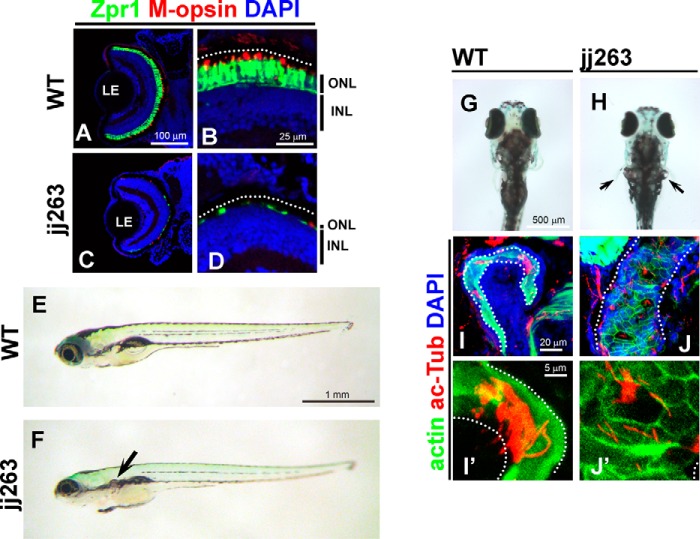
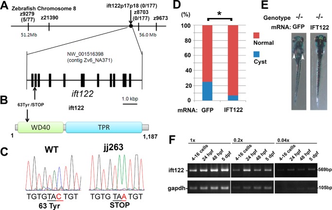
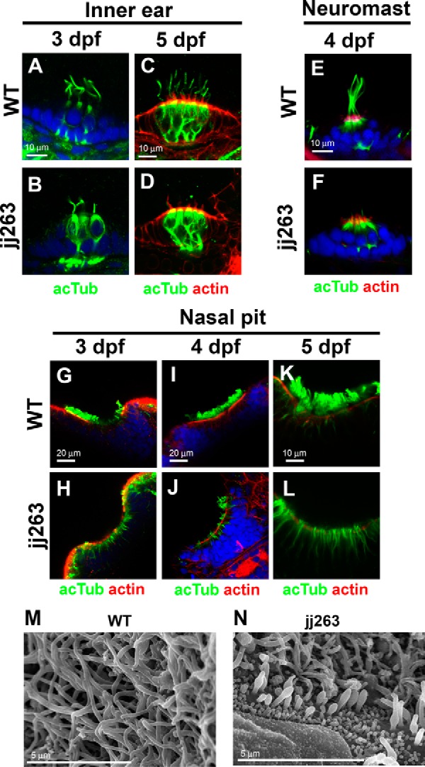
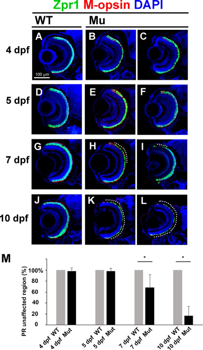
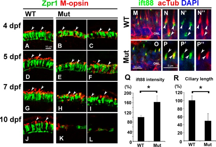
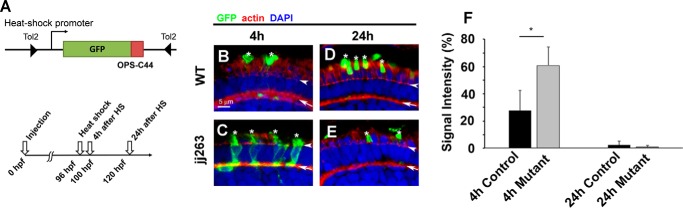
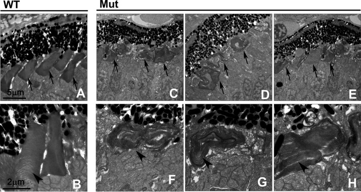
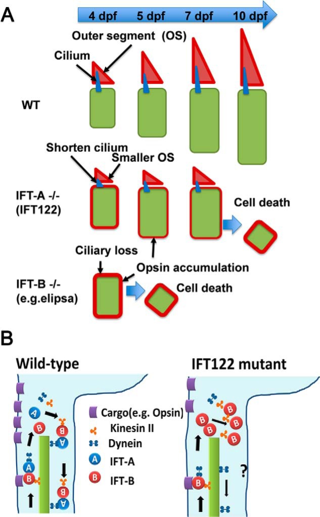
In the retina, aberrant opsin transport from cell bodies to outer segments leads to retinal degenerative diseases such as retinitis pigmentosa. Opsin transport is facilitated by the intraflagellar transport (IFT) system that mediates the bidirectional movement of proteins within cilia. In contrast to functions of the anterograde transport executed by IFT complex B (IFT-B), the precise functions of the retrograde transport mediated by IFT complex A (IFT-A) have not been well studied in photoreceptor cilia. Here, we analyzed developing zebrafish larvae carrying a null mutation in ift122 encoding a component of IFT-A. ift122 mutant larvae show unexpectedly mild phenotypes, compared with those of mutants defective in IFT-B. ift122 mutants exhibit a slow onset of progressive photoreceptor degeneration mainly after 7 days post-fertilization. ift122 mutant larvae also develop cystic kidney but not curly body, both of which are typically observed in various ciliary mutants. ift122 mutants display a loss of cilia in the inner ear hair cells and nasal pit epithelia. Loss of ift122 causes disorganization of outer segment discs. Ectopic accumulation of an IFT-B component, ift88, is observed in the ift122 mutant photoreceptor cilia. In addition, pulse-chase experiments using GFP-opsin fusion proteins revealed that ift122 is required for the efficient transport of opsin and the distal elongation of outer segments. These results show that IFT-A is essential for the efficient transport of outer segment proteins, including opsin, and for the survival of retinal photoreceptor cells, rendering the ift122 mutant a unique model for human retinal degenerative diseases.

Keywords: cilia; hair cell; photoreceptor; retinal degeneration; rhodopsin.


Figures

FIGURE 1.
FIGURE 1.
jj263 mutant larvae exhibited photoreceptor degeneration and cystic kidney without curly body axis phenotype. A–D, jj263 mutant larvae showed photoreceptor degeneration in the retina. Retinal sections at 10 dpf of wild-type (A and B) and jj263 mutant (C and D) larvae were immunostained with anti-Zpr1 (an-arrestin, double cone photoreceptors, green) and anti-M-opsin (a cone outer segment marker, red) antibodies. Nuclei were stained with DAPI (blue). In the wild-type retina, cone photoreceptors are localized in the ONL and form the outer segments on apical sides of the ONL. In the jj263 mutant larvae, photoreceptor cells in the ONL were severely degenerated. In contrast, the INL in the jj263 mutant larvae was almost normal. Dotted lines indicate the boundary of retinal pigment epithelia. E–H, lateral (E and F) and dorsal (G and H) views of wild-type (E and G) and jj263 mutant (F and H) larvae at 6 dpf. A cystic kidney phenotype was observed in the jj263 mutant larvae (F and H, arrow). The curly body axis phenotype, a common feature of ciliary mutants, was not observed in the jj263 mutant larvae. I–J′, pronephric cilia are disorganized and shortened in the jj263 mutant larvae. Pronephric cilia in the wild-type (I and I′) and mutant (J and J′) larvae were immunostained with an acetylated α-tubulin antibody (red). Pronephric epithelia in pronephric tubules were stained with phalloidin (green). Nuclei were stained with DAPI (blue). Dotted lines indicate boundaries of pronephric tubules. ONL, outer nuclear layer; INL, inner nuclear layer; LE, lens.
FIGURE 2.
FIGURE 2.
jj263 locus encodes the zebrafish ift122. A, genetic map of the jj263 locus and the exon/intron structure of the ift122 gene. The ift122 locus was mapped on zebrafish chromosome 8 in the vicinity of a genetic marker Z8703. B, domain structure of the zebrafish ift122 protein and the mutation of the jj263 allele. The N-terminal WD40 repeats and the C-terminal TPR domain were indicated. The position of the ift122jj263 mutation is indicated with an arrow. This mutation produces a premature stop codon resulting in a 62-amino acid truncated product of ift122. C, sequencing trace data for the wild-type (left) and jj263 allele (right). The tyrosine residue at position 63 was changed to stop codon in the jj263 mutant. D and E, rescue by injection of IFT122 mRNA into embryos from crosses between jj263 heterozygotes. D, percentages of larvae that display pronephric cysts at 5 dpf following injection of GFP mRNA or IFT122 mRNA into embryos. E, images of ift122 mutant larvae at 5 dpf following injection of GFP (left panel) or IFT122 mRNA (right panel). Pronephric cyst formation is partially eliminated in homozygous ift122 mutants by injection of IFT122 mRNA (right panel). Arrowheads point to pronephric cysts. F, RT-PCR analysis of ift122 expression in embryos at several developmental stages. Maternal ift122 transcript was detected in embryos at 4–16-cell stage. A housekeeping gene, glyceraldehyde-3-phosphate dehydrogenase (gapdh), was used as a loading control. Three different concentrations of cDNA templates (1×, 0.2×, and 0.04×) were used.
FIGURE 3.
FIGURE 3.
Ciliary defects in sensory organs of the ift122 mutant. A–F, hair cell kinocilium in the inner ear (A–D) and neuromasts (E and F) of wild-type (A, C, and E) and jj263 (B, D, and F) embryos at 3 dpf (A and B), 4 dpf (E and F), and 5 dpf (C and D). Cilia are immunostained with an anti-acetylated α-tubulin antibody (green). Stereocilia of hair cells were stained with phalloidin (actin staining, red). Nuclei were stained with DAPI (blue). Kinocilia of hair cells were disorganized in the ift122 mutant inner ear and neuromast. G–L, cilia in the wild-type (G, I, and K) and ift122 mutant (H, J, and L) nasal pit at 3 dpf (G and H), 4 dpf (I and J), and 5 dpf (K and L). Cilia are immunostained with an anti-acetylated α-tubulin antibody (green). Apical surface of epithelia were labeled with phalloidin (actin staining, red). Nuclei were stained with DAPI (blue). Cilia do not develop in mutant nasal pit at 3 dpf. M and N, ultrastructural analysis of nasal cilia in wild-type and ift122 mutant larvae at 4 dpf. Swollen cilia were observed in the ift122 mutant.
FIGURE 4.
FIGURE 4.
Slow progressive photoreceptor degeneration in the ift122 mutant retina. A–L, ift122 mutant larvae showed slow photoreceptor degeneration in the retina. Retinal sections at 4 dpf (A–C), 5 dpf (D–F), 7 dpf (G–I), and 10 dpf (J–L) of wild-type (A, D, G, and J) and ift122 mutant (B, C, E, F, H, I, K, and L) larvae were stained with anti-Zpr1 (double cone photoreceptors, green), and M-opsin (a cone outer segment marker, red) antibodies. Nuclei were stained with DAPI (blue). No photoreceptor degeneration was observed in the ift122 mutant retina between 4 and 5 dpf. Only small portions of the photoreceptor layer degenerate at 7 dpf (H and I, dotted lines). Most of the photoreceptor layer degenerated by 10 dpf in the ift122 mutant retina (K and L). M, we measured the depth of the degenerated photoreceptor region and calculated the size of the unaffected region. Significant photoreceptor degeneration was observed at 7 and 10 dpf. n = 9–18.
FIGURE 5.
FIGURE 5.
Defect of the opsin transport machinery in the ift122 mutant retina. A–L, photoreceptor cells at 4 dpf (A–C), 5 dpf (D–F), 7 dpf (G–I), and 10 dpf (J–L) in wild-type (A, D, G, and J) and ift122 mutant (B, C, E, F, H, I, K, and L) larvae were immunostained with anti-Zpr1 (green) and anti-M-opsin (red) antibodies. In the 4 dpf wild type, the M-opsin was localized at the outer segments (A). In contrast, the M-opsin signal was widely distributed in photoreceptor cell bodies in ift122 mutant larvae at 4 dpf (B and C). At 5 dpf, ectopic accumulation of M-opsin signal in photoreceptor cell bodies was still observed (E and F), but enrichment of the M-opsin in the outer segment was also observed in mutant photoreceptors. At 7 dpf, the cell bodies of mutant photoreceptors were smaller compared with those of wild-type photoreceptors (H and I). At 10 dpf, only degenerated photoreceptors were observed in most of the photoreceptor layer in the mutant retina (K and L). Arrows indicate outer segments. M–P″, retinal sections were immunostained with an anti-Ift88 antibody (green) in the wild-type (M–N″) and mutant larvae (O–P″) at 4 dpf. Photoreceptor cilia are immunostained with an anti-acetylated α-tubulin antibody (red). Accumulations of the ift88 signals are observed in the photoreceptor cilia of mutant larvae. Q and R, quantification of length and ift88 signal intensity in photoreceptor cilia of ift122 mutant larvae at 4 dpf. Accumulation of ift88 at ciliary tips was significantly increased in ift122 photoreceptor cilia (Q). Photoreceptor cilia stained with acetylated α-tubulin were significantly shorter in ift122 mutant retina (R). Nuclei were stained with DAPI (blue). ONL, outer nuclear layer.
FIGURE 6.
FIGURE 6.
Pulse-chase experiment using GFP-opsin fusion protein in ift122-deficient photoreceptor cells. A, schematic diagram of the heat-shock promoter-driven GFP-opsin expression construct used in this study. Time course of heat-shock induction of GFP-opsin in the larvae (lower panel). B–E, representative confocal images of transverse cryosections through the central retina of wild-type and ift122 mutant larvae at 4 and 24 h after heat-shock induction. The subcellular distribution of the GFP-opsin (green) was evaluated. Sections are counterstained with phalloidin (red) to visualize the outer limiting membrane and the outer plexiform layer and DAPI (blue) to visualize cell nuclei. Signal intensity in cell bodies was measured between the outer limiting membrane and the outer plexiform layer. Asterisks indicate outer segments; arrowheads indicate the outer limiting membrane, and arrows indicate the outer plexiform layer. F, graph representing signal intensities of GFP-opsin in photoreceptor cell bodies of wild-type and ift122 mutant larvae at 4 and 24 h after heat-shock induction. Percentages of GFP signal intensity in the photoreceptor cell body relative to the entire cell are shown.
FIGURE 7.
FIGURE 7.
Ultrastructural analysis of photoreceptor outer segments in the ift122 mutant retina. A–H, electron microscopic images of sections in the wild-type (A and B) and ift122 mutant (C–H) photoreceptor cells at 4 dpf. EM images of sections perpendicular to outer segments in wild-type and mutant photoreceptors. Severely disorganized outer segment discs were observed in mutant photoreceptor cells. Arrows indicate outer segments and arrowheads in enlarged images point to outer segment discs.
FIGURE 8.
FIGURE 8.
Hypothetical model of ift122 function in photoreceptor cell. A, upper panel, at 4 dpf elongation of photoreceptor OS begins in the wild-type retina. Opsin produced in photoreceptor cell bodies is transported to the OS (red triangles) and accumulated in the OS. Middle panel, in ift122 mutant photoreceptors, smaller OS and shorter cilia were observed at 4 dpf. A certain amount of opsin is transported to the OS; however, probably because of insufficient transport capacity of IFT, opsins are accumulated in the photoreceptor cell bodies. The moderate ectopic opsin accumulation causes the late onset photoreceptor cell death around 7–10 dpf. Lower panel, in the IFT complex B mutant retina (e.g. elipsa) photoreceptors, cilia, and OS are not formed, because the anterograde IFT system is completely disrupted. Opsin accumulates in the photoreceptor cell bodies in the IFT-B mutant retina. The ectopic opsin accumulation causes early photoreceptor cell death around 5 dpf. B, left panel, in wild-type photoreceptor cells, the IFT complex B mediates efficient cargo transport from inner segments to outer segments along ciliary axonemes and are recycled by retrograde transport machinery. Right panel, in ift122 mutant photoreceptors, the IFT complex B is accumulated in the tip of axonemes of connecting cilia due to the loss of IFT recycling. Even under such conditions, certain amount of cargos are transported to outer segments and small outer segments are formed in ift122 mutant photoreceptors.

References

    1. Fliegauf M., Benzing T., and Omran H. (2007) When cilia go bad: cilia defects and ciliopathies. Nat. Rev. Mol. Cell Biol. 8, 880–893 - PubMed
    1. Gerdes J. M., Davis E. E., and Katsanis N. (2009) The vertebrate primary cilium in development, homeostasis, and disease. Cell 137, 32–45 - PMC - PubMed
    1. Nigg E. A., and Raff J. W. (2009) Centrioles, centrosomes, and cilia in health and disease. Cell 139, 663–678 - PubMed
    1. Kennedy B., and Malicki J. (2009) What drives cell morphogenesis: a look inside the vertebrate photoreceptor. Dev. Dyn. 238, 2115–2138 - PMC - PubMed
    1. Rosenbaum J. L., and Witman G. B. (2002) Intraflagellar transport. Nat. Rev. Mol. Cell Biol. 3, 813–825 - PubMed