. 2016 Oct 11;113(41):E6248-E6255.
doi: 10.1073/pnas.1605575113. Epub 2016 Sep 28.

Functional receptor molecules CD300lf and CD300ld within the CD300 family enable murine noroviruses to infect cells

Affiliations

Functional receptor molecules CD300lf and CD300ld within the CD300 family enable murine noroviruses to infect cells

Kei Haga et al. Proc Natl Acad Sci U S A. .

Abstract

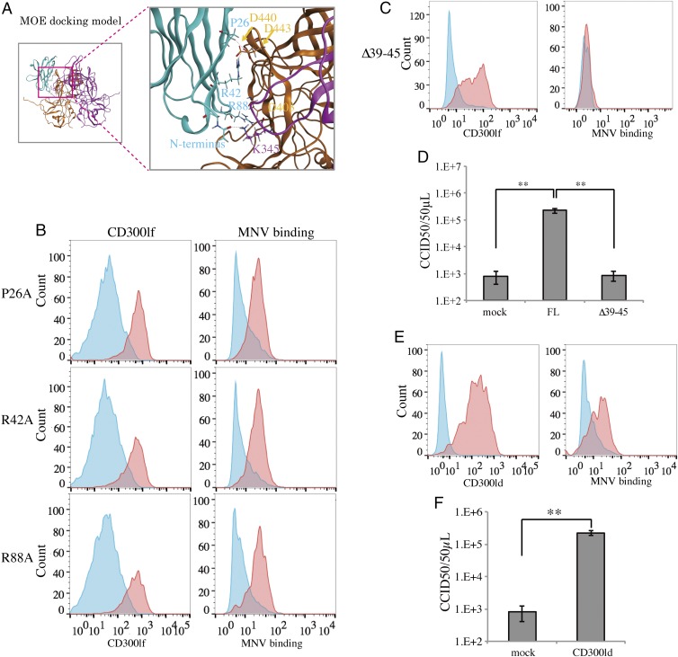
Norovirus is the leading cause of acute gastroenteritis worldwide. Since the discovery of human norovirus (HuNoV), an efficient and reproducible norovirus replication system has not been established in cultured cells. Although limited amounts of virus particles can be produced when the HuNoV genome is directly transfected into cells, the HuNoV cycle of infection has not been successfully reproduced in any currently available cell-culture system. Those results imply that the identification of a functional cell-surface receptor for norovirus might be the key to establishing a norovirus culture system. Using a genome-wide CRISPR/Cas9 guide RNA library, we identified murine CD300lf and CD300ld as functional receptors for murine norovirus (MNV). The treatment of susceptible cells with polyclonal antibody against CD300lf significantly reduced the production of viral progeny. Additionally, ectopic CD300lf expression in nonsusceptible cell lines derived from other animal species enabled MNV infection and progeny production, suggesting that CD300lf has potential for dictating MNV host tropism. Furthermore, CD300ld, which has an amino acid sequence in the N-terminal region of its extracellular domain that is highly homologous to that of CD300lf, also functions as a receptor for MNV. Our results indicate that direct interaction of MNV with two cell-surface molecules, CD300lf and CD300ld, dictates permissive noroviral infection.

Keywords: CD300 molecules; CD300ld; CD300lf; murine norovirus; norovirus proteinaceous receptors.


Conflict of interest statement

The authors declare no conflict of interest.

Figures

Fig. 1.
Fig. 1.
CD300lf depletion decreases MNV production in RAW264.7 cells. (A) Microscopic images of RAW264.7 cells taken 48 h post inoculation with (+) or without (−) MNV. (Upper) Differential interference contrast (DIC) images. (Lower) Fluorescent images after Hoechst 33342 staining. The cells were transduced with a lentiviral gRNA library targeting sites throughout the murine genome (library) or with a lentiviral vector carrying gRNA specific to CD300lf (CD300lf KO). (B) Flow cytometric analysis of RAW/Cas9 cells transduced with lentiviral vector carrying the library (Left) or CD300lf KO (Right). Red and blue histograms indicate the cells that reacted or did not react with anti-CD300lf polyclonal antibody (α-mCD300lf), respectively. (C) MNV production in cells transfected with the library or CD300lf KO. Stably transfected cells were infected with MNV (MOI of 1) and incubated for 48 h. The titer is shown on a logarithmic scale. (D) Detection of VP1 and RdRp in MNV-infected RAW264.7 cells transfected with the library or CD300lf KO. Actin was used as a loading control. (E) MNV-S7 production in RAW264.7 cells. The cells were untreated or pretreated with neuraminidase (Neu; 50 mU/mL), antibodies to murine CD300lf (α-mCD300lf) or human CD300f (α-hCD300lf), or combinations thereof for 2 h at 37 °C. After pretreatment, the cells were infected with MNV (MOI of 0.05). The MNV titer in the cell supernatant at 48 hpi is shown on a logarithmic scale. Error bars represent the SD from three wells for each sample (**P < 0.01).
Fig. S1.
Fig. S1.
Treatment with neuraminidase or anti-mCD300lf antibody did not attenuate the MNV binding to RAW264.7 cells. (A) MNV binding to RAW264.7 cells treated with neuraminidase or goat anti-mCD300lf antibody. RAW264.7 cells were treated with [MNV(+) Neu; orange histogram] or without [MNV(+) untreated; red histogram] neuraminidase (50 mU/mL) or goat polyclonal antibodies to murine CD300lf [200 ng/2 × 105 cells; MNV(+) α-mCD300lf; green histogram] for 2 h at 37 °C. After washing, the cells were incubated with MNV (10 µg) for 30 min at 4 °C. MNV binding was detected by using rabbit anti-MNV polyclonal antibody, and PE-conjugated donkey anti-rabbit IgG was used as secondary antibody. (B) Confirmation of glycosidase activity. Fetuin (10 µg), a glycoprotein containing sialylated N-linked and O-linked glycans that is generally used as a positive control for endoglycosidase enzymes, was treated with or without neuraminidase (50 mU/mL) for 2 h at 37 °C. Deglycosylation of fetuin was confirmed by SDS-PAGE/Coomassie Brilliant Blue (CBB) staining analysis.
Fig. S2.
Fig. S2.
Anti-mCD300lf polyclonal antibody inhibits the production of MNV-1 progeny. MNV-1 production in RAW264.7 cells that were untreated or pretreated with neuraminidase (Neu; 50 mU/mL), antibodies to murine CD300lf (α-mCD300lf) or human CD300lf (α-hCD300lf), or a combination thereof. The cells were pretreated with the inhibitors for 2 h at 37 °C and then infected with MNV (MOI of 0.05). The MNV titer in the cell supernatant at 48 hpi is shown on a logarithmic scale. Error bars represent the SD from three wells for each sample (**P < 0.01).
Fig. 2.
Fig. 2.
HEK293T cells expressing CD300lf are susceptible to MNV infection. Flow cytometry was performed with HEK293T cells mock transfected or transduced with a CD300lf expression vector. (A) Immunofluorescence staining with [α-mCD300lf(+); red histogram] or without [α-mCD300lf(−); blue histogram] anti-CD300lf antibody. (B) The cells were incubated in the absence [MNV(−); blue histogram] or presence [MNV(+); red histogram] of MNV. The cells bound to the MNV were immunostained by using an anti-MNV VP1 antibody. The x-axes represent the extent of CD300lf expression (A) and MNV binding (B). (C) Detection of VP1 and RdRp after MNV infection in the cells described earlier. Actin was evaluated as the loading control. (D) MNV production in HEK293T/CD300lf cells. The MNV titer in the supernatant of infected cells (MOI of 1) is shown. (E) Mammalian cell lines CRFK, CHO, COS7, and NIH 3T3 transduced with CD300lf became susceptible to MNV. Flow cytometry of the four cell lines was performed to measure CD300lf expression. Red and blue histograms indicate CD300lf-transduced and mock cells, respectively. (F) MNV production in CD300lf-expressing cell lines was examined by the detection of RdRp and VP1 expression by Western blotting. (G) CCID50/50 μL of viral progeny produced from each CD300lf-expressing cell line at 48 hpi. Error bars represent the SD from three wells for each sample.
Fig. 3.
Fig. 3.
The N-terminal region of CD300lf is important for MNV infection in HEK293T cells. (A) HEK293T cells cotransduced with GFP and FL, ∆130–170, ∆Cterm, Δ18–51, or Δcpd were examined for CD300lf expression (Left) and MNV binding (Right). (Left) Overlay histograms of the GFP-gated cells expressing [αCD300lf(+); red histogram] or not expressing [αCD300lf(−); blue histogram] CD300lf. (Right) Overlay histograms of the MNV binding [GFP(+), red histogram; GFP(−), blue histogram]. (B) HEK293T cells expressing the CD300lf variants or no construct (mock) were infected with MNV. Viral protein expression was analyzed by Western blotting. RdRp and VP1 in CD300lf cells expressing FL, ∆130–170, ∆Cterm, Δ18–51, or Δcpd were probed with anti-RdRp and anti-VP1 antibodies, respectively. Actin was used as a loading control. (C) MNV titer in the supernatant of HEK293T cells expressing FL, ∆130–170, ∆Cterm, Δ18–51, or Δcpd at 48 hpi. Error bars represent the SD from three wells for each sample (**P < 0.01).
Fig. S3.
Fig. S3.
Schematic diagrams of CD300lf and four mutant constructs. The constructs encoding (A) FL CD300lf cDNA coding region and the deletion mutants (B) Δ130–170, (C) ΔCterm, (D) Δ18–51, and (E) Δcpd are shown.
Fig. S4.
Fig. S4.
Amino acid sequence alignment of FL CD300lf and three of the deletion-mutant constructs. Dashed lines represent deleted amino acid residues. The alignment was produced using ClustalW and GENETYX version 18.
Fig. 4.
Fig. 4.
Docking model of the MNV-S7 capsid P-domain dimer and the CD300lf extracellular domain. (A) The docking simulation was performed by using the Dock application in MOE software. The overall structure of the docking model (Left) and an enlarged view of the interface of MNV-S7 and CD300lf (Right) are shown. The orange and magenta ribbons and the cyan ribbon are MNV-S7 and CD300lf, respectively. The colored sticks show the residues that form the hydrogen bonds (gold dotted line) or ionic interactions between MNV-S7 and CD300lf. (B, C, and E) HEK293T cells cotransduced with GFP and CD300lf mutants were analyzed for CD300lf expression (Left) and MNV binding (Right). (Left) Overlaid histograms of the GFP-gated cells expressing [αCD300lf(+); red histogram] and not expressing [αCD300lf(−); blue histogram] CD300lf. (Right) Overlaid histograms of the MNV binding [GFP(+), red histogram; GFP(−), blue histogram]. (D and F) MNV titer in the supernatant of HEK293T cells mock-transduced or transduced with the constructs expressing ∆39–45 or CD300ld at 48 hpi. Error bars represent the SD from three wells for each sample (**P < 0.01).
Fig. S5.
Fig. S5.
Amino acid sequence of CD300lf indicated with secondary structure and 3D structure. The open rectangle, shadowed rectangle, gray letters, underlines, and bold letters represent the signal peptide, transmembrane domain, cytoplasmic domain, β-chain, and 3/10 helix, respectively. The CD300lf 3D structure was reconstructed from the PDB file (ID code 1ZOX). The α-helices and β-chains are colored purple and yellow, respectively.
Fig. S6.
Fig. S6.
Cryo-EM image of MNV-S7 and model fitting. A cryo-EM map of the MNV-S7 infectious particle was reconstructed at 7.9-Å resolution. The capsid structure of the C/C dimer was clipped. The crystal structure of MNV 1 protruding domain (PDB ID code 3LQ6) was fitted into the cryo-EM map of the C/C dimer using “fit in map” in University of California, San Francisco, Chimera (30).
Fig. S7.
Fig. S7.
Amino acid sequence alignment between CD300lf and CD300ld. The amino acid sequences of mouse CD300lf (AB292061), CD300lf (LC131461), and CD300ld (LC132714) are shown with known motifs and secondary structure highlighted. The open rectangular box shows the signal peptide. The green and red letters show β-chains and 3/10 helices, respectively. Asterisks represent identical amino acid residues among the three proteins. Dashes indicate gaps.

References

    1. Kapikian AZ, et al. Visualization by immune electron microscopy of a 27-nm particle associated with acute infectious nonbacterial gastroenteritis. J Virol. 1972;10(5):1075–1081. - PMC - PubMed
    1. Jones MK, et al. Enteric bacteria promote human and mouse norovirus infection of B cells. Science. 2014;346(6210):755–759. - PMC - PubMed
    1. Ruvoën-Clouet N, Ganière JP, André-Fontaine G, Blanchard D, Le Pendu J. Binding of rabbit hemorrhagic disease virus to antigens of the ABH histo-blood group family. J Virol. 2000;74(24):11950–11954. - PMC - PubMed
    1. Hutson AM, Atmar RL, Graham DY, Estes MK. Norwalk virus infection and disease is associated with ABO histo-blood group type. J Infect Dis. 2002;185(9):1335–1337. - PubMed
    1. Kambhampati A, Payne DC, Costantini V, Lopman BA. Host genetic susceptibility to enteric viruses: A systematic review and metaanalysis. Clin Infect Dis. 2016;62(1):11–18. - PMC - PubMed

Publication types

MeSH terms